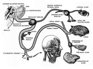
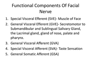
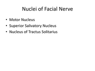
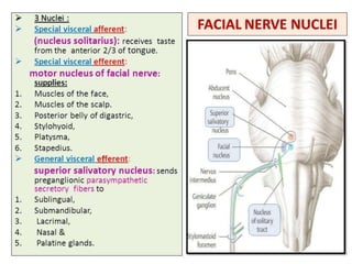
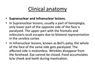
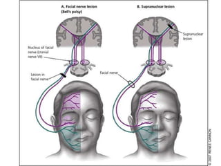
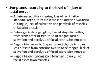
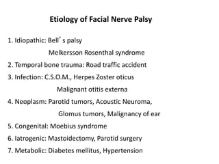
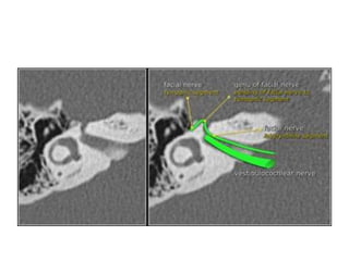
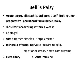
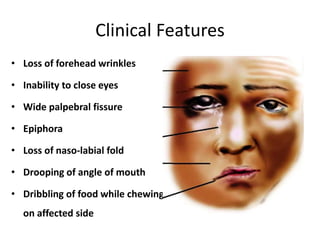
Surgical Anatomy of
Facial Nerve
Otology could be a dull way of life without the seventh
cranial nerve arrogantly swerving through the temporal
bone to the muscles of facial expression.
John Groves
Functional Components Of Facial
Nerve
1. Special Visceral Efferent (SVE)- Muscle of Face
2. General Visceral Efferent (GVE)- Secretomotor to
Submandibular and Sublingual Salivary Gland,
the Lacrimal gland, gland of nose, palate and
pharynx.
3. General Visceral Afferent (GVA)
4. Special Visceral Afferent (SVA)- Taste Sensation
5. General Somatic Afferent (GSA)
Nuclei of Facial Nerve
• Motor Nucleus
• Superior Salivatory Nucleus
• Nucleus of Tractus Solitarius
Courses and Relation
Supranuclear Anatomy
• Cortex and internal capsule:
Voluntary responses of the facial
muscles arises from efferent
discharge from motor face area
of the cerebral cortex.
• The motor face area is situated
on the pre central and post central
gyrus.
7
8
•Discharges from the facial motor
area are carried through fascicles
of the Corticobulbar tract to the
internal capsule, then through the
upper midbrain to the lower
brainstem, where they synapse in
the facial nerve nucleus located in
pons.
Posterior view of the brainstem
Intracranial
• The Intracranial portion of the facial nerve runs from the
brain stem to the fundus of internal auditory meatus.
• Length of Intracranial Segment is Approx. 24 mm
• Facial Nerve attaches to brain stem by 2 roots- Motor and
Sensory
• Sensory Root is also called Nervus Intermedius
• The two roots of facial nerve are attached to the lateral
part of lower border of pons just medial to eighth cranial
nerve.
• The two roots run laterally and forward, with the eight
nerve to reach the Internal Acoustic Meatus.
• Facial Nerve in intracranial course is covered only by a
thin layer of pia mater and not by perineurium.
• The lack of perineurium makes it extremely vulnerable
in cerebellopontine angle tumor surgery.
• The facial nerve enters the foramen of the IAM in its
anterosuperior segment and runs a distance of 5-12
mm to the fundus.
• Crista falciformis- Horizontal Plane
• Bill’s Bar- Septum between Facial Nerve and Superior
Vestibular Nerve
Intratemporal Segment
• It runs from the entrance of the facial
(Fallopian) canal at the fundus of the IAM to
the stylomastoid foramen.
• The length of this portion is 28-30 mm.
• It’s divided into 3 parts-
– Labyrinthine
– Tympanic
– Mastoid
Labyrinthine Segment
• It’s the shortest (3-5 mm) and thinnest part of the facial nerve
within the fallopian canal.
• Narrowest part is at its entrance (0.68mm diameter)
• Periosteum is thicker here than in rest of facial canal
• There are no anastomosing arterial arcades in this area and it is the
part of the facial nerve most vulnerable to ischemia.
• During the trans labyrinthine approach the labyrinthine segment is
at risk while drilling alone the superior semicircular canal
• The ampullated ends of the superior and lateral semicircular canals
are identified and are known as the cat’s eyes
• Labyrinthine segment is also that part of the facial nerve most likely
to be injured in temporal bone fracture
Tympanic Segment
• The Tympanic segment of facial nerve (8-11 mm)
is straight and lies beneath the lateral
semicircular canal and oval window
• Proximal end passes just above and medial to
cochleariform process and tensor tympani
tendon.
• Cochleariform process is consistent landmark in
identifying facial nerve
• Tympanic Segment is vulnerable in middle ear
surgery, especially around oval window
Mastoid Segment
• It start from second genu to stylomastoid foramen
• The pyramidal eminence is another useful landmark for
the second genu where the facial nerve make the sharp
turn downward
• A useful landmark for the course of the mastoid
segment is the digastric ridge
• The Mastoid Segment of nerve has three branches-
– Nerve to Stapedius Muscle
– Chorda Tympani Nerve
• It’s used as landmark when performing posterior tympanotomy
and serves as lateral margin of facial recess
– Sensory Auricular Branch
Extra cranial Segment
• Once the nerve exits from the stylomastoid
foramen, it lies below the tympanic plate and
lateral to the base of styloid
• It gives 3 branches after coming out from
stylomastoid foramen-
– Posterior Auricular Nerve- passes upward and behind
the ear to supply occipital belly of occipitofrontalis.
– Nerve to Stylohyoid
– Nerve to Posterior Belly of Digastric
• The main trunk of the facial nerve divides into
five major division in Parotid Gland-
– Temporal
– Zygomatic
– Buccal
– Marginal Mandibular
– Cervical
Temporal branch
• Emerge from the parotid
gland at its upper pole
slightly in front of the
superficial temporal
artery
• Anterior temporal :
frontalis, superior part of
orbicularis oculi,
corrugator supercilii,
procerus
• Posterior temporal :
anterior and superior
auricular muscles
Zygomatic branch
• Leave the parotid
gland on its
anterosuperior border
• Crosses the body of
Zygomatic bone
• Supply part of
orbicularis oculi
Buccal branch
• Emerge at the anterior
border of parotid
• Upper Buccal: muscles of
upper lip and the muscles
of the nose
• Lower Buccal : Buccinator
and Risorius
• Orbicularis Oris
• It run parallel and 1 cm
below the Zygomatic arch
and often along inferior
aspect of parotid duct
Marginal mandibular
• Runs parallel to lower
border of the mandible
• Cross Facial vein and
Facial artery
• Supplies muscles of lower
lip (Depressor anguli oris
and Depressor labii
inferioris) and mental
muscles
• Located 1-2 cm below the
inferior ramus of
mandible
Cervical
Supplies Platysma
For middle ear and mastoid surgery
1. Processes cochleariform: small bony
protuberance, geniculate ganglion
anterior
2. Short process of Incus: nerve medial
3. Lateral/Horizontal SCC: nerve runs
below
4. Oval window: above the oval window
5. Pyramid: nerve runs behind
6. Tympanomastoid suture: nerve runs
behind
7. Digastric ridge: nerve at anterior end
Surgical landmarks of facial nerve
For parotid surgery:
1. Tympano-mastoid suture: 6-8 mm
deep to this suture
2. Groove between mastoid &
bony EAC: bisected by facial nerve
3. Tragal pointer: 1 cm antero-infero-
medial is facial nerve
4. Styloid process: lateral lies facial
nerve
5. Posterior belly of digastric:
superior & parallel lies facial nerve
Clinical anatomy
• Supranuclear and Infranuclear lesions.
• In Supranuclear lesions; usually a part of hemiplegia,
only lower part of the opposite side of the face is
paralyzed. The upper part with the frontalis and
orbicularis oculi escapes due to bilateral representation
in the cerebra cortex
• In Infranuclear lesions, known as Bell’s palsy, the whole
of the face of the same side gets paralyzed. The
affected side is motionless. Wrinkles disappear from
the forehead. Eye cannot be closed. Food accumulates
b/w cheek and teeth during mastication.
• Symptoms according to the level of injury of
facial nerve-
– At internal auditory meatus; loss of lacrimation,
stapedial reflex, taste from most of anterior two-third
of tongue, lack of salivation and paralysis of muscles
of facial expression
– Below geniculate ganglion; loss of stapedial reflex,
taste from anterior two-third of tongue, lack of
salivation and paralysis of facial expression muscles
– Region b/w nerve to Stapedius and chorda tympani :
loss of taste from anterior two-third of tongue, lack of
salivation and paralysis of facial expression muscles.
– Region below stylomastoid foramen : paralysis of
facial expression muscles.
1. Idiopathic: Bell’s palsy
Melkersson Rosenthal syndrome
2. Temporal bone trauma: Road traffic accident
3. Infection: C.S.O.M., Herpes Zoster oticus
Malignant otitis externa
4. Neoplasm: Parotid tumors, Acoustic Neuroma,
Glomus tumors, Malignancy of ear
5. Congenital: Moebius syndrome
6. Iatrogenic: Mastoidectomy, Parotid surgery
7. Metabolic: Diabetes mellitus, Hypertension
Etiology of Facial Nerve Palsy
Grade Name Characteristics
I Neuropraxia Partial block of axoplasm
II Axonotemesis Injury to axon
III Neurotemesis Injury to endoneurium or
myelin sheath
IV Partial
transection
V Complete
transection
Sunderland’s Classification (1951)
Grade Description Characteristics
I Normal Normal facial function
II Mild dysfunction Slight weakness seen only on
close inspection
III Moderate dysfunction Obvious asymmetry; complete
eye closure
IV Moderately severe
dysfunction
Obvious asymmetry;
incomplete eye closure
V Severe dysfunction Only minimal motion seen;
asymmetry at rest
VI Total paralysis No movement
House Brackmann Classification (Post-Injury)
Diagnosis
• Topodiagnostic Tests
• Electrophysiological Test
– Minimum Nerve Excitability (NET)
– Maximum Stimulation Test (MST)
– Electroneuronography (ENoG)
– Electromyography (EMG)
• CT scan Temporal Bone
• MRI brain
• Surgical exploration
Topodiagnostic tests
• Topodiagnostic evaluation refers to the functional
testing of an individual facial nerve branch in an
attempt to locate the level of dysfunction or injury
– Schirmer Test
– Stapedial Reflex
– Electrogustometry
– Salivary Flow Testing
Schirmer test
• Greater Superficial Petrosal
Nerve assessed
• Strip of paper are placed in
inferior conjunctival fornix
for 5 min and the length of
paper moistened is
compared between eyes
• >75% unilateral decrease
in lacrimation, or a
bilateral decrease in
lacrimation (less than 10
mm for both side at 5 min)
Stapedial Reflex
• Nerve to Stapedius
Muscle Assessed
• Psychoacoustic
Audiometry
Electrogustometry
• Chorda Tympani is
assessed
• The tongue is
stimulated electrically
to produce a metallic
taste and the two sides
are compared
• Threshold of the test is
compared between
sides
Salivary flow Testing
• Chorda Tympani is
assessed
• Warthin’s ducts are
cannulated and salivary
flow is measured over time
following a gustatory
stimulus
• Reduction of 25% is
considered abnormal
Electrophysiological Test
• Minimum Nerve Excitability Test (NET)
– Compares transcutaneous current threshold required
to elicit minimal muscle contraction between 2 sides
• Maximal Stimulation Test (MST)
– Compares muscle contraction at maximal nerve
stimulation between two sides
• Electroneuronography (ENoG)
– Between 3 days to 3 weeks
• Electromyography (EMG)
CT Scan
Magnetic Resonance Imaging
Bell’s Palsy
• Acute onset, idiopathic, unilateral, self-limiting, non-
progressive, peripheral facial nerve palsy
• 85% start recovering within 3 weeks
• Etiology:
1. Viral: Herpes simplex, Herpes Zoster
2. Ischemia of facial nerve: exposure to cold,
emotional stress, nerve compression
3. Hereditary 4. Autoimmune
Clinical Features
• Loss of forehead wrinkles
• Inability to close eyes
• Wide palpebral fissure
• Epiphora
• Loss of naso-labial fold
• Drooping of angle of mouth
• Dribbling of food while chewing
on affected side
Treatment
• Prednisolone (1mg/kg in 2 doses): for 2 - 3 weeks
• Acyclovir: 200-400 mg 5 times per day X 7days
• Eye care: Voluntary closure @ 2 / min. Ciplox eye drops 2
hourly & ointment H.S. Eye cover at night.
• Physiotherapy: moist heat + facial massage + facial
muscle exercise
• Electrical stimulation of facial nerve & muscle
• Facial nerve decompression: Controversial

Facial nerve

  • 1.
    Surgical Anatomy of FacialNerve Otology could be a dull way of life without the seventh cranial nerve arrogantly swerving through the temporal bone to the muscles of facial expression. John Groves
  • 3.
    Functional Components OfFacial Nerve 1. Special Visceral Efferent (SVE)- Muscle of Face 2. General Visceral Efferent (GVE)- Secretomotor to Submandibular and Sublingual Salivary Gland, the Lacrimal gland, gland of nose, palate and pharynx. 3. General Visceral Afferent (GVA) 4. Special Visceral Afferent (SVA)- Taste Sensation 5. General Somatic Afferent (GSA)
  • 4.
    Nuclei of FacialNerve • Motor Nucleus • Superior Salivatory Nucleus • Nucleus of Tractus Solitarius
  • 6.
  • 7.
    Supranuclear Anatomy • Cortexand internal capsule: Voluntary responses of the facial muscles arises from efferent discharge from motor face area of the cerebral cortex. • The motor face area is situated on the pre central and post central gyrus. 7
  • 8.
    8 •Discharges from thefacial motor area are carried through fascicles of the Corticobulbar tract to the internal capsule, then through the upper midbrain to the lower brainstem, where they synapse in the facial nerve nucleus located in pons. Posterior view of the brainstem
  • 9.
    Intracranial • The Intracranialportion of the facial nerve runs from the brain stem to the fundus of internal auditory meatus. • Length of Intracranial Segment is Approx. 24 mm • Facial Nerve attaches to brain stem by 2 roots- Motor and Sensory • Sensory Root is also called Nervus Intermedius • The two roots of facial nerve are attached to the lateral part of lower border of pons just medial to eighth cranial nerve. • The two roots run laterally and forward, with the eight nerve to reach the Internal Acoustic Meatus.
  • 10.
    • Facial Nervein intracranial course is covered only by a thin layer of pia mater and not by perineurium. • The lack of perineurium makes it extremely vulnerable in cerebellopontine angle tumor surgery. • The facial nerve enters the foramen of the IAM in its anterosuperior segment and runs a distance of 5-12 mm to the fundus. • Crista falciformis- Horizontal Plane • Bill’s Bar- Septum between Facial Nerve and Superior Vestibular Nerve
  • 13.
    Intratemporal Segment • Itruns from the entrance of the facial (Fallopian) canal at the fundus of the IAM to the stylomastoid foramen. • The length of this portion is 28-30 mm. • It’s divided into 3 parts- – Labyrinthine – Tympanic – Mastoid
  • 14.
    Labyrinthine Segment • It’sthe shortest (3-5 mm) and thinnest part of the facial nerve within the fallopian canal. • Narrowest part is at its entrance (0.68mm diameter) • Periosteum is thicker here than in rest of facial canal • There are no anastomosing arterial arcades in this area and it is the part of the facial nerve most vulnerable to ischemia. • During the trans labyrinthine approach the labyrinthine segment is at risk while drilling alone the superior semicircular canal • The ampullated ends of the superior and lateral semicircular canals are identified and are known as the cat’s eyes • Labyrinthine segment is also that part of the facial nerve most likely to be injured in temporal bone fracture
  • 16.
    Tympanic Segment • TheTympanic segment of facial nerve (8-11 mm) is straight and lies beneath the lateral semicircular canal and oval window • Proximal end passes just above and medial to cochleariform process and tensor tympani tendon. • Cochleariform process is consistent landmark in identifying facial nerve • Tympanic Segment is vulnerable in middle ear surgery, especially around oval window
  • 18.
    Mastoid Segment • Itstart from second genu to stylomastoid foramen • The pyramidal eminence is another useful landmark for the second genu where the facial nerve make the sharp turn downward • A useful landmark for the course of the mastoid segment is the digastric ridge • The Mastoid Segment of nerve has three branches- – Nerve to Stapedius Muscle – Chorda Tympani Nerve • It’s used as landmark when performing posterior tympanotomy and serves as lateral margin of facial recess – Sensory Auricular Branch
  • 20.
    Extra cranial Segment •Once the nerve exits from the stylomastoid foramen, it lies below the tympanic plate and lateral to the base of styloid • It gives 3 branches after coming out from stylomastoid foramen- – Posterior Auricular Nerve- passes upward and behind the ear to supply occipital belly of occipitofrontalis. – Nerve to Stylohyoid – Nerve to Posterior Belly of Digastric
  • 21.
    • The maintrunk of the facial nerve divides into five major division in Parotid Gland- – Temporal – Zygomatic – Buccal – Marginal Mandibular – Cervical
  • 23.
    Temporal branch • Emergefrom the parotid gland at its upper pole slightly in front of the superficial temporal artery • Anterior temporal : frontalis, superior part of orbicularis oculi, corrugator supercilii, procerus • Posterior temporal : anterior and superior auricular muscles
  • 24.
    Zygomatic branch • Leavethe parotid gland on its anterosuperior border • Crosses the body of Zygomatic bone • Supply part of orbicularis oculi
  • 25.
    Buccal branch • Emergeat the anterior border of parotid • Upper Buccal: muscles of upper lip and the muscles of the nose • Lower Buccal : Buccinator and Risorius • Orbicularis Oris • It run parallel and 1 cm below the Zygomatic arch and often along inferior aspect of parotid duct
  • 26.
    Marginal mandibular • Runsparallel to lower border of the mandible • Cross Facial vein and Facial artery • Supplies muscles of lower lip (Depressor anguli oris and Depressor labii inferioris) and mental muscles • Located 1-2 cm below the inferior ramus of mandible
  • 27.
  • 28.
    For middle earand mastoid surgery 1. Processes cochleariform: small bony protuberance, geniculate ganglion anterior 2. Short process of Incus: nerve medial 3. Lateral/Horizontal SCC: nerve runs below 4. Oval window: above the oval window 5. Pyramid: nerve runs behind 6. Tympanomastoid suture: nerve runs behind 7. Digastric ridge: nerve at anterior end Surgical landmarks of facial nerve
  • 29.
    For parotid surgery: 1.Tympano-mastoid suture: 6-8 mm deep to this suture 2. Groove between mastoid & bony EAC: bisected by facial nerve 3. Tragal pointer: 1 cm antero-infero- medial is facial nerve 4. Styloid process: lateral lies facial nerve 5. Posterior belly of digastric: superior & parallel lies facial nerve
  • 30.
    Clinical anatomy • Supranuclearand Infranuclear lesions. • In Supranuclear lesions; usually a part of hemiplegia, only lower part of the opposite side of the face is paralyzed. The upper part with the frontalis and orbicularis oculi escapes due to bilateral representation in the cerebra cortex • In Infranuclear lesions, known as Bell’s palsy, the whole of the face of the same side gets paralyzed. The affected side is motionless. Wrinkles disappear from the forehead. Eye cannot be closed. Food accumulates b/w cheek and teeth during mastication.
  • 32.
    • Symptoms accordingto the level of injury of facial nerve- – At internal auditory meatus; loss of lacrimation, stapedial reflex, taste from most of anterior two-third of tongue, lack of salivation and paralysis of muscles of facial expression – Below geniculate ganglion; loss of stapedial reflex, taste from anterior two-third of tongue, lack of salivation and paralysis of facial expression muscles – Region b/w nerve to Stapedius and chorda tympani : loss of taste from anterior two-third of tongue, lack of salivation and paralysis of facial expression muscles. – Region below stylomastoid foramen : paralysis of facial expression muscles.
  • 33.
    1. Idiopathic: Bell’spalsy Melkersson Rosenthal syndrome 2. Temporal bone trauma: Road traffic accident 3. Infection: C.S.O.M., Herpes Zoster oticus Malignant otitis externa 4. Neoplasm: Parotid tumors, Acoustic Neuroma, Glomus tumors, Malignancy of ear 5. Congenital: Moebius syndrome 6. Iatrogenic: Mastoidectomy, Parotid surgery 7. Metabolic: Diabetes mellitus, Hypertension Etiology of Facial Nerve Palsy
  • 34.
    Grade Name Characteristics INeuropraxia Partial block of axoplasm II Axonotemesis Injury to axon III Neurotemesis Injury to endoneurium or myelin sheath IV Partial transection V Complete transection Sunderland’s Classification (1951)
  • 35.
    Grade Description Characteristics INormal Normal facial function II Mild dysfunction Slight weakness seen only on close inspection III Moderate dysfunction Obvious asymmetry; complete eye closure IV Moderately severe dysfunction Obvious asymmetry; incomplete eye closure V Severe dysfunction Only minimal motion seen; asymmetry at rest VI Total paralysis No movement House Brackmann Classification (Post-Injury)
  • 36.
    Diagnosis • Topodiagnostic Tests •Electrophysiological Test – Minimum Nerve Excitability (NET) – Maximum Stimulation Test (MST) – Electroneuronography (ENoG) – Electromyography (EMG) • CT scan Temporal Bone • MRI brain • Surgical exploration
  • 37.
    Topodiagnostic tests • Topodiagnosticevaluation refers to the functional testing of an individual facial nerve branch in an attempt to locate the level of dysfunction or injury – Schirmer Test – Stapedial Reflex – Electrogustometry – Salivary Flow Testing
  • 38.
    Schirmer test • GreaterSuperficial Petrosal Nerve assessed • Strip of paper are placed in inferior conjunctival fornix for 5 min and the length of paper moistened is compared between eyes • >75% unilateral decrease in lacrimation, or a bilateral decrease in lacrimation (less than 10 mm for both side at 5 min)
  • 39.
    Stapedial Reflex • Nerveto Stapedius Muscle Assessed • Psychoacoustic Audiometry
  • 40.
    Electrogustometry • Chorda Tympaniis assessed • The tongue is stimulated electrically to produce a metallic taste and the two sides are compared • Threshold of the test is compared between sides
  • 41.
    Salivary flow Testing •Chorda Tympani is assessed • Warthin’s ducts are cannulated and salivary flow is measured over time following a gustatory stimulus • Reduction of 25% is considered abnormal
  • 42.
    Electrophysiological Test • MinimumNerve Excitability Test (NET) – Compares transcutaneous current threshold required to elicit minimal muscle contraction between 2 sides • Maximal Stimulation Test (MST) – Compares muscle contraction at maximal nerve stimulation between two sides • Electroneuronography (ENoG) – Between 3 days to 3 weeks • Electromyography (EMG)
  • 43.
  • 45.
  • 47.
    Bell’s Palsy • Acuteonset, idiopathic, unilateral, self-limiting, non- progressive, peripheral facial nerve palsy • 85% start recovering within 3 weeks • Etiology: 1. Viral: Herpes simplex, Herpes Zoster 2. Ischemia of facial nerve: exposure to cold, emotional stress, nerve compression 3. Hereditary 4. Autoimmune
  • 48.
    Clinical Features • Lossof forehead wrinkles • Inability to close eyes • Wide palpebral fissure • Epiphora • Loss of naso-labial fold • Drooping of angle of mouth • Dribbling of food while chewing on affected side
  • 49.
    Treatment • Prednisolone (1mg/kgin 2 doses): for 2 - 3 weeks • Acyclovir: 200-400 mg 5 times per day X 7days • Eye care: Voluntary closure @ 2 / min. Ciplox eye drops 2 hourly & ointment H.S. Eye cover at night. • Physiotherapy: moist heat + facial massage + facial muscle exercise • Electrical stimulation of facial nerve & muscle • Facial nerve decompression: Controversial