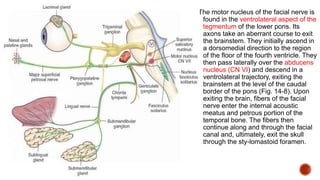
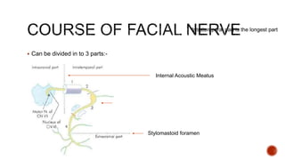
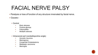
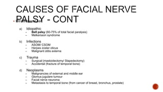
 Facial nerve is a mixed nerve.
 Consists of 2 parts:
 Facial nerve proper (motor)
 Nervus intermedius (sensory)
 Facial nerve has 3 nuclei:-
 Main motor nuclei (SVE)
 Upper part of nuleus
 Innervates forehead muscle
 Receives fibre from both hemispheres
 Lower part of nucleus
 Innervates lower face muscle
 Receives fibre from only one hemisphere
The motor nucleus of the facial nerve is
found in the ventrolateral aspect of the
tegmentum of the lower pons. Its
axons take an aberrant course to exit
the brainstem. They initially ascend in
a dorsomedial direction to the region
of the floor of the fourth ventricle. They
then pass laterally over the abducens
nucleus (CN VI) and descend in a
ventrolateral trajectory, exiting the
brainstem at the level of the caudal
border of the pons (Fig. 14-8). Upon
exiting the brain, fibers of the facial
nerve enter the internal acoustic
meatus and petrous portion of the
temporal bone. The fibers then
continue along and through the facial
canal and, ultimately, exit the skull
through the sty-lomastoid foramen.
The diagram show the
petrous portion of
temporal bone as
mentioned in the
previous slide.
Temporal bone consist
of squamo, petrous,
mastoid, tympanic
parts and styloid
process
 Parasympathetic Nuclei
 Superior Salivatory Nuclei
 Lacrimal Nuclei
 Emotional response
 Reflex lacrimation secondary to
irritation of cornea & conjunctiva
 Sensory Nuclei
 Can be divided in to 3 parts:-
Stylomastoid foramen
Internal Acoustic Meatus
Intratemporal part is the longest part
1. Greater superficial petrosal nerve
 taste to soft palate
 preganglionic parasympathetic for lacrimal gland and glands of nasal & oral cavities
2. Nerve to stapedius
3. Chorda tympani
 preganglionic parasympathetic for submandibular & sublingual glands
 taste from anterior 2/3 tongue
4. Communicating branch
 Concha
 Retroauricular groove
 Posterior meatus
 Outer surface of tympanic membrane
1. Posterior auricular nerve
 Muscle of pinna
 Occipital belly of occipitofrontalis
2. Muscular branches
 Stylohyoid
 Posterior belly of diagastric
3. Peripheral branches
 Paralysis or loss of function of any structure innervated by facial nerve.
 Causes:-
 Central
 Brain abscess
 Pontine gliomas
 Poliomyelitis
 Multiple sclerosis
 Intracranial part (cerebellopontine angle)
 Acoustic neuroma
 Meningioma
 Congenital Cholesteatoma
 Metastatic carcinoma
 Meningitis
 Intratemporal part
a) Idiopathic
– Bell palsy (60-75% of total facial paralysis)
– Melkersson syndrome
b) Infections
– ASOM/ CSOM
– Herpes zoster oticus
– Malignant otitis externa
c) Trauma
– Surgical (mastoidectomy/ Stapedectomy)
– Accidental (fracture of temporal bone)
d) Neoplasms
– Malignancies of external and middle ear
– Glomus jugulare tumour
– Facial nerve neuroma
– Metastasis to temporal bone (from cancer of breast, bronchus, prostate)
 Extracranial part
 Malignancy of parotid
 Surgery of parotid
 Accidental injury in parotid
region
 Neonatal facial injury (obstetrical
forceps)
 Systemic disease
 DM
 Hypothyroidism
 Uraemia
 Polyarteritis nodosa
 Wegener’s granulomatosis
 Sarcoidosis
 Leprosy
 Leukaemia
 Demyelinating disease
 Bell palsy
 Melkersson syndrome
 60% to 75% of facial paralysis
 Idiopathic, peripheral facial paralysis or paresis of acute onset
 Affect both sexes equally
 Affect all age group but incidence higher with increasing age
 6-8% of patient has positive family history
 Higher risk in diabetics (angiopathy) and pregnant lady (retention of fluids)
1. Viral infection
 herpes simplex, herpes zoster, EBV
2. Vascular ischemia
 primary (induced by cold, emotional stress)
 secondary (result of primary ischemia which causes increased capillary permeability leading to
exudation of fluid, oedema and compression of microcirculation of the nerve.
3. Hereditary
 Narrow fallopian canal (makes the nerve susceptible to early compression with the slightest oedema)
4. Autoimmune disorder
 T-lymphocyte changes have been observed.
 Sudden onset
 Unable to close eyes, eyeball turns up and out (Bell’s Phenomenon)
 Drooping of mouth
 Epiphora ( tears flow down from the eye )
 Noise intolerance ( stapedial paralysis )
 Loss of taste ( chorda tympani involvement )
 Exclusional diagnosis
 Requires
 complete history, ontological, head and neck examination.
 X-ray
 Blood test eg. Total count, peripheral smear, sedimentation rate, blood sugar and serology
 Nerve excitability tests (done daily or on alternate days and compared with the normal side to
monitor nerve degeneration)
 General
Reassurance
Analgesics to relieve ear pain
Eye protection to prevent exposure keratitis and corneal ulcer
- artificial tears (methylcellulose drops every 1-2hrs)
- eye paddling
-spectacles if outdoor
Physiotherapy or massage of the facial muscles
 Medical
Steroids (e.g. prednisolone)
 Surgical
Nerve decompression (relieves pressure on the nerve and thus improves the
microcirculation of the nerve)
 85% to 90% fully recover
 10% to 15% has incomplete recover and left with some stigmata of degeneration
 Recurrent facial palsy may not fully recover
 Prognosis is good in incomplete bell palsy (95% complete recovery) and in those
where clinical recovery starts within 3 weeks of onset (75% complete recovery)
Idiopathic disorder
Consists of a triad of:-
Facial paralysis
Swelling of the lips
Fissured tongue
Paralysis may be recurrent
Treatment is the same as Bell’s Palsy
Herpes Zoster Oticus
Acute Otitis Media
Chronic Otitis Media
Malignant Otitis Externa
• Herpes zoster oticus (HZ oticus) is a viral infection of the inner, middle, and
external ear. HZ oticus manifests as severe otalgia (ear pain) and associated
cutaneous vesicular eruption, usually of the external canal and pinna.
• When associated with facial paralysis, the infection is called Ramsay Hunt
syndrome.
• Reactivation of latent varicella-zoster virus (VZV) that has remained dormant within
sensory ganglia (commonly the geniculate ganglion) of the facial nerve
• Precipitating factor :
• Immunocompromised (carcinoma, radiation therapy, chemotherapy, HIV infection )
• Physical stress
• Emotional stress
 Severe otalgia
 Painful, burning blisters in and around the ear, on the face, in the mouth or
on the tongue
 Vertigo, nausea, vomiting
 Hearing loss, hyperacusis, tinnitus
 Eye pain, lacrimation
 Associated symptoms :
 Anaesthesia of face
 Hearing impairment
 Giddiness
due to involvement of CN V & CN
VIII
* Other viral infection also can cause facial
paralysis, such as herpes simplex / Epstein-Barr
virus
 Generally supportive including warm compresses,
narcotic analgesics and antibiotic for secondary bacterial
infection.
 Antiviral (acyclovir) = play a role in limiting the severity
and duration of symptoms if given early in the course of
the illness + increased rate of facial nerve function
recovery and prevented further nerve degeneration.
 Corticosteroids (prednisone) = used to relieve acute
pain, decrease vertigo, and limit the occurrence of
postherpetic neuralgia
 Facial nerve is normally well protected in its bony canal.
 Sometimes, the bony canal is dehiscent, and the nerve lies just
under the middle ear mucosa. It is in these cases that inflammation
of middle ear spreads to epi- and perineurium, causing facial
paralysis.
 Facial nerve function fully recovers if acute otitis media is
controlled with systemic antibiotics.
 Surgical Treatment : Myringotomy or cortical mastoidectomy
A surgical procedure of the tympanic membrane,
performed by making a small incision with a
myringotomy knife through the layers of tympanic
membrane which permits direct access to the middle ear
space and allows the release of middle-ear fluid.
 Either result from cholesteatoma (abnormal skin growth in the
middle ear behind the eardrum, consists of squamous epithelium) or
from penetrating granulation tissue.
 Cholesteatoma destroys bony canal and then causes pressure on
the nerve, further aided by edema of associated inflammatory
process.
 Facial paralysis is insidious but slowly progressive.
Treatment :
 Caused by pseudomonas infection
 Usually in diabetic elderly/those on
immunosuppressant drugs
 Early manifestation resemble otitis externa
but there is excruciating pain and
granulations in the meatus
 Facial paralysis is common (cranial nerve
involvement indicates a poor prognosis)
 May spread to skull base and jugular
foramen  multiple nerve involvement
Anteriorly, infection can spreads to temporomandibular fossa
Posteriorly to the mastoid
Medially to middle ear and petrous bone
Treatment :
High dose of IV antibiotics for 6-8weeks or longer
Diabetes must be controlled
Surgical debridement of devitalised tissue/bone
 Fracture of temporal bone
 Ear or mastoid surgery
 Parotid surgery and trauma to face
 May be longitudinal, transverse or mixed fractures. Transverse
fracture more commonly cause facial nerve palsy
 Facial palsy is more often seen in transverse fractures (50%)
 Paralysis is due to intraneural hematoma, compression by a
bony spicule or transection of nerve.
 Facial nerve paralysis
 Hearing loss
 Vertigo
 Dizziness
 Otorrhagia (hemorrhage from the ear)
 CSF otorrhea (Ear discharge)
 Tympanic membrane perforation
 Hemotympanum (The presence of blood in the middle ear)
 Delayed onset:
 Treat conservatively like Bell’s Palsy
 Caused edema of the surrounding structure
 Immediate onset:
 Surgical intervention in the form or decompression, re-anastomosis of cut
ends or cable nerve graft.
 Injured during stapedectomy,
tympanoplasty, or mastoid surgery.
 Sometimes, nerve is paralysed due to
pressure of packing on the exposed
nerve and this should be relieved first.
 Paralysis may be immediate or delay
 Treatment same as fractured temporal
bone.
 Facial nerve may be injured in surgery of parotid tumours or
deliberately excised in malignant tumours.
 Accidental injuries in the parotid region can also cause facial
paralysis.
 Application of obstetrical forceps may also result in facial paralysis
in the neonate due to pressure on the extratemporal part of nerve.
i) Intratemporal Neoplasms
Carcinoma of external or middle ear, glomus tumour,
rhabdomyosarcoma and metastatic tumours of temporal bone.
Facial nerve neuroma can occur anywhere along the course of the
nerve and produce paralysis of gradual or sudden onset
Investigation : High resolution CT scan and gadolinium-enhanced
MRI is very useful for facial nerve tumour
Management : Excision and nerve grafting
ii) Tumours of Parotid
 Facial paralysis with tumour of the parotid almost always
implies malignancy
1. Incomplete recovery
facial asymmetry, epiphora(excessive tearing), drooling and difficulty in taking
food
2. Exposure keratitis
eyes cannot be closed  tear film evaporates  dryness, exposure keratitis,
corneal ulcer
Treatment : artificial tears (methylcellulose drops) every 1-2 hours, eye
ointment and proper cover for eye at night
3. Synkinesis (mass movement)
Due to cross innervation of fibres
When patient wishes to close the eye, corner of mouth also twitches or vice
versa
4. Tics & spasms
 Faulty regeneration of fibres. Involuntary movements are seen
on the affected side of the face .
5. Contractures
Fibrosis of atrophied muscle or fixed contraction of a group of muscles
6. Crocodile tears (gustatory lacrimation)
Unilateral lacrimation with mastication
Due to faulty regeneration of parasympathetic fibres which now supply lacrimal
gland instead of the salivary gland
Treated by section of greater superficial petrosal nerve or tympanic neurectomy
7. Frey’s syndrome (gustatory sweating)
Sweating and flushing of skin over parotid area during mastication
 It results from parotid surgery
8. Psychological and social problems
Drooling during eating/drinking
Impairment of speech
Facial nerve palcy

Facial nerve palcy

  • 2.
     Facial nerveis a mixed nerve.  Consists of 2 parts:  Facial nerve proper (motor)  Nervus intermedius (sensory)
  • 3.
     Facial nervehas 3 nuclei:-  Main motor nuclei (SVE)  Upper part of nuleus  Innervates forehead muscle  Receives fibre from both hemispheres  Lower part of nucleus  Innervates lower face muscle  Receives fibre from only one hemisphere
  • 4.
    The motor nucleusof the facial nerve is found in the ventrolateral aspect of the tegmentum of the lower pons. Its axons take an aberrant course to exit the brainstem. They initially ascend in a dorsomedial direction to the region of the floor of the fourth ventricle. They then pass laterally over the abducens nucleus (CN VI) and descend in a ventrolateral trajectory, exiting the brainstem at the level of the caudal border of the pons (Fig. 14-8). Upon exiting the brain, fibers of the facial nerve enter the internal acoustic meatus and petrous portion of the temporal bone. The fibers then continue along and through the facial canal and, ultimately, exit the skull through the sty-lomastoid foramen.
  • 5.
    The diagram showthe petrous portion of temporal bone as mentioned in the previous slide. Temporal bone consist of squamo, petrous, mastoid, tympanic parts and styloid process
  • 7.
     Parasympathetic Nuclei Superior Salivatory Nuclei  Lacrimal Nuclei  Emotional response  Reflex lacrimation secondary to irritation of cornea & conjunctiva  Sensory Nuclei
  • 8.
     Can bedivided in to 3 parts:- Stylomastoid foramen Internal Acoustic Meatus Intratemporal part is the longest part
  • 10.
    1. Greater superficialpetrosal nerve  taste to soft palate  preganglionic parasympathetic for lacrimal gland and glands of nasal & oral cavities 2. Nerve to stapedius 3. Chorda tympani  preganglionic parasympathetic for submandibular & sublingual glands  taste from anterior 2/3 tongue 4. Communicating branch  Concha  Retroauricular groove  Posterior meatus  Outer surface of tympanic membrane
  • 11.
    1. Posterior auricularnerve  Muscle of pinna  Occipital belly of occipitofrontalis 2. Muscular branches  Stylohyoid  Posterior belly of diagastric 3. Peripheral branches
  • 16.
     Paralysis orloss of function of any structure innervated by facial nerve.  Causes:-  Central  Brain abscess  Pontine gliomas  Poliomyelitis  Multiple sclerosis  Intracranial part (cerebellopontine angle)  Acoustic neuroma  Meningioma  Congenital Cholesteatoma  Metastatic carcinoma  Meningitis
  • 17.
     Intratemporal part a)Idiopathic – Bell palsy (60-75% of total facial paralysis) – Melkersson syndrome b) Infections – ASOM/ CSOM – Herpes zoster oticus – Malignant otitis externa c) Trauma – Surgical (mastoidectomy/ Stapedectomy) – Accidental (fracture of temporal bone) d) Neoplasms – Malignancies of external and middle ear – Glomus jugulare tumour – Facial nerve neuroma – Metastasis to temporal bone (from cancer of breast, bronchus, prostate)
  • 18.
     Extracranial part Malignancy of parotid  Surgery of parotid  Accidental injury in parotid region  Neonatal facial injury (obstetrical forceps)  Systemic disease  DM  Hypothyroidism  Uraemia  Polyarteritis nodosa  Wegener’s granulomatosis  Sarcoidosis  Leprosy  Leukaemia  Demyelinating disease
  • 19.
     Bell palsy Melkersson syndrome
  • 20.
     60% to75% of facial paralysis  Idiopathic, peripheral facial paralysis or paresis of acute onset  Affect both sexes equally  Affect all age group but incidence higher with increasing age  6-8% of patient has positive family history  Higher risk in diabetics (angiopathy) and pregnant lady (retention of fluids)
  • 21.
    1. Viral infection herpes simplex, herpes zoster, EBV 2. Vascular ischemia  primary (induced by cold, emotional stress)  secondary (result of primary ischemia which causes increased capillary permeability leading to exudation of fluid, oedema and compression of microcirculation of the nerve. 3. Hereditary  Narrow fallopian canal (makes the nerve susceptible to early compression with the slightest oedema) 4. Autoimmune disorder  T-lymphocyte changes have been observed.
  • 22.
     Sudden onset Unable to close eyes, eyeball turns up and out (Bell’s Phenomenon)  Drooping of mouth  Epiphora ( tears flow down from the eye )  Noise intolerance ( stapedial paralysis )  Loss of taste ( chorda tympani involvement )
  • 23.
     Exclusional diagnosis Requires  complete history, ontological, head and neck examination.  X-ray  Blood test eg. Total count, peripheral smear, sedimentation rate, blood sugar and serology  Nerve excitability tests (done daily or on alternate days and compared with the normal side to monitor nerve degeneration)
  • 24.
     General Reassurance Analgesics torelieve ear pain Eye protection to prevent exposure keratitis and corneal ulcer - artificial tears (methylcellulose drops every 1-2hrs) - eye paddling -spectacles if outdoor Physiotherapy or massage of the facial muscles  Medical Steroids (e.g. prednisolone)  Surgical Nerve decompression (relieves pressure on the nerve and thus improves the microcirculation of the nerve)
  • 25.
     85% to90% fully recover  10% to 15% has incomplete recover and left with some stigmata of degeneration  Recurrent facial palsy may not fully recover  Prognosis is good in incomplete bell palsy (95% complete recovery) and in those where clinical recovery starts within 3 weeks of onset (75% complete recovery)
  • 26.
    Idiopathic disorder Consists ofa triad of:- Facial paralysis Swelling of the lips Fissured tongue Paralysis may be recurrent Treatment is the same as Bell’s Palsy
  • 28.
    Herpes Zoster Oticus AcuteOtitis Media Chronic Otitis Media Malignant Otitis Externa
  • 29.
    • Herpes zosteroticus (HZ oticus) is a viral infection of the inner, middle, and external ear. HZ oticus manifests as severe otalgia (ear pain) and associated cutaneous vesicular eruption, usually of the external canal and pinna. • When associated with facial paralysis, the infection is called Ramsay Hunt syndrome. • Reactivation of latent varicella-zoster virus (VZV) that has remained dormant within sensory ganglia (commonly the geniculate ganglion) of the facial nerve • Precipitating factor : • Immunocompromised (carcinoma, radiation therapy, chemotherapy, HIV infection ) • Physical stress • Emotional stress
  • 30.
     Severe otalgia Painful, burning blisters in and around the ear, on the face, in the mouth or on the tongue  Vertigo, nausea, vomiting  Hearing loss, hyperacusis, tinnitus  Eye pain, lacrimation  Associated symptoms :  Anaesthesia of face  Hearing impairment  Giddiness due to involvement of CN V & CN VIII
  • 32.
    * Other viralinfection also can cause facial paralysis, such as herpes simplex / Epstein-Barr virus
  • 33.
     Generally supportiveincluding warm compresses, narcotic analgesics and antibiotic for secondary bacterial infection.  Antiviral (acyclovir) = play a role in limiting the severity and duration of symptoms if given early in the course of the illness + increased rate of facial nerve function recovery and prevented further nerve degeneration.  Corticosteroids (prednisone) = used to relieve acute pain, decrease vertigo, and limit the occurrence of postherpetic neuralgia
  • 34.
     Facial nerveis normally well protected in its bony canal.  Sometimes, the bony canal is dehiscent, and the nerve lies just under the middle ear mucosa. It is in these cases that inflammation of middle ear spreads to epi- and perineurium, causing facial paralysis.  Facial nerve function fully recovers if acute otitis media is controlled with systemic antibiotics.  Surgical Treatment : Myringotomy or cortical mastoidectomy
  • 35.
    A surgical procedureof the tympanic membrane, performed by making a small incision with a myringotomy knife through the layers of tympanic membrane which permits direct access to the middle ear space and allows the release of middle-ear fluid.
  • 36.
     Either resultfrom cholesteatoma (abnormal skin growth in the middle ear behind the eardrum, consists of squamous epithelium) or from penetrating granulation tissue.  Cholesteatoma destroys bony canal and then causes pressure on the nerve, further aided by edema of associated inflammatory process.  Facial paralysis is insidious but slowly progressive.
  • 37.
  • 38.
     Caused bypseudomonas infection  Usually in diabetic elderly/those on immunosuppressant drugs  Early manifestation resemble otitis externa but there is excruciating pain and granulations in the meatus  Facial paralysis is common (cranial nerve involvement indicates a poor prognosis)  May spread to skull base and jugular foramen  multiple nerve involvement
  • 39.
    Anteriorly, infection canspreads to temporomandibular fossa Posteriorly to the mastoid Medially to middle ear and petrous bone Treatment : High dose of IV antibiotics for 6-8weeks or longer Diabetes must be controlled Surgical debridement of devitalised tissue/bone
  • 40.
     Fracture oftemporal bone  Ear or mastoid surgery  Parotid surgery and trauma to face
  • 41.
     May belongitudinal, transverse or mixed fractures. Transverse fracture more commonly cause facial nerve palsy  Facial palsy is more often seen in transverse fractures (50%)  Paralysis is due to intraneural hematoma, compression by a bony spicule or transection of nerve.
  • 44.
     Facial nerveparalysis  Hearing loss  Vertigo  Dizziness  Otorrhagia (hemorrhage from the ear)  CSF otorrhea (Ear discharge)  Tympanic membrane perforation  Hemotympanum (The presence of blood in the middle ear)
  • 45.
     Delayed onset: Treat conservatively like Bell’s Palsy  Caused edema of the surrounding structure  Immediate onset:  Surgical intervention in the form or decompression, re-anastomosis of cut ends or cable nerve graft.
  • 46.
     Injured duringstapedectomy, tympanoplasty, or mastoid surgery.  Sometimes, nerve is paralysed due to pressure of packing on the exposed nerve and this should be relieved first.  Paralysis may be immediate or delay  Treatment same as fractured temporal bone.
  • 47.
     Facial nervemay be injured in surgery of parotid tumours or deliberately excised in malignant tumours.  Accidental injuries in the parotid region can also cause facial paralysis.  Application of obstetrical forceps may also result in facial paralysis in the neonate due to pressure on the extratemporal part of nerve.
  • 49.
    i) Intratemporal Neoplasms Carcinomaof external or middle ear, glomus tumour, rhabdomyosarcoma and metastatic tumours of temporal bone. Facial nerve neuroma can occur anywhere along the course of the nerve and produce paralysis of gradual or sudden onset Investigation : High resolution CT scan and gadolinium-enhanced MRI is very useful for facial nerve tumour Management : Excision and nerve grafting ii) Tumours of Parotid  Facial paralysis with tumour of the parotid almost always implies malignancy
  • 50.
    1. Incomplete recovery facialasymmetry, epiphora(excessive tearing), drooling and difficulty in taking food 2. Exposure keratitis eyes cannot be closed  tear film evaporates  dryness, exposure keratitis, corneal ulcer Treatment : artificial tears (methylcellulose drops) every 1-2 hours, eye ointment and proper cover for eye at night 3. Synkinesis (mass movement) Due to cross innervation of fibres When patient wishes to close the eye, corner of mouth also twitches or vice versa 4. Tics & spasms  Faulty regeneration of fibres. Involuntary movements are seen on the affected side of the face .
  • 51.
    5. Contractures Fibrosis ofatrophied muscle or fixed contraction of a group of muscles 6. Crocodile tears (gustatory lacrimation) Unilateral lacrimation with mastication Due to faulty regeneration of parasympathetic fibres which now supply lacrimal gland instead of the salivary gland Treated by section of greater superficial petrosal nerve or tympanic neurectomy 7. Frey’s syndrome (gustatory sweating) Sweating and flushing of skin over parotid area during mastication  It results from parotid surgery 8. Psychological and social problems Drooling during eating/drinking Impairment of speech