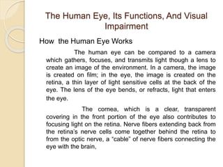
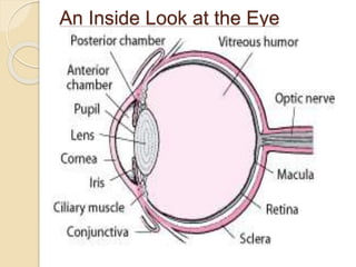
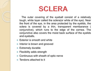
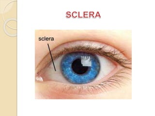
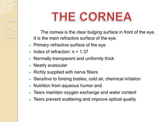
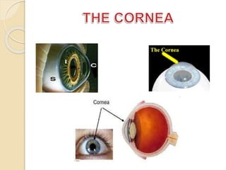
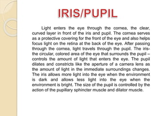
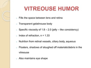
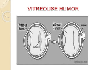
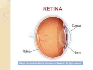
structure of eye ball,eyeball is a specialized sense organ that helps us to understand our environment. It is a sensory unit composed of three parts: receptor, sensory pathway, and a brain center
The main parts of the human eye are The Conjunctiva,
Sclera,Choroid,
Cornea, Iris, Pupil,
Anterior Chamber,
Posterior Chamber, Aqueous humor, Lens, Vitreous humor, Retina,Macula and Optic nerve.
Following points are explain in this article:
Introduction of eye
Eyelids & tears
Morphology of eye-ball
Components of eye
Functions of eye parts
Diagrams
he sense organs — eyes, ears, tongue, skin, and nose — help to protect the body. The human sense organs contain receptors that relay information through sensory neurons to the appropriate places within the nervous system.
Each sense organ contains different receptors.
General receptors are found throughout the body because they are present in skin, visceral organs (visceral meaning in the abdominal cavity), muscles, and joints.
Special receptors include chemoreceptors (chemical receptors) found in the mouth and nose, photoreceptors (light receptors) found in the eyes, and mechanoreceptors found in the ears.
structure and fuction of eyes and ears,types of memory,sharpe memory,attentionUmarKhan422
The external covering of the eyeball comprises of a generally intense, white layer called the sclera (or white of the eye). Close to the front of the eye, in the zone secured by the eyelids, the sclera is secured by a slim, straightforward layer (conjunctiva), which rushes to the edge of the cornea.
anatomy of an eye, internal structure of eye,layers of an eye ball, features of eye ball, cornea, sclera, retina, choroid, ciliary body lense, macula, blind spot, yellow spot, clinical aspect of eye , drainage of aqueous humor, cataract, glaucoma, causes of cataract and treatment.
Multi-source connectivity as the driver of solar wind variability in the heli...Sérgio Sacani
The ambient solar wind that flls the heliosphere originates from multiple
sources in the solar corona and is highly structured. It is often described
as high-speed, relatively homogeneous, plasma streams from coronal
holes and slow-speed, highly variable, streams whose source regions are
under debate. A key goal of ESA/NASA’s Solar Orbiter mission is to identify
solar wind sources and understand what drives the complexity seen in the
heliosphere. By combining magnetic feld modelling and spectroscopic
techniques with high-resolution observations and measurements, we show
that the solar wind variability detected in situ by Solar Orbiter in March
2022 is driven by spatio-temporal changes in the magnetic connectivity to
multiple sources in the solar atmosphere. The magnetic feld footpoints
connected to the spacecraft moved from the boundaries of a coronal hole
to one active region (12961) and then across to another region (12957). This
is refected in the in situ measurements, which show the transition from fast
to highly Alfvénic then to slow solar wind that is disrupted by the arrival of
a coronal mass ejection. Our results describe solar wind variability at 0.5 au
but are applicable to near-Earth observatories.
Earliest Galaxies in the JADES Origins Field: Luminosity Function and Cosmic ...Sérgio Sacani
We characterize the earliest galaxy population in the JADES Origins Field (JOF), the deepest
imaging field observed with JWST. We make use of the ancillary Hubble optical images (5 filters
spanning 0.4−0.9µm) and novel JWST images with 14 filters spanning 0.8−5µm, including 7 mediumband filters, and reaching total exposure times of up to 46 hours per filter. We combine all our data
at > 2.3µm to construct an ultradeep image, reaching as deep as ≈ 31.4 AB mag in the stack and
30.3-31.0 AB mag (5σ, r = 0.1” circular aperture) in individual filters. We measure photometric
redshifts and use robust selection criteria to identify a sample of eight galaxy candidates at redshifts
z = 11.5 − 15. These objects show compact half-light radii of R1/2 ∼ 50 − 200pc, stellar masses of
M⋆ ∼ 107−108M⊙, and star-formation rates of SFR ∼ 0.1−1 M⊙ yr−1
. Our search finds no candidates
at 15 < z < 20, placing upper limits at these redshifts. We develop a forward modeling approach to
infer the properties of the evolving luminosity function without binning in redshift or luminosity that
marginalizes over the photometric redshift uncertainty of our candidate galaxies and incorporates the
impact of non-detections. We find a z = 12 luminosity function in good agreement with prior results,
and that the luminosity function normalization and UV luminosity density decline by a factor of ∼ 2.5
from z = 12 to z = 14. We discuss the possible implications of our results in the context of theoretical
models for evolution of the dark matter halo mass function.
THE IMPORTANCE OF MARTIAN ATMOSPHERE SAMPLE RETURN.Sérgio Sacani
The return of a sample of near-surface atmosphere from Mars would facilitate answers to several first-order science questions surrounding the formation and evolution of the planet. One of the important aspects of terrestrial planet formation in general is the role that primary atmospheres played in influencing the chemistry and structure of the planets and their antecedents. Studies of the martian atmosphere can be used to investigate the role of a primary atmosphere in its history. Atmosphere samples would also inform our understanding of the near-surface chemistry of the planet, and ultimately the prospects for life. High-precision isotopic analyses of constituent gases are needed to address these questions, requiring that the analyses are made on returned samples rather than in situ.
Professional air quality monitoring systems provide immediate, on-site data for analysis, compliance, and decision-making.
Monitor common gases, weather parameters, particulates.
Comparing Evolved Extractive Text Summary Scores of Bidirectional Encoder Rep...University of Maribor
Slides from:
11th International Conference on Electrical, Electronics and Computer Engineering (IcETRAN), Niš, 3-6 June 2024
Track: Artificial Intelligence
https://www.etran.rs/2024/en/home-english/
Richard's aventures in two entangled wonderlandsRichard Gill
Since the loophole-free Bell experiments of 2020 and the Nobel prizes in physics of 2022, critics of Bell's work have retreated to the fortress of super-determinism. Now, super-determinism is a derogatory word - it just means "determinism". Palmer, Hance and Hossenfelder argue that quantum mechanics and determinism are not incompatible, using a sophisticated mathematical construction based on a subtle thinning of allowed states and measurements in quantum mechanics, such that what is left appears to make Bell's argument fail, without altering the empirical predictions of quantum mechanics. I think however that it is a smoke screen, and the slogan "lost in math" comes to my mind. I will discuss some other recent disproofs of Bell's theorem using the language of causality based on causal graphs. Causal thinking is also central to law and justice. I will mention surprising connections to my work on serial killer nurse cases, in particular the Dutch case of Lucia de Berk and the current UK case of Lucy Letby.
4. The Human Eye, Its Functions, And Visual
Impairment
How the Human Eye Works
The human eye can be compared to a camera
which gathers, focuses, and transmits light though a lens to
create an image of the environment. In a camera, the image
is created on film; in the eye, the image is created on the
retina, a thin layer of light sensitive cells at the back of the
eye. The lens of the eye bends, or refracts, light that enters
the eye.
The cornea, which is a clear, transparent
covering in the front portion of the eye also contributes to
focusing light on the retina. Nerve fibers extending back from
the retina’s nerve cells come together behind the retina to
from the optic nerve, a “cable” of nerve fibers connecting the
eye with the brain,
5. The optic nerve transmits messages about what we
see from the eye to the brain. Like a camera, the human eye
controls the amount of light that enters the eye through the
lens under various lighting conditions.
The orbit is the bony cavity that contains the
eyeball, muscles, and blood vessels, as well as the structures
that produce and drain tears. Each orbit is a pear-shaped
structure that is formed by several bones.
7. The outer covering of the eyeball consist of a relatively
tough, white layer called the sclera(or white of the eye). Near
the front of the eye, in the area protected by the eyelids, the
sclera is covered by a thin, transparent membarance (
conjunctive), which runs to the edge of the cornea. The
conjunctive also covers the moist back surface of the eyelids
and eyeballs.
Exterior is smooth and white
Interior is brown and grooved
Extremely durable
Flexibility adds strength
Continuous with sheath of optic nerve
Tendons attached to it
8.
9. The cornea is the clear bulging surface in front of the eye.
It is the main refractive surface of the eye.
Primary refractive surface of the eye
Index of refraction: n = 1.37
Normally transparent and uniformly thick
Nearly avascular
Richly supplied with nerve fibers
Sensitive to foreing bodies, cold air, chemical irritation
Nutrition from aqueous humor and
Tears maintain oxygen exchange and water content
Tears prevent scattering and improve optical quality
10.
11. The anterior segment is divided into two chambers. The
front (anterior) chamber extends from the cornea to the iris. The
back (posterior) chamber extends from the iris to the lens.
Normally, the equeous humor is produced in the posterior
chamber, flows slowly through the pipil into the anterior
chamber, and then drains out of the eyeball through out flow
channels located where the iris meets the cornea.
The anterior chamber is between the cornea and the iris
The posterior chamber is between the iris and the lens
Contains the aqueous humor
Index of refraction : n =1.33
Specific viscosity of the aqueous just over 1.0 (like water, hence
the name)
Pressure of 15-18 mm of mercury maintains shape of eye and
spacing of the elements
12. Light enters the eye through the cornea, the clear,
curved layer in front of the iris and pupil. The cornea serves
as a protective covering for the front of the eye and also helps
focus light on the retina at the back of the eye. After passing
through the cornea, light travels through the pupil. The iris-the
circular, colored area of the eye that surrounds the pupil –
controls the amount of light that enters the eye. The pupil
dilates and constricts like the aperture of a camera lens as
the amount of light in the immediate surroundings changes.
The iris allows more light into the eye when the environment
is dark and allows less light into the eye when the
environment is bright. The size of the pupil is controlled by the
action of the pupillary sphincter muscle and dilator muscle.
13. Iris is heavily pigmented
Sphincter muscle to constrict or dilate the pupil
Pupil is the hole through which light passes
Pupil diameter ranges from about 3 - 7 mm
Area of 7 – 38 square mm
Eye color (brown, green, blue, etc.) dependent on amount
and distribution of the pigment melanin
14.
15. Transparent body enclosed in an elastic capsule
Made up of proteins and water
Consists of layers, like an onion, with firm nucleus, soft cortex
Gradient refractive index (1.38 -1.40)
Young person can change shape of the lens via ciliary muscles
Contraction of muscle cause lens to bulge
At roughly age 50 the lens can no longer change shape
16.
17. o Fills the space between lens and retina
o Transparent gelatinouse body
o Specific viscosity of 1.8 – 2.0 (jelly – like consistency)
o Index of refraction, n = 1.33
o Nutrition from retinal vessels, ciliary body, aqueous
o Floaters, shadows of sloughed off materials/debris in the
vitreouse
o Also maintains eye shape
18.
19. The light has to pass through many layers of cells
before finally reaching the photoreceptors. The
photoreceptors are where the light is absorbed and
transformed into the electrochemical signals used by the
nervous system. This change is called transduction
The interior of the eyeball is the “inner” side and the
exterior is the “ outer” side. The nuclear layers contain cell
bodies. The plexiform layers contain the connection between
cells in the retina.
20.
21. The location where the optic nerve is bundled and leaves the
retina is known as the optic disk. There are no photoreceptors at
the location of the optic disk and hence there is a blind spot. The
scientific term for a blind spot is a scotoma. So the blind spot
due to the disk is a natural permanent scotoma in normal vision.
Here is a demonstration of the natural permanent scotoma.
22.
23. The fovea is the location on the retina of central gaze.
when you look directly, or fixate, at a stimulus you the retina
locus of this central fixation is the fovea. There are only cones in
the human fovea (no rods). They are thinner, elongated, any
very tightly packed. Because of this, the fovea is the location of
highest visual acuity and best color vision.
24.
25. Covering the fovea is a pigment called the macula. It is
thought that macula serves a protective filter over the foviea that
absorbs blue and ultraviolet radiation. This pigment varies from
observer to observer and is a source of individual variation in
color vision. Usually we do not notice the filtering of the macula
but under special conditions we can notice its presence causing
what is known as Maxwell’s spot.