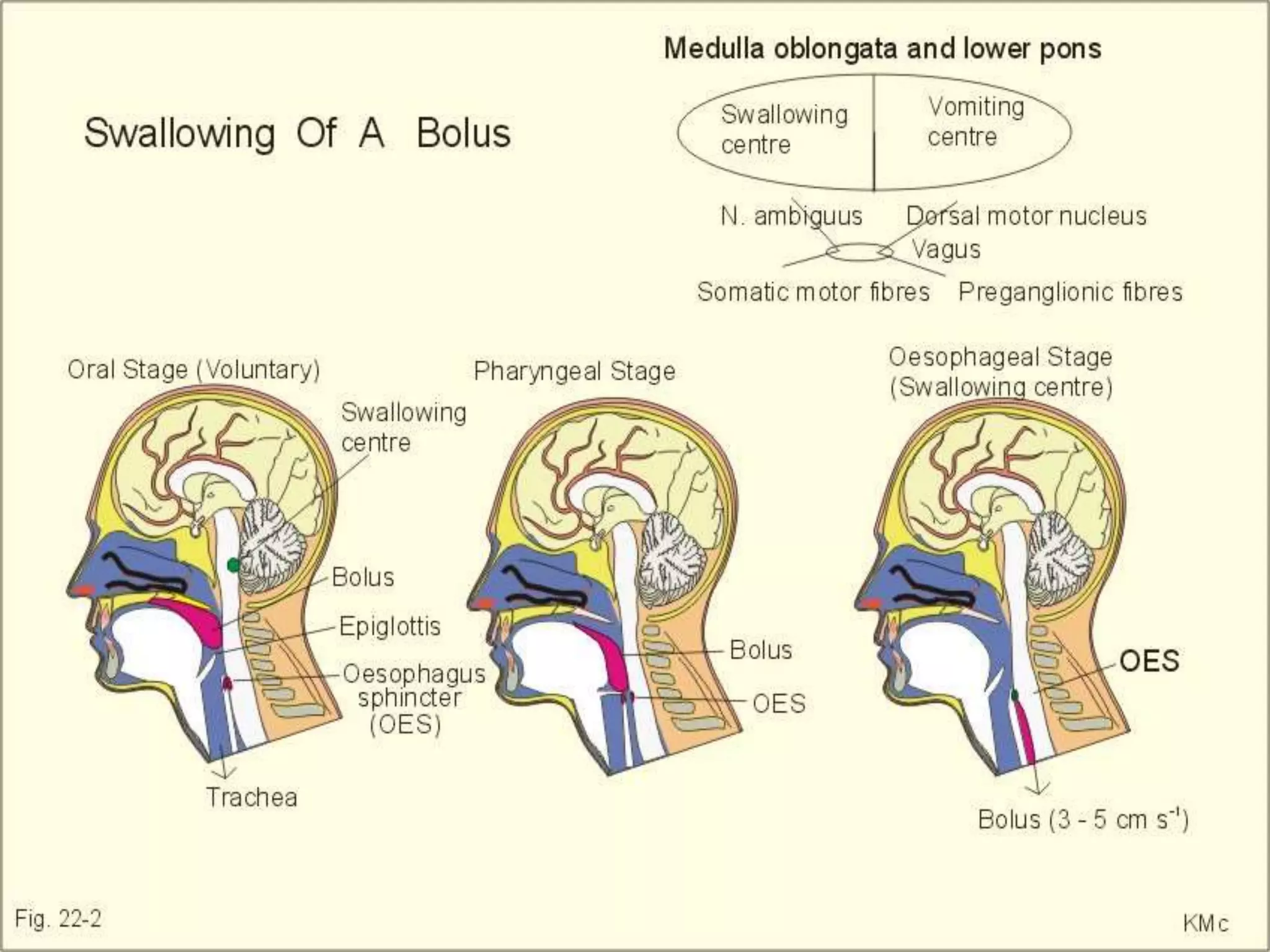
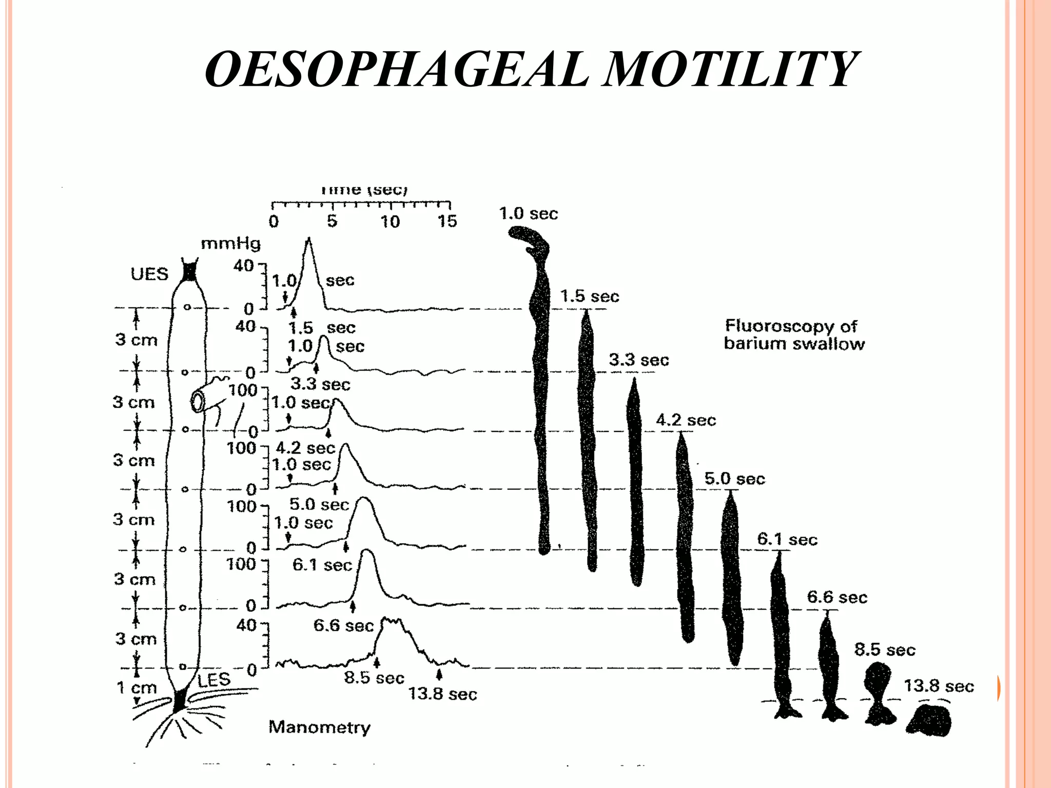
This document provides an overview of deglutition (swallowing) physiology and esophageal manometry. It describes the three phases of swallowing (oral, pharyngeal, esophageal) and the muscles involved in each phase. It also outlines the neural control of swallowing and the brainstem nuclei involved. Regarding esophageal manometry, it describes the different catheter types, indications for the test, and provides a detailed outline of the components and steps to perform esophageal manometry including identifying high pressure zones and measuring lower esophageal sphincter relaxation.
2. DEGLUTITION
Deglutition is the act of swallowing, through which a food or
liquid bolus is transported from the mouth through the pharynx
and esophagus into the stomach.
Normal deglutition is a smooth coordinated process that
involves a complex series of voluntary and involuntary
neuromuscular contractions and typically is divided into three
distinct phases:
Oral
Pharyngeal
Esophageal
4. ORAL PREPARATORY PHASE
This phase is where the food is readied for swallowing
by reducing & mixing it with saliva, by the muscles of
jaw and oral cavity.
Jaw is closed by jaw elevator muscles ( temporalis,
masseter & medial pterygoid).
Lips maintain a seal under the action of Orbicularis
oris.
Food is returned from the vestibule by contraction of
buccinators.
5. Through out this phase, the soft palate is lowered &
Ant and Post pillars approx. under the action of
palatoglossus & palato pharyngeus muscles.
Thus, the oral cavity is sealed post. & the airway
remains open.
Bolus is progressively accumulated on the posterior
surface of the tongue, by several cycles of upward and
downward movement on the tongue surface.
6. When the bolus consistency ( sensed by mechano-receptors in
the oral cavity) is suitable for swallowing, the oral phase proper
begins.
7.
8. ORAL PHASE PROPER
The first event is mandibular elevation
Although the mouth does not have to be completely
closed, it is hard to swallow with an open mouth.
Mandibular elevation assists the suprahyoid muscles
in raising the hyoid bone
Next, the tip of the tongue is elevated towards the
hard palate by the action of genioglossus muscle
9. Blade of the tongue then moves up due to contraction
of intrinsic muscles.
These movements are accompanied by lifting the floor
of the mouth under the action of stylohyoid.
As the bolus reaches the back of the tongue ,the soft
palate is elevated by tensor and levator veli palatini to
protect the nasopharynx.
10.
11. PHARYNGEAL PHASE
As the bolus enters the oropharynx, it makes
contact with faucial pillars or with the mucosa
overlying the posterior pharynx, the region
which is sensory innervated by glossopharyngeal
nerve.
Hereafter swallowing becomes reflexive
Pharyngeal phase consists of a sequence of
events that ensures that the airway is protected
during bolus transport.
12. Diaphragmatic contraction is inhibited making
simultaneous breathing and swallowing impossible.
Soft palate is elevated to ensure closure of the
nasopharynx.
Vocal cords start to close to protect the airway, either
do the vestibular folds.
The larynx is closed by the contraction of muscles of
laryngeal inlet(AEF, interarytenoid and thyro
epiglottic) resembling a draw string purse.
13.
14. The larynx is closed under the contraction of
suprahyoid muscles, in order to narrow the laryngeal
inlet and moving it towards the pharyngeal surface of
epiglottis.
As the bolus moves in to oropharynx, the epiglottis
moves downwards.
This downward movement occurs in 2 distinct stages.
1.movement from vertical to horizontal position
2.movement from horizontal to below horizontal in
order to cover the narrow laryngeal inlet.
15. The first epiglottis movement is passive, due to the
forces generated by compression of the pre epiglottic
adipose fat and ligamentous attachment of epiglottis.
The second movement occurs by a combination of
passive and active(contraction of thyroepiglottic and
hyoepiglottic) components.
16.
17. The bolus enters the pharynx which is widened,
resembling the engulfing of prey by a snake.
Widening is partly due to relaxation of constrictor
muscles and partly due to anterior movement. Of the
pharynx under the action of suprahyoid muscles.
As the food passes over the post. Part of the epiglottis,
it is diverted into the pyriform fossae. Solids tend to
go straight over the epiglottis, whereas liquids are
diverted laterally.
18. OESOPHAGEAL PHASE
The crico pharyngeus muscle relaxes so the
upper oesophageal sphincter opens, bolus is
passed on into & through the sphincter &
oesophagus by peristalsis.
Tensor & Levator veli palatini relax, lowering
the soft palate, laryngeal inlet & vestibule open-
> hyoid & larynx drops -> at the very end stage
of swallowing, the glottis open.
19.
20. NEURAL CONTROL
Neural control of swallowing involves a number of
different regions of the CNS, extending from the
motor nuclei within the brainstem, up to the cortex.
The act of swallowing is regulated by sensory
feedback.
21. The initiation of swallowing can either be as a
voluntary act, or a reflex as the result of stimulation of
the mucosa in the oral cavity. The latter may occur
during saliva accumulation or by presence of food or
liquid.
Due to anatomical & physiological close relationship
between swallowing, ventilation & mastication, there
is extensive overlap in the brainstem areas controlling
these functions.
22. The voluntary initiation of swallowing involves b/l
areas of frontal, pre-frontal & parietal cortices. Frontal
swallowing centre is associated with motor control of
swallowing. This centre includes-
Lower pre-central & post inferior frontal gyri
- oral phase
Middle frontal & anterior inferior frontal gyri
- pharyngeal & oesophageal
23. Voluntary sensory control of swallowing is mediated
by the parietal cortex.
Swallowing control is asymmetrical with projections
from one hemisphere being larger than the other,
independent of handedness.
This explains why damage to the hemisphere that is
source of greater projection to the swallowing centres
in the brain stem will cause initial difficulty, and
recovery the occurs as the intact projection from the
undamaged hemisphere is re organized.
24. Descending pathways project from the frontal
swallowing areas of cortex to the medullary
swallowing centers within the medulla
There are a number of nucleus in the medulla
,involved in control of swallowing.
Swallowing is initiated by the touch or pressure
sensation from the posterior part of the oral cavity or
oropharynx.
The nuclei receiving afferent input include -
Nucleus tractus solitaries
Spinal trigeminal nucleus
25. The efferent pathways from the medulla and pons to
the swallowing muscles include –
1.nucleus ambigus->for muscles of the palate,
pharynx and the larynx.
2.motor nuclei of hypoglossal->tongue
3.motor nuclei of trigeminal -> jaw
4.motor nuclei of facial->lips
26.
27.
28. ESOPHAGEAL MANOMETRY
A test to assess motor function of the upper
oesophageal sphincter (UES), oesophageal body
and lower oesophageal sphincter (LES).
When does it help?
Functional disorder is suspected
Unrevealing morphological studies
Part of pre-operative evaluation
30. All GI manometry setups consist of two hardware
components:
A pressure sensor/transducer, which is
able to sense changes in intra luminal pressure and
convert what is detected into an electrical signal.
A recording device that amplifies the signal and stores it.
Two types of sensing/transducer devices are currently
used for oesophageal manometry-
water-perfused catheters coupled to volume-displacement
transducers.
solid-state strain gauge transducers
31. WATER-PERFUSED CATHETERS COUPLED TO
VOLUME-DISPLACEMENT
TRANSDUCERS
This type of catheter comprises a bundle of thin plastic tubes
each with an outward facing side-hole. There are typically 3-
8pressure-sensing side holes spaced along the length of the
catheter and radially orientated, thereby allowing simultaneous
measurement of pressures at multiple locations. The
tubes are continuously perfused with bubble-free water as a
non-compressible medium and the pressure in each tube is
monitored by a volume-displacement transducer. Water flow
through the side holes is impeded by oesophageal contraction
33. SOLID-STATE STRAIN GAUGES
This type of catheter is composed of a linear arrangement of
miniature, solid-state strain gauges spatially and radially
orientated along a flexible tube. The signal from each strain
gauge provides a direct measure of intra luminal pressure.
These catheters are technically easier to use and less cumbersome
than traditional water-perfused systems, but are more
expensive both to buy and repair. There have been no studies
to compare the relative running costs of the two alternative
systems. Absolute pressure values and normal ranges
obtained with water-perfused versus solid state systems are
not identical and the choice of laboratory reference range
should reflect the type of catheter assembly
35. HIGH RESOLUTION MANOMETRY (HRM)
Miniaturisation of solid state pressure sensors has
allowed the
development of high resolution manometry (HRM),
employing catheters with multiple sensors (up to 36)
distributed
longitudinally and radially.(3,4) This allows topographical
analyses with the generation of 2- and 3-dimensional
contour
plots based on simultaneous pressure readings taken at
multiple sensors within the sphincters and oesophageal
body.
These catheters have the potential to reduce the need for
repositioning, thereby shortening the duration of the
36. Simultaneous assessment of sphincters and body with
a single series of swallows is possible with the
catheter in a single, fixed position. The increased
resolution and better radial information promised by
HRM should reduce the problems of asymmetry and
artefact inherent in existing systems.(3,4).
This type of equipment is not widely available in the
UK and the present guidance relates to traditional
water-perfused and solid state catheters
37. INDICATIONS FOR OESOPHAGEAL
MANOMETRY
1) To diagnose suspected primary oesophageal motility
disorders (eg. achalasia and diffuse oesophageal spasm)
2) To diagnose suspected secondary oesophageal motility
disorders occurring in association with systemic
diseases (eg. systemic sclerosis)
3) To guide the accurate placement of pH electrodes for
ambulatory pH monitoring studies.
38. (B) Equipment preparation
(1) Calibrate the equipment and document it on
recording
(2) Record the catheter type and configuration
(3) Check to assure functioning of the recording
device prior to intubation
(a) Are the recording devices turned on?
(b) Are all of the appropriate connections made and
documented?
39. 4) As part of the pre-operative assessment of some
patients undergoing anti-reflux procedures.
5) To reassess oesophageal function in patients who
have been treated for a primary oesophageal
disorder (eg. sub-optimal clinical response to
pneumatic balloon dilatation) or undergone anti-
reflux surgery (eg. dysphagia following
fundoplication).
40. OUTLINE OF COMPONENTS OF
OESOPHAGEAL MANOMETRY
(A) Patient preparation
(1) (NPO) >4–6h
(2) Ideally, the patient should be off all
medications that may affect oesophageal motor
function for 24 h (b-adrenergic antagonists,
nitrates, calcium-channel antagonists, anti
cholinergic agents, prokinetic agents, nicotine,
narcotics and caffeine)
(3) Make a record of medications that the patient is
using complete the study. The use of sedation
should be documented
41. (4) Sedation should be used as part of the patient
preparation for oesophageal manometry only if it
is absolutely needed to
(5) Local anaesthesia may or may not be used. Its
use should be documented
(6) Accurate detection of swallowing is desirable,
and can be achieved by concurrent, online, intra
luminal recording of swallowing
(7) A respiratory monitor is helpful but optional. It
allows reliable identification of respiratory artefact
42. (C) Performing the study
(1) The manometry catheter may be placed via the nares
or mouth –document method. Trans-nasal placement
of the manometry probe should be used if trans-
nasal placement of a pH probe will be carried out
subsequently.
(2) The patient should be in the recumbent position after
the catheter is passed.
43. (3) Wait 5–10 min to allow the patient to accommodate
to the catheter and the solid-state sensors to reach
body temperature
(4) If a perfused catheter system is being used, the
patient should be placed so that all of the pressure
transducers are at the same level as the mid-axillary
line of the recumbent patient
(5) At least the most distal (preferably three of the most
distal) recording site(s) should be in the stomach and
their intragastric(subdiaphragmatic) location verified.
If a Dent sleeve catheter is being used, the sleeve and
the recording port just above it should be positioned in
the stomach
44. 6) Identification of the high-pressure zone. This part of the study
is performed as the catheter is withdrawn in a stepwise fashion,
thestation pull-through technique(Fig. 7).
(a) The station pull-through is performed by pulling the catheter
back in 0.5–1.0 cm steps
(b) The distances of the recording sites from the incisors or nares
should be documented on the recording as the station
pullthrough is being accomplished
(c) At each step swallows and deep inspirations can be used to
identify the lower oesophageal sphincter (LES)
(d) The high-pressure zone (HPZ) (Fig. 7)
(i) Record the distance of the HPZ from the incisors or nares
(ii) The length of the high-pressure zone can be measured
(e) The pressure inversion point(PIP) –the location at which
pressure converts from positive to negative deflection on
inspiration
45. (7) The LES –a zone of high pressure at the gastroesophageal junction
that normally relaxes with swallowing
(a) Measure LES resting pressure; position the recording port(s) or Dent
sleeve within the HPZ and record the mean baseline
LES pressure
(i) LES pressure ¼pressure of HPZ)gastric pressure
(ii) It is best if pressures in the HPZ and stomach are recorded
simultaneously at least in two sensors or two passes with
one sensor
(b) Examine LES relaxation
(i) Use at least five wet swallows of‡3 cc (preferably 5 cc) of water at
room temperature
(ii) At least 20 s should elapse between swallows
(iii) Measure the residual LES pressure relative to intragastric pressure–
the minimum LES pressure during LES relaxation
produced by swallowing
(iv) Recognize pressure overshoot after LES relaxation
46. (8) The oesophageal body
(a) ‡3 pressure sensors positioned 3–5 cm apart
should be located above the LES
(b) Both the distal (lower) and proximal (upper)
oesophageal body regions should be examined
(c) At least 10 wet swallows should be performed in
the lower and upper oesophagus
(d) Swallows should occur at intervals 20–30 s
(e) If no peristalsis is seen, have the patient cough
to check the sensors
47. (9) The upper oesophageal sphincter–
measurement of the motor activity of the UES is not
part of the minimal study but may be
useful when disorders of the striated muscle
segment are possible.
(a) Identify a region of increased resting pressure in
the upper oesophagus that relaxes with swallowing
(b) Verify relaxation subjectively
(c) Document the position from the nares or incisors
48. CLASSIFICATION OF PRIMARY
OESOPHAGEAL
MOTILITY DISORDERS
• Inadequate LOS relaxation
Achalasia
Atypical disorders of LOS relaxation
• Uncoordinated contraction
Diffuse oesophageal spasm
• Hypertensive contraction
Nutcracker oesophagus
Hypertensive LOS
• Hypotensive contraction
Ineffective oesophageal motility
Hypotensive LO