Downloaded 210 times

![SEROUS OTITIS MEDIA: EXTRUDED REUTTER BOBBIN TUBE WITH COLLAR OF DRIED SEROUS TRANSUDATE
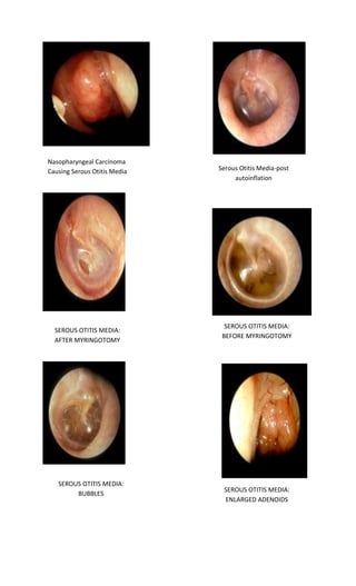
Serous OM Old 2
Serous B Before Autoinflation
Severe Acute Diffuse Otitis Externa
Acute Localized Otitis Externa [Furuncle]](https://image.slidesharecdn.com/tympanicmembranedr-141109103407-conversion-gate02/85/Tympanic-membrane-dr-fadil-19-320.jpg)
![Acute Localized Otitis Externa [Furuncle]
Acute Diffuse Otitis Externa [swimmer's ear]](https://image.slidesharecdn.com/tympanicmembranedr-141109103407-conversion-gate02/85/Tympanic-membrane-dr-fadil-20-320.jpg)

The document discusses the normal tympanic membrane and various conditions that can affect it. It provides over 50 images of normal tympanic membranes, as well as membranes affected by conditions like perforation, trauma, infection, effusion, and more. The tympanic membrane, also known as the eardrum, can be photographed via the external auditory canal or by drilling a hole perpendicular to gain different optical perspectives.


