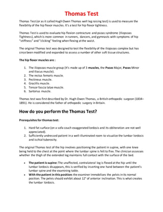
Thomas Test is used to evaluate hip flexion contracture and psoas syndrome (Iliopsoas Tightness), which is more common in runners, dancers, and gymnasts with symptoms of hip “stiffness” and “clicking” feeling when flexing at the waist.
Thomas Test is used to evaluate hip flexion contracture and psoas syndrome (Iliopsoas Tightness), which is more common in runners, dancers, and gymnasts with symptoms of hip “stiffness” and “clicking” feeling when flexing at the waist.
Tendoachilles rupture and its managementRohan Vakta
Achilles tendon is the strongest tendon of body. There are many causes of its rupture. It can be acute or chronic rupture. Management of chronic rupture by semitendinosus tendon is mentioned here.
Tendoachilles rupture and its managementRohan Vakta
Achilles tendon is the strongest tendon of body. There are many causes of its rupture. It can be acute or chronic rupture. Management of chronic rupture by semitendinosus tendon is mentioned here.
Lower Limb Human Anatomy ( Muscles )
by DR RAI M. AMMAR
www.facebook.com/drraiammar
www.twitter.com/drraiammar
www.instagram.com/drraiammar
www.linkedin.com/in/drraiammar
www.themedicall.com/blog/auther/drraiammar/
For Any Book or Notes Visit Our Website:
www.allmedicaldata.wordpress.com
www.drraiammar.blogspot.com
YOUTUBE CHANNEL :
https://www.youtube.com/channel/UCu-oR9V3OdFNTJW5yqXWXxA
ANY QUESTION ??
Get in touch with us at Any of the Above Social Media or Email at
drraiammar@gmail.com
allmedicaldata@gmail.com
Postural deviations of spine by Dr. NidhiNidhiVedawala
Types of Postural deviation ,Spinal deviation -Lordosis,Forward head posture,Sway back,Flat back,Kyphosis and Scoliosis....Each deformity's causes and correction...Physiotherapy Treatment.
Oswestry Disability Index or Oswestry low back pain disability questionnaire is a self-administered questionnaire divided into ten sections designed to assess limitations of various activities of daily living.
Each section is scored on a 0 – 5 scale, 5 representing the greatest disability. The index is calculated by dividing the summed score by the total possible score, which is then multiplied by 100 and expressed as a percentage.
Oswestry low back pain disability questionnaire has questions about: Pain Intensity, Standing, Personal Care, Sleeping, Lifting, Sex Life, Walking, social life, Sitting and Traveling.
Neck Disability Index (NDI) is a 10 item questions that measures a patient's neck pain related disability, it was first published in 1991 by Dr. Howard Vernon and was based on the Oswestry Low Back Pain Disability Questionnaire.
The 10 Questions of NDI include activities of daily living, such as: personal care, lifting, reading, work, driving, sleeping, recreational activities, pain intensity, concentration and headache.
The items are scored in descending order with the top statement = 0 and the bottom statement = 5
All subsections are added together for a cumulative score. The higher the score, the greater the disability.
Homan's Sign is a screening test used to check for deep vein thrombosis (DVT) of the calf. It’s sometimes called dorsiflexion sign.
It was first describes by John Homans in 1941 who was an American surgeon.
Read More: https://orthofixar.com/special-test/homans-sign/
A variety of hip muscles surround the hip joint, and act to accelerate, decelerate, and stabilize the hip joint. About 21 muscles cross the hip, providing both tri-planar movement and stability between the femur and the acetabulum.
https://orthofixar.com/anatomy/hip-muscles-anatomy/
Thomas Test (or as it called Hugh Owen Thomas well leg raising test) is used to measure the flexibility of the hip flexor muscles. It’s used to test for hip flexion contracture and psoas syndrome, which is more common in runners, dancers, and gymnasts with symptoms of hip “stiffness” and “clicking” feeling when flexing at the waist.
For more, See:
https://www.orthofixar.com/special-test/thomas-test/
How to Split Bills in the Odoo 17 POS ModuleCeline George
Bills have a main role in point of sale procedure. It will help to track sales, handling payments and giving receipts to customers. Bill splitting also has an important role in POS. For example, If some friends come together for dinner and if they want to divide the bill then it is possible by POS bill splitting. This slide will show how to split bills in odoo 17 POS.
Welcome to TechSoup New Member Orientation and Q&A (May 2024).pdfTechSoup
In this webinar you will learn how your organization can access TechSoup's wide variety of product discount and donation programs. From hardware to software, we'll give you a tour of the tools available to help your nonprofit with productivity, collaboration, financial management, donor tracking, security, and more.
How to Create Map Views in the Odoo 17 ERPCeline George
The map views are useful for providing a geographical representation of data. They allow users to visualize and analyze the data in a more intuitive manner.
How to Make a Field invisible in Odoo 17Celine George
It is possible to hide or invisible some fields in odoo. Commonly using “invisible” attribute in the field definition to invisible the fields. This slide will show how to make a field invisible in odoo 17.
The French Revolution, which began in 1789, was a period of radical social and political upheaval in France. It marked the decline of absolute monarchies, the rise of secular and democratic republics, and the eventual rise of Napoleon Bonaparte. This revolutionary period is crucial in understanding the transition from feudalism to modernity in Europe.
For more information, visit-www.vavaclasses.com
The Indian economy is classified into different sectors to simplify the analysis and understanding of economic activities. For Class 10, it's essential to grasp the sectors of the Indian economy, understand their characteristics, and recognize their importance. This guide will provide detailed notes on the Sectors of the Indian Economy Class 10, using specific long-tail keywords to enhance comprehension.
For more information, visit-www.vavaclasses.com
Synthetic Fiber Construction in lab .pptxPavel ( NSTU)
Synthetic fiber production is a fascinating and complex field that blends chemistry, engineering, and environmental science. By understanding these aspects, students can gain a comprehensive view of synthetic fiber production, its impact on society and the environment, and the potential for future innovations. Synthetic fibers play a crucial role in modern society, impacting various aspects of daily life, industry, and the environment. ynthetic fibers are integral to modern life, offering a range of benefits from cost-effectiveness and versatility to innovative applications and performance characteristics. While they pose environmental challenges, ongoing research and development aim to create more sustainable and eco-friendly alternatives. Understanding the importance of synthetic fibers helps in appreciating their role in the economy, industry, and daily life, while also emphasizing the need for sustainable practices and innovation.
This is a presentation by Dada Robert in a Your Skill Boost masterclass organised by the Excellence Foundation for South Sudan (EFSS) on Saturday, the 25th and Sunday, the 26th of May 2024.
He discussed the concept of quality improvement, emphasizing its applicability to various aspects of life, including personal, project, and program improvements. He defined quality as doing the right thing at the right time in the right way to achieve the best possible results and discussed the concept of the "gap" between what we know and what we do, and how this gap represents the areas we need to improve. He explained the scientific approach to quality improvement, which involves systematic performance analysis, testing and learning, and implementing change ideas. He also highlighted the importance of client focus and a team approach to quality improvement.
1. Thomas Test
Thomas Test (or as it called Hugh Owen Thomas well leg raising test) is used to measure the
flexibility of the hip flexor muscles. It's a test for hip flexor tightness.
Thomas Test is used to evaluate hip flexion contracture and psoas syndrome (Iliopsoas
Tightness), which is more common in runners, dancers, and gymnasts with symptoms of hip
“stiffness” and “clicking” feeling when flexing at the waist.
The original Thomas test was designed to test the flexibility of the iliopsoas complex but has
since been modified and expanded to assess a number of other soft tissue structures.
The hip flexor muscles are :
1. The iliopsoas muscle group (It’s made up of 3 muscles, the Psoas Major, Psoas Minor
and Iliacus muscle).
2. The rectus femoris muscle.
3. Pectineus muscle.
4. Gracillis muscle.
5. Tensor fascia latae muscle.
6. Sartorius muscle.
Thomas test was first described by Dr. Hugh Owen Thomas, a British orthopedic surgeon (1834–
1891). He is considered the father of orthopedic surgery in Britain.
How do you perform the Thomas Test?
Prerequisites for thomas test:
1. Hard fat surface (on a sofa couch exaggerated lordosis and its obliteration are not well
appreciated).
2. Sufficiently undressed patient in a well-illuminated room to visualize the lumbar lordosis
and ischial tuberosity.
The original Thomas test of the hip involves positioning the patient in supine, with one knee
being held to the chest at the point where the lumbar spine is felt to flex. The clinician assesses
whether the thigh of the extended leg maintains full contact with the surface of the bed.
The patient is supine: The unaffected, contralateral leg is flexed at the hip until the
lumbar lordosis disappears, this is verified by inserting one hand between the patient’s
lumbar spine and the examining table.
With the patient in this position: the examiner immobilizes the pelvis in its normal
position. The pelvis should exhibit about 12° of anterior inclination. This is what creates
the lumbar lordosis.
2. Another way to do Thomas test: With patient supine with both hips flexed and maintaining
one hip in flexion (to keep the pelvis fixed in corrected position),the patient is asked to actively
extend the limb as much as he/she can. Thomas test Positive if unable to touch posterior thigh
with examination table.
The angle between the thigh and the hard surface gives an idea of the flexion contracture at the
hip.
An increased flexion contracture in the hip can be compensated for by an increase in lumbar
lordosis, in which case the patient only appears to assume a normal position.
Starting position For the left hip assesment
3. Negative Thomas Test - Normal Left Hip Original Thomas Test
What does a positive Thomas Test mean?
The thomas test positive if the thigh is raised off the surface of the table. A positive test
indicates a decrease in flexibility in the rectus femoris or iliopsoas muscles or both.
In normal hip (Negative Thomas test), extension is only possible up to the neutral
position (0°); the thigh lies at on the surface of the examining table. Further flexion can
tilt the pelvis further upright. So long as the leg being examined remains in contact with
the examining table, the angle of pelvic tilt achieved corresponds to the maximum
hyperextension of the hip.
The flexion contracture can be quantified by measuring the angle that the flexed,
affected leg forms with the examining table.
One of the limitations of thomas test is that it merely determines the amount of hip extension
possible at any given degree of pelvic flexion. Another problem is that there are better methods
of measuring the flexibility of the iliopsoas complex. For example, positioning the patient in
prone, stabilizing the pelvis, and then extending the thigh. The precise point at which the pelvis
begins to rise marks the end of the hip motion and the beginning of pelvic and spine motion.
The causes of false positive Thomas test include:
1. Wrong technique is the most common cause,
2. Fixed pelvic obliquity in scoliosis and polio,
3. Exaggerated lordosis in obese individuals,
4. 4. Malformed pelvis.
Positive Thomas Test - Flexion contracture of the left hip
How reliable is the Thomas test?
Neither the original Thomas Test nor the suggested variations have ever been substantiated for
reliability, sensitivity, or specificity1:
Sensitivity: 31 %
Specificity: 57 %
Modified Thomas Test
A modified thomas test is commonly used to help eliminate the effect of the lumbar curve.
For the modified Thomas Test , the patient is positioned in sitting at the end of the bed. From
this position, the patient is asked to lie back, while bringing both knees against the chest. Once
in this position, the patient is asked to perform a posterior pelvic tilt. While the contralateral
hip is held in maximum hip flexion by the patient's hands, the tested limb is lowered over the
end of the bed toward the floor.
What does a positive modified Thomas Test indicate?
5. If normal, the thigh should be parallel with the bed, in neutral rotation, and neither abducted
nor adducted, with the lower leg being perpendicular to the thigh and in neutral rotation. There
should be 100–110 degrees of knee flexion present with the thigh in line with the table.
If the thigh is raised compared to the table, a decrease in the flexibility of the iliopsoas muscle
complex should be suspected.
If the rectus femoris is adaptively shortened, the amount of knee extension should increase
with the application of overpressure into hip extension.
If the decrease in flexibility lies with the iliopsoas, attempts to correct the hip position should
result in an increase in the external rotation of the thigh.
The application of overpressure into knee flexion can also be used. If the increase in knee
flexion produces an increase in hip flexion (the thigh rises higher off the bed), the rectus
femoris is implicated, whereas if the overpressure produces no change in the degree of hip
flexion, the iliopsoas is implicated.
The data illustrated that reliable assessment using the modified Thomas test may be
influenced by:
1. variations in the application of assessment criteria among examiners,
2. the scoring method used,
3. the consistency and accuracy of establishing surface landmarks,
4. the population from which the sample was selected.
6. Modified Thomas Test
Notes
The Tomas test is not useful in bilateral pathology, as the sound limb needs to be
maneuvered, and patients with knee pathologies restricting flexion.
Hip extension is important for the action of various athletic activities. A restriction of hip
extension has been thought to lead to an over striding gait and increased impact forces
during running, which may increase the risk of tibial stress fracture.
A restriction of the hip extension may be associated with contracture in the hip flexor
muscles. A postural hypothesis related to hamstring strains is that contracted hip flexors
lead to an anterior pelvic tilt, which may predispose runner athletes to hamstring
strains.
For individuals with low back pain that is sensitive to spinal extension, contracted hip
flexors may lead these individuals to perform spinal movements that lead to increased
spinal extension, as the individual lacks movement options due to their hip extension
limitations.
Thomas test can also be used to assess the flexibility of the Tensor fascia latae (TFL), if
the hip of the tested leg is maximally adducted while monitoring the ipsilateral the
anterior superior iliac spine (ASIS)for motion. There should be 20 degrees of hip
adduction available.
Flexion contracture of the hip may result from psoas spasmsecondary to inflammation
or pus in the region of its sheath in the pelvis. This is seen, for example, in appendicitis,
appendix abscess or other pelvic inflammatory disease. Examination of the abdomen is
essential.
7. Two things must be remembered when interpreting the results of Thomas Test:
1. The criteria are arbitrary and have been shown to vary between genders and limb
dominance and to depend on the types and the levels of activity undertaken by the
individual.
2. The apparent tightness might simply be normal tissue tension, producing a deviation of
the leg because of an increased flexibility of the antagonists.
The principle behind Thomas test:
Fixed saggital plane deformity is compensated by pelvic extension (flexion deformity) and vice
versa. This is produced by lumbar lordosis which shares the compensation. The combined effect
allows a patient to walk with feet touching the ground in conjunction with knee flexion. While
we flex the normal hip the deformity in pelvis first gets corrected (something akin to squaring
pelvis) then lumbar lordosis is corrected simultaneously revealing the deformity.
Related Anatomy
Iliopsoas Muscle:
The iliopsoas muscle, formed by the iliacus and psoas major muscles, is the most
powerful hip flexor, while also functioning as a weak adductor and external rotator of
the hip.
The iliopsoas attaches to the hip joint capsule, thereby giving it some support.
Since the muscle spans both the axial and appendicular components of the skeleton, it
also functions as a trunk flexor, and affords an important element to the vertical
stability of the lumbar spine, especially when the hip is in full extension and passive
tension is greatest in the muscle.
Theoretically, a sufficiently strong and isolated bilateral contraction of any hip flexor
muscle will either rotate the femur toward the pelvis, the pelvis (and possibly the trunk)
toward the femur, or both actions simultaneously.
Rectus Femoris Muscle:
The rectus femoris muscle, one of the four quadriceps muscles, is a two-joint muscle
that arises from two tendons: one, the anterior or straight, from the anterior inferior
iliac spine (AIIS); the other, the posterior or reflected, from a groove above the brim of
the acetabulum.
The rectus femoris combines movements of flexion at the hip and extension at the knee.
It functions more effectively as a hip flexor when the knee is flexed, as when a person
kicks a ball.
Pectineus Muscle:
8. The pectineus is an adductor, flexor, and internal rotator of the hip. Like the iliopsoas,
the pectineus attaches to and supports the joint capsule of the hip.
Gracilis Muscle:
The gracilis , the longest of the hip adductors, is also the most superficial and medial of
the hip adductor muscles.
gracilis functions to adduct and flex the thigh and flex and internally rotate the leg.
Tensor Fascia Latae Muscle:
The TFL envelops the muscles of the thigh.
The TFL counteracts the backward pull of the gluteus maximus on the iliotibial band
(ITB).
The TFL also flexes, abducts, and externally rotates the hip.
The trochanteric bursa is found deep to this muscle, as it passes over the greater
trochanter.
The attachment of the TFL via the ITB to the anterolateral tibia provides a flexion
moment in knee flexion and
an extension moment in knee extension.
Sartorius Muscle:
The sartorius muscle is the longest muscle in the body.
The sartorius is responsible for flexion, abduction, and external rotation of the hip, and
some degree of knee flexion.
Muscle Origin Insertion Nerve
Iliopsoas muscle Transverseprocesses of L1-L5
vertebra
Lesser trochanter Femoral Nerve
The rectus femoris muscle Anterior inferior iliac spine
AIIS, acetabular rim
Patella and tibial
tubercle
Femoral Nerve
Pectineus muscle Pectineal lineof pubis Pectineal lineof
femur
Femoral and
obturator Nerve
Gracillis muscle Inferior symphysis/ pubic arch Proximal medial
tibia
Obturator Anterior
Nerve
Tensor fasciae latae muscle
(tensor fasciae femoris)
Anterior iliaccrest Iliotibial band Superior gluteal
Nerve
Sartorius muscle Anterior superior iliac spine
ASIS
Proximal medial
tibia
Femoral Nerve
9. Progressive Fibrosis of the Quadriceps:
Progressive fibrosis of the quadriceps muscle is a condition in which extension contracture of
the knee develops in early childhood as a result of fibrosis of one or more components of the
quadriceps muscle. The condition is more common in girls than in boys.
The exact cause of progressive fibrosis of the quadriceps is not known. Gunn2 first proposed
that it was a sequela of multiple injections of antibiotics into the thigh muscles during early
infancy.
The pathophysiology of progressive fibrosis is speculative. It has been proposed that the
volume of drug injected in
very young infants compresses the capillaries and muscle fibers and causes muscle ischemia,
which leads to fibrotic changes. Local necrosis may occur as a result of focal disruption of fibers
at the site of injection. The irritative nature of the injected drug may also play a role in
producing fibrosis.
Clinical Symptoms:
1. The clinical hallmark of progressive fibrosis of the quadriceps is painless, progressive
limitation of both active and passive knee flexion with an extension contracture. The
vastus intermedius is most commonly involved. Fibrosis
occurs more distally than proximally, within and between the muscle fibers.
2. A dimple in the skin may be present because of the rigid, fibrous septa that extend
between the skin and the deep fascia; the dimple deepens with forced flexion of the
knee.
3. Range of motion is painless within the available arc.
4. The involved muscle is atrophic, with subcutaneous hardness and limitation of motion.
5. Genu recurvatum may develop in severe cases.
6. The patella is high riding. Habitual dislocation of the patella may occur in chronic cases.
7. Knee flexion in these patients is accomplished through lateral dislocation of the patella.
With the patella held within the groove of the femur, the knee cannot be flexed. In
these patients the vastus lateralis is usually involved. This condition differs from
congenital lateral dislocation of the patella in that it is an acquired contracture resulting
from progressive fibrosis.
Treatment
Two different surgical releases have been advocated for the treatment of quadriceps fibrosis:
1. The first is surgical release of the extension contracture by proximal division of the
fibrotic muscular bands, which is often combined with transverse division of the iliotibial
10. tract. This approach is preferred in patients younger than 10 years in whom no
radiographic changes are present in the distal end of the femur.
2. The other surgical approach is V-Y quadricepsplasty to lengthen the extensor
mechanism as a whole when the fibrosis is extensive. Postoperative extensor lag may be
present but resolves with time in most cases. The extensor lag is more prevalent
following V-Y plasty than after proximal release of the fibrotic bands.
When the fibrosis is chronic and genu recurvatum is present, skeletal changes may develop in
the distal end of the femur where the articular surface points anteriorly. In such cases it may be
necessary to perform distal femoral flexion osteotomy to gain knee flexion and maintain joint
congruity.
Hip Flexion contracture
A flexion contracture at the hip is a common occurrence. Hip flexion contractures can result
from:
1. adaptive shortening of the iliopsoas muscle or rectus femoris muscles;
2. contracture of the anterior hip capsuloligamentous complex.
These changes to the soft tissue and connective tissues around the hip can result from OA,
injury, or sustained postures involving hip flexion. The resulting anterior rotation of the pelvis
shifts the weight-bearing of the hip to a thinner region of hyaline cartilage, in both the femur
and the acetabulum, and places the hip extensors in a state of low-level tension.
Flexion contractures can be diagnosed using the Thomas test.
The intervention for the contracture is based on the cause. Adaptive shortening of the
contractile tissues may be addressed using muscle energy, passive stretching, and myofascial
techniques. Stretching of the capsuloligamentous complex is accomplished by grade III
distraction mobilizations and by prolonged stretching.
11. Reference
1. Thomas Test - Orthofixar
2. The modified Thomas test is not a valid measure of hip extension unless pelvic tilt is
controlled | Andrew D. Vigotsky, Gregory J. Lehman, Chris Beardsley, Bret Contreras,
Bryan Chung, Erin H. Feser PeerJ. 2016; 4: e2325. Published online 2016 Aug 11. doi:
10.7717/peerj.2325 PMCID: PMC4991856.
3. Gunn DR: Contracture of the quadriceps muscle. A discussion on the etiology and
relationship to recurrent dislocation of the patella, J Bone Joint Surg Br 46: 492, 1964.
4. Clapis, Davis & Davis (2007) Clapis PA, Davis SM, Davis RO. Reliability of inclinometer
and goniometric measurements of hip extension flexibility using the modified Thomas
test. Physiotherapy Theory and Practice. 2007;24:135–141. doi:
10.1080/09593980701378256.
5. Harvey D: Assessment of the flexibility of elite athletes using the modified Thomas test.
Br J Sports Med 32:68–70, 1998.
6. Peeler JD, Anderson JE. Reliability limits of the modified Thomas test for assessing rectus
femoris muscle flexibility about the knee joint. J Athl Train. 2008 Sep-Oct;43(5):470-6.
doi: 10.4085/1062-6050-43.5.470. PMID: 18833309; PMCID: PMC2547866.
7. Lee LW, Kerrigan DC, Della Croce U. Dynamic implications of hip flexion contractures.
Am J Phys Med Rehabil. 1997 Nov-Dec;76(6):502-8. doi: 10.1097/00002060-199711000-
00013. PMID: 9431270.
8. Magee D.J. Orthopedic Physical Assessment. 4th ed. Vol. 2002. Philadelphia, PA: WB
Saunders; Hip; pp. 607–660.
9. Kendall F.P, McCreary E.K, Provance P.G, Rodgers M.M, Romani W.A. Muscles: Testing
and Function, With Posture and Pain. 5th ed. Vol. 2005. Baltimore, MD: Lippincott
Williams & Wilkins; Lower extremity; pp. 359–464.
10. Peeler J, Anderson J.E. Reliability of the Thomas test for assessing range of motion about
the hip. Phys Ther Sport. 2007;8(1):14–21.
11. Cibere J, Thorne A, Bellamy N, Greidanus N, Chalmers A, Mahomed N, Shojania K, Kopec
J, Esdaile JM. Reliability of the hip examination in osteoarthritis: effect of
standardization. Arthritis Rheum. 2008 Mar 15;59(3):373-81. doi: 10.1002/art.23310.
PMID: 18311750.
12. Clinical Tests for the Musculoskeletal System 3rd Ed. Book.
13. Mark Dutton, Pt . Dutton’s Orthopaedic Examination, Evaluation, And Intervention, 3rd
Edition Book.
14. Millers Review of Orthopaedics, 7th Edition Book.