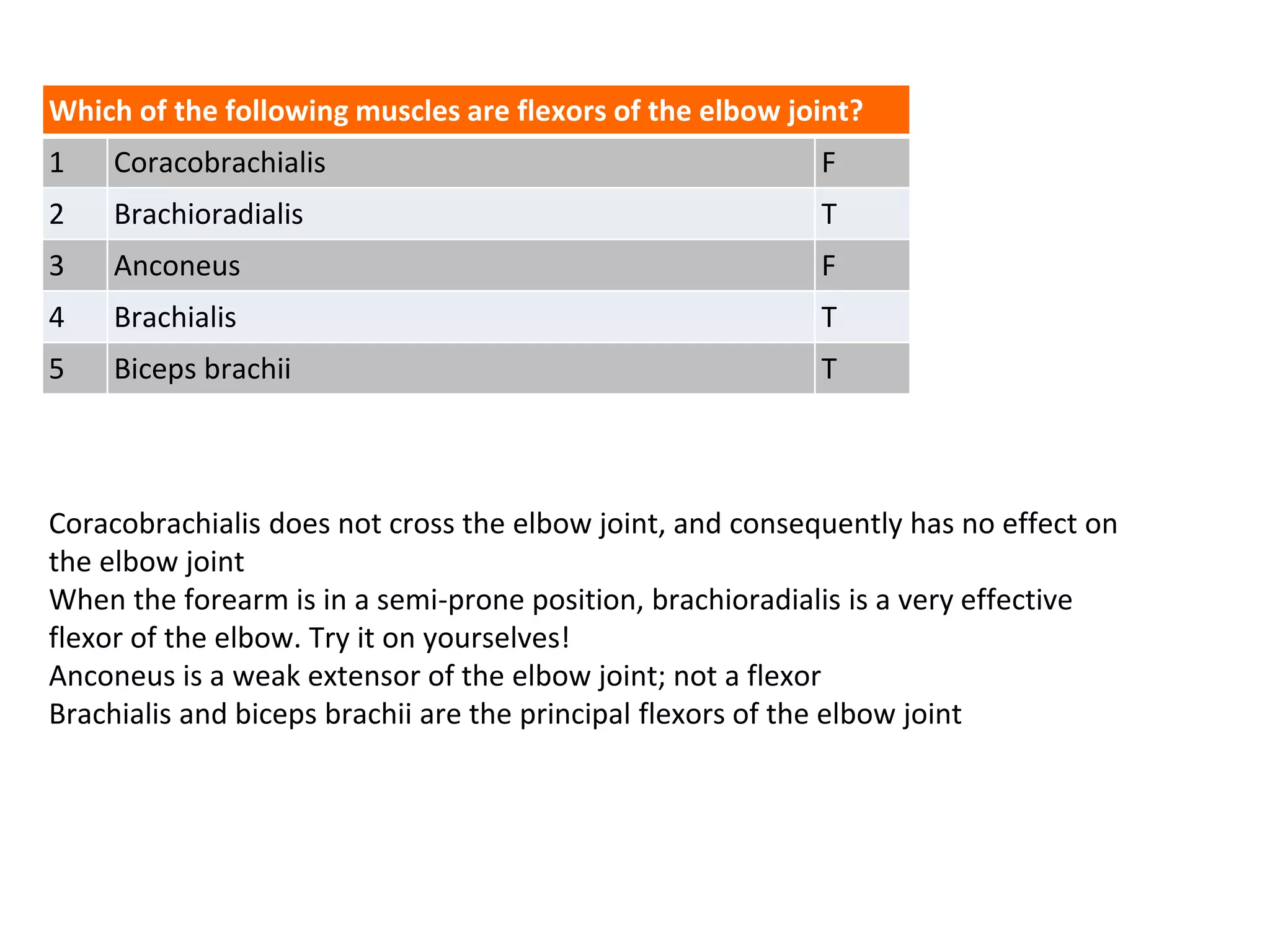
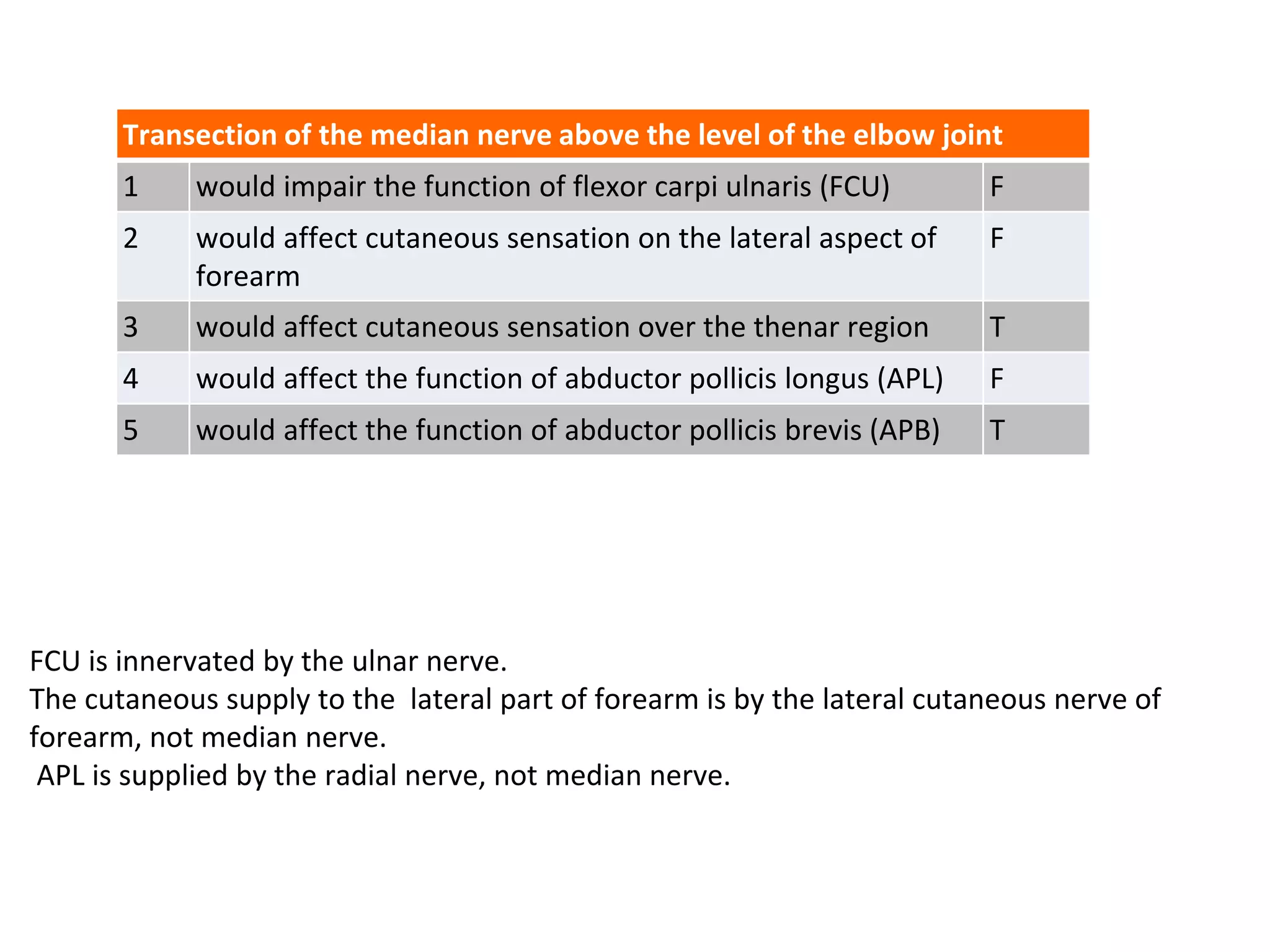
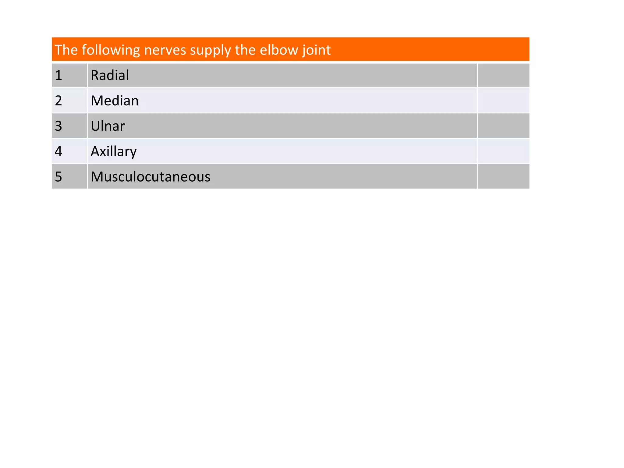
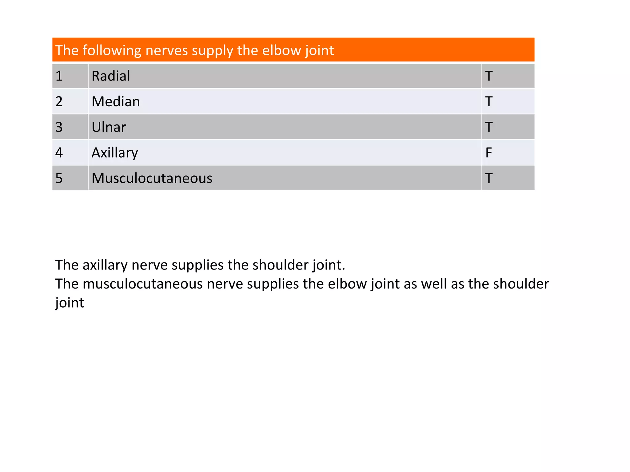
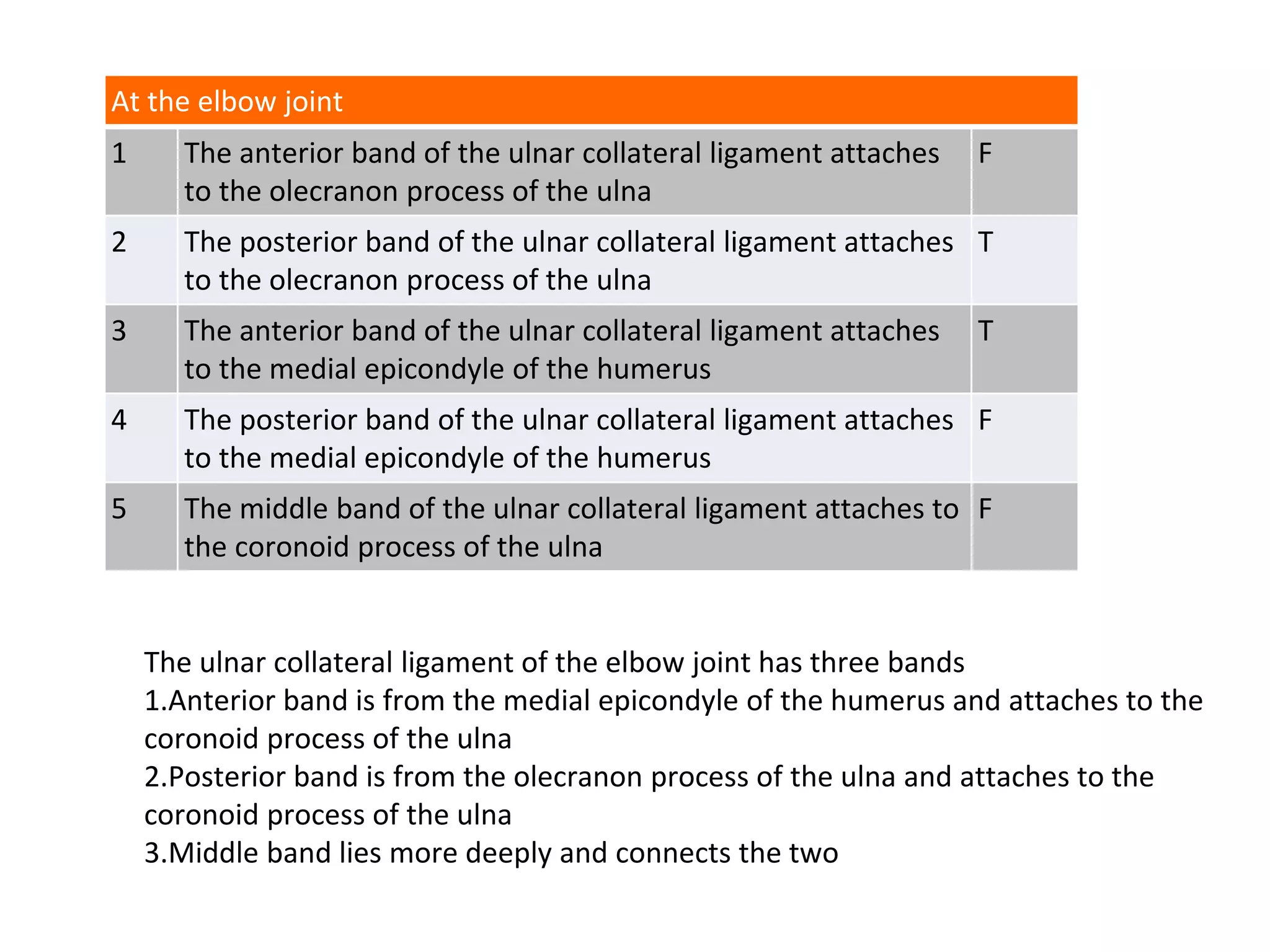
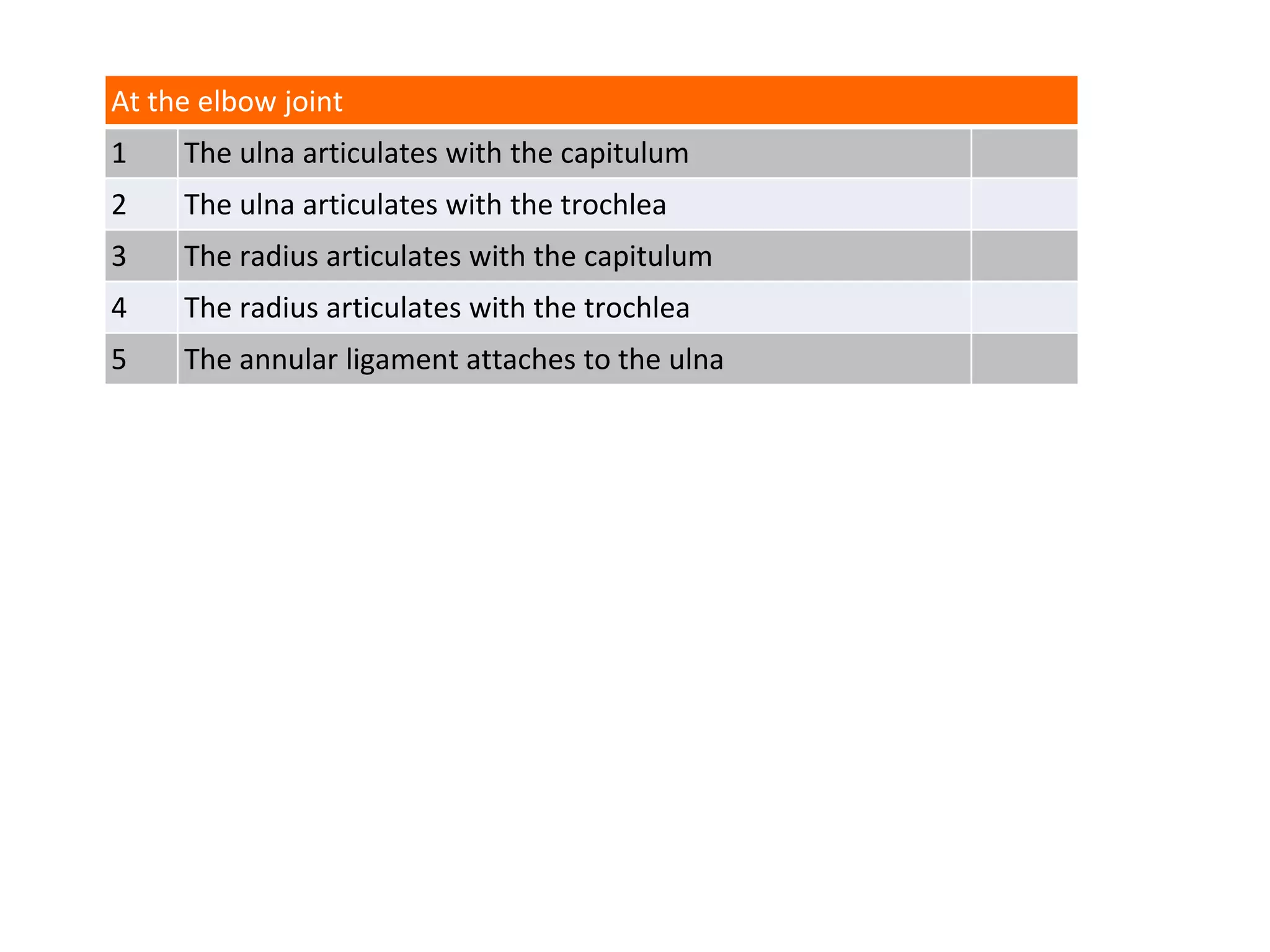
Questions
Elbow Joint
At the elbow joint
1 the radius articulates with the capitulum of the humerus
2 the lateral collateral ligament of the elbow joint is attached to the
neck of radius
3 the medial (ulnar) collateral ligament of the elbow joint is attached
to the medial epicondyle of humerus
4 the brachialis muscle is attached to the capsule of the elbow joint
5 the superior radio-ulnar joint shares a synovial cavity with the
elbow joint
The lateral collateral ligament has no direct attachment to the radius. It is attached to
the annular ligament which surrounds the head and neck of radius.
At the elbow joint
1 the radius articulates with the capitulum of the humerus T
2 the lateral collateral ligament of the elbow joint is attached to the
neck of radius
F
3 the medial (ulnar) collateral ligament of the elbow joint is attached
to the medial epicondyle of humerus
T
4 the brachialis muscle is attached to the capsule of the elbow joint T
5 the superior radio-ulnar joint shares a synovial cavity with the
elbow joint
T
In the cubital fossa
1 the ulnar nerve lies lateral to the brachial artery
2 the tendon of biceps brachii lies medial to the
brachial artery
3 the median nerve lies medial to the brachial artery
4 the bicipital aponeurosis crosses deep to the brachial
artery
5 the median nerve is seen to enter the pronator teres
muscle
The ulnar nerve is not a content of the cubital fossa.
The biceps tendon lies lateral to the brachial artery.
The bicipital aponeurosis (also known as the lacertus fibrosus) runs obliquely downwards
and medially superficial to the brachial artery and median nerve)
The median nerve leaves the cubital fossa by coursing distally between the two heads of
pronator teres.
In the cubital fossa
1 the ulnar nerve lies lateral to the brachial artery F
2 the tendon of biceps brachii lies medial to the
brachial artery
F
3 the median nerve lies medial to the brachial artery T
4 the bicipital aponeurosis crosses deep to the brachial
artery
F
5 the median nerve is seen to enter the pronator teres
muscle
T
Transection of the radial nerve at the level of the humeral epicondyles will cause
1 loss of sensation in the nailbeds of index and middle fingers
2 wrist drop
3 paralysis of extensor pollicis longus
4 loss of cutaneous sensation on dorsum of ulnar border of hand
5 loss of extension of the elbow joint
The nailbeds of the index and middle fingers are innervated by the median nerve; not the
radial nerve.
Wrist drop occurs due to paralysis of virtually all the muscles in the extensor compartment
of forearm, including the wrist extensors
The dorsum of the ulnar aspect of hand is supplied by the dorsal branches of the ulnar
nerve, not by the radial nerve
The radial nerve’s branches to triceps brachii (the extensor of the elbow) are given off in the
proximal part of the arm. Consequently an injury to the radial nerve at the level of the
epicondyles will not compromise elbow extension.
Transection of the radial nerve at the level of the humeral epicondyles will cause
1 loss of sensation in the nailbeds of index and middle fingers F
2 wrist drop T
3 paralysis of extensor pollicis longus T
4 loss of cutaneous sensation on dorsum of ulnar border of hand F
5 loss of extension of the elbow joint F
Which of the following muscles are flexors of the elbow joint?
1 Coracobrachialis
2 Brachioradialis
3 Anconeus
4 Brachialis
5 Biceps brachii
Coracobrachialis does not cross the elbow joint, and consequently has no effect on
the elbow joint
When the forearm is in a semi-prone position, brachioradialis is a very effective
flexor of the elbow. Try it on yourselves!
Anconeus is a weak extensor of the elbow joint; not a flexor
Brachialis and biceps brachii are the principal flexors of the elbow joint
Which of the following muscles are flexors of the elbow joint?
1 Coracobrachialis F
2 Brachioradialis T
3 Anconeus F
4 Brachialis T
5 Biceps brachii T
Transection of the median nerve above the level of the elbow joint
1 would impair the function of flexor carpi ulnaris (FCU)
2 would affect cutaneous sensation on the lateral aspect of
forearm
3 would affect cutaneous sensation over the thenar region
4 would affect the function of abductor pollicis longus (APL)
5 would affect the function of abductor pollicis brevis (APB)
FCU is innervated by the ulnar nerve.
The cutaneous supply to the lateral part of forearm is by the lateral cutaneous nerve of
forearm, not median nerve.
APL is supplied by the radial nerve, not median nerve.
Transection of the median nerve above the level of the elbow joint
1 would impair the function of flexor carpi ulnaris (FCU) F
2 would affect cutaneous sensation on the lateral aspect of
forearm
F
3 would affect cutaneous sensation over the thenar region T
4 would affect the function of abductor pollicis longus (APL) F
5 would affect the function of abductor pollicis brevis (APB) T
A distal humeral fracture causing complete transection of the ulnar
nerve at the level of the medial epicondyle will produce
1 impairment of flexion of the distal interphalangeal joint of
index finger
2 cutaneous sensory loss over the medial aspect of the hand
3 weakness of pinch between index finger and thumb
4 wrist drop
5 wasting of all intrinsic muscles of the hand
Weakness of pinch is a consequence of paralysis of the adductor pollicis and 1st
dorsal interosseous
Wrist drop is caused by a radial nerve injury
A distal humeral fracture causing complete transection of the ulnar
nerve at the level of the medial epicondyle will produce
1 impairment of flexion of the distal interphalangeal joint of
index finger
F
2 cutaneous sensory loss over the medial aspect of the hand T
3 weakness of pinch between index finger and thumb T
4 wrist drop F
5 wasting of all intrinsic muscles of the hand F
The following nerves supply the elbow joint
1 Radial
2 Median
3 Ulnar
4 Axillary
5 Musculocutaneous
The following nerves supply the elbow joint
1 Radial T
2 Median T
3 Ulnar T
4 Axillary F
5 Musculocutaneous T
The axillary nerve supplies the shoulder joint.
The musculocutaneous nerve supplies the elbow joint as well as the shoulder
joint
At the elbow joint
1 The anterior band of the ulnar collateral ligament attaches
to the olecranon process of the ulna
2 The posterior band of the ulnar collateral ligament attaches
to the olecranon process of the ulna
3 The anterior band of the ulnar collateral ligament attaches
to the medial epicondyle of the humerus
4 The posterior band of the ulnar collateral ligament attaches
to the medial epicondyle of the humerus
5 The middle band of the ulnar collateral ligament attaches to
the coronoid process of the ulna
At the elbow joint
1 The anterior band of the ulnar collateral ligament attaches
to the olecranon process of the ulna
F
2 The posterior band of the ulnar collateral ligament attaches
to the olecranon process of the ulna
T
3 The anterior band of the ulnar collateral ligament attaches
to the medial epicondyle of the humerus
T
4 The posterior band of the ulnar collateral ligament attaches
to the medial epicondyle of the humerus
F
5 The middle band of the ulnar collateral ligament attaches to
the coronoid process of the ulna
F
The ulnar collateral ligament of the elbow joint has three bands
1.Anterior band is from the medial epicondyle of the humerus and attaches to the
coronoid process of the ulna
2.Posterior band is from the olecranon process of the ulna and attaches to the
coronoid process of the ulna
3.Middle band lies more deeply and connects the two
The radial collateral ligament has
1 1 band
2 2 bands
3 3 bands
4 4 bands
5 5 bands
The radial collateral ligament has
1 1 band T
2 2 bands F
3 3 bands F
4 4 bands F
5 5 bands F
The radial collateral ligament of the elbow joint is a single band that attaches to the
lateral epicondyle of the humerus just below the common extensor origin and it
blends with the annular ligament.
At the elbow joint
1 The ulna articulates with the capitulum
2 The ulna articulates with the trochlea
3 The radius articulates with the capitulum
4 The radius articulates with the trochlea
5 The annular ligament attaches to the ulna
At the elbow joint
1 The ulna articulates with the capitulum F
2 The ulna articulates with the trochlea T
3 The radius articulates with the capitulum T
4 The radius articulates with the trochlea F
5 The annular ligament attaches to the ulna T

Exam Questions Elbow Joint

  • 1.
  • 2.
    At the elbowjoint 1 the radius articulates with the capitulum of the humerus 2 the lateral collateral ligament of the elbow joint is attached to the neck of radius 3 the medial (ulnar) collateral ligament of the elbow joint is attached to the medial epicondyle of humerus 4 the brachialis muscle is attached to the capsule of the elbow joint 5 the superior radio-ulnar joint shares a synovial cavity with the elbow joint
  • 3.
    The lateral collateralligament has no direct attachment to the radius. It is attached to the annular ligament which surrounds the head and neck of radius. At the elbow joint 1 the radius articulates with the capitulum of the humerus T 2 the lateral collateral ligament of the elbow joint is attached to the neck of radius F 3 the medial (ulnar) collateral ligament of the elbow joint is attached to the medial epicondyle of humerus T 4 the brachialis muscle is attached to the capsule of the elbow joint T 5 the superior radio-ulnar joint shares a synovial cavity with the elbow joint T
  • 4.
    In the cubitalfossa 1 the ulnar nerve lies lateral to the brachial artery 2 the tendon of biceps brachii lies medial to the brachial artery 3 the median nerve lies medial to the brachial artery 4 the bicipital aponeurosis crosses deep to the brachial artery 5 the median nerve is seen to enter the pronator teres muscle
  • 5.
    The ulnar nerveis not a content of the cubital fossa. The biceps tendon lies lateral to the brachial artery. The bicipital aponeurosis (also known as the lacertus fibrosus) runs obliquely downwards and medially superficial to the brachial artery and median nerve) The median nerve leaves the cubital fossa by coursing distally between the two heads of pronator teres. In the cubital fossa 1 the ulnar nerve lies lateral to the brachial artery F 2 the tendon of biceps brachii lies medial to the brachial artery F 3 the median nerve lies medial to the brachial artery T 4 the bicipital aponeurosis crosses deep to the brachial artery F 5 the median nerve is seen to enter the pronator teres muscle T
  • 6.
    Transection of theradial nerve at the level of the humeral epicondyles will cause 1 loss of sensation in the nailbeds of index and middle fingers 2 wrist drop 3 paralysis of extensor pollicis longus 4 loss of cutaneous sensation on dorsum of ulnar border of hand 5 loss of extension of the elbow joint
  • 7.
    The nailbeds ofthe index and middle fingers are innervated by the median nerve; not the radial nerve. Wrist drop occurs due to paralysis of virtually all the muscles in the extensor compartment of forearm, including the wrist extensors The dorsum of the ulnar aspect of hand is supplied by the dorsal branches of the ulnar nerve, not by the radial nerve The radial nerve’s branches to triceps brachii (the extensor of the elbow) are given off in the proximal part of the arm. Consequently an injury to the radial nerve at the level of the epicondyles will not compromise elbow extension. Transection of the radial nerve at the level of the humeral epicondyles will cause 1 loss of sensation in the nailbeds of index and middle fingers F 2 wrist drop T 3 paralysis of extensor pollicis longus T 4 loss of cutaneous sensation on dorsum of ulnar border of hand F 5 loss of extension of the elbow joint F
  • 8.
    Which of thefollowing muscles are flexors of the elbow joint? 1 Coracobrachialis 2 Brachioradialis 3 Anconeus 4 Brachialis 5 Biceps brachii
  • 9.
    Coracobrachialis does notcross the elbow joint, and consequently has no effect on the elbow joint When the forearm is in a semi-prone position, brachioradialis is a very effective flexor of the elbow. Try it on yourselves! Anconeus is a weak extensor of the elbow joint; not a flexor Brachialis and biceps brachii are the principal flexors of the elbow joint Which of the following muscles are flexors of the elbow joint? 1 Coracobrachialis F 2 Brachioradialis T 3 Anconeus F 4 Brachialis T 5 Biceps brachii T
  • 10.
    Transection of themedian nerve above the level of the elbow joint 1 would impair the function of flexor carpi ulnaris (FCU) 2 would affect cutaneous sensation on the lateral aspect of forearm 3 would affect cutaneous sensation over the thenar region 4 would affect the function of abductor pollicis longus (APL) 5 would affect the function of abductor pollicis brevis (APB)
  • 11.
    FCU is innervatedby the ulnar nerve. The cutaneous supply to the lateral part of forearm is by the lateral cutaneous nerve of forearm, not median nerve. APL is supplied by the radial nerve, not median nerve. Transection of the median nerve above the level of the elbow joint 1 would impair the function of flexor carpi ulnaris (FCU) F 2 would affect cutaneous sensation on the lateral aspect of forearm F 3 would affect cutaneous sensation over the thenar region T 4 would affect the function of abductor pollicis longus (APL) F 5 would affect the function of abductor pollicis brevis (APB) T
  • 12.
    A distal humeralfracture causing complete transection of the ulnar nerve at the level of the medial epicondyle will produce 1 impairment of flexion of the distal interphalangeal joint of index finger 2 cutaneous sensory loss over the medial aspect of the hand 3 weakness of pinch between index finger and thumb 4 wrist drop 5 wasting of all intrinsic muscles of the hand
  • 13.
    Weakness of pinchis a consequence of paralysis of the adductor pollicis and 1st dorsal interosseous Wrist drop is caused by a radial nerve injury A distal humeral fracture causing complete transection of the ulnar nerve at the level of the medial epicondyle will produce 1 impairment of flexion of the distal interphalangeal joint of index finger F 2 cutaneous sensory loss over the medial aspect of the hand T 3 weakness of pinch between index finger and thumb T 4 wrist drop F 5 wasting of all intrinsic muscles of the hand F
  • 14.
    The following nervessupply the elbow joint 1 Radial 2 Median 3 Ulnar 4 Axillary 5 Musculocutaneous
  • 15.
    The following nervessupply the elbow joint 1 Radial T 2 Median T 3 Ulnar T 4 Axillary F 5 Musculocutaneous T The axillary nerve supplies the shoulder joint. The musculocutaneous nerve supplies the elbow joint as well as the shoulder joint
  • 16.
    At the elbowjoint 1 The anterior band of the ulnar collateral ligament attaches to the olecranon process of the ulna 2 The posterior band of the ulnar collateral ligament attaches to the olecranon process of the ulna 3 The anterior band of the ulnar collateral ligament attaches to the medial epicondyle of the humerus 4 The posterior band of the ulnar collateral ligament attaches to the medial epicondyle of the humerus 5 The middle band of the ulnar collateral ligament attaches to the coronoid process of the ulna
  • 17.
    At the elbowjoint 1 The anterior band of the ulnar collateral ligament attaches to the olecranon process of the ulna F 2 The posterior band of the ulnar collateral ligament attaches to the olecranon process of the ulna T 3 The anterior band of the ulnar collateral ligament attaches to the medial epicondyle of the humerus T 4 The posterior band of the ulnar collateral ligament attaches to the medial epicondyle of the humerus F 5 The middle band of the ulnar collateral ligament attaches to the coronoid process of the ulna F The ulnar collateral ligament of the elbow joint has three bands 1.Anterior band is from the medial epicondyle of the humerus and attaches to the coronoid process of the ulna 2.Posterior band is from the olecranon process of the ulna and attaches to the coronoid process of the ulna 3.Middle band lies more deeply and connects the two
  • 18.
    The radial collateralligament has 1 1 band 2 2 bands 3 3 bands 4 4 bands 5 5 bands
  • 19.
    The radial collateralligament has 1 1 band T 2 2 bands F 3 3 bands F 4 4 bands F 5 5 bands F The radial collateral ligament of the elbow joint is a single band that attaches to the lateral epicondyle of the humerus just below the common extensor origin and it blends with the annular ligament.
  • 20.
    At the elbowjoint 1 The ulna articulates with the capitulum 2 The ulna articulates with the trochlea 3 The radius articulates with the capitulum 4 The radius articulates with the trochlea 5 The annular ligament attaches to the ulna
  • 21.
    At the elbowjoint 1 The ulna articulates with the capitulum F 2 The ulna articulates with the trochlea T 3 The radius articulates with the capitulum T 4 The radius articulates with the trochlea F 5 The annular ligament attaches to the ulna T