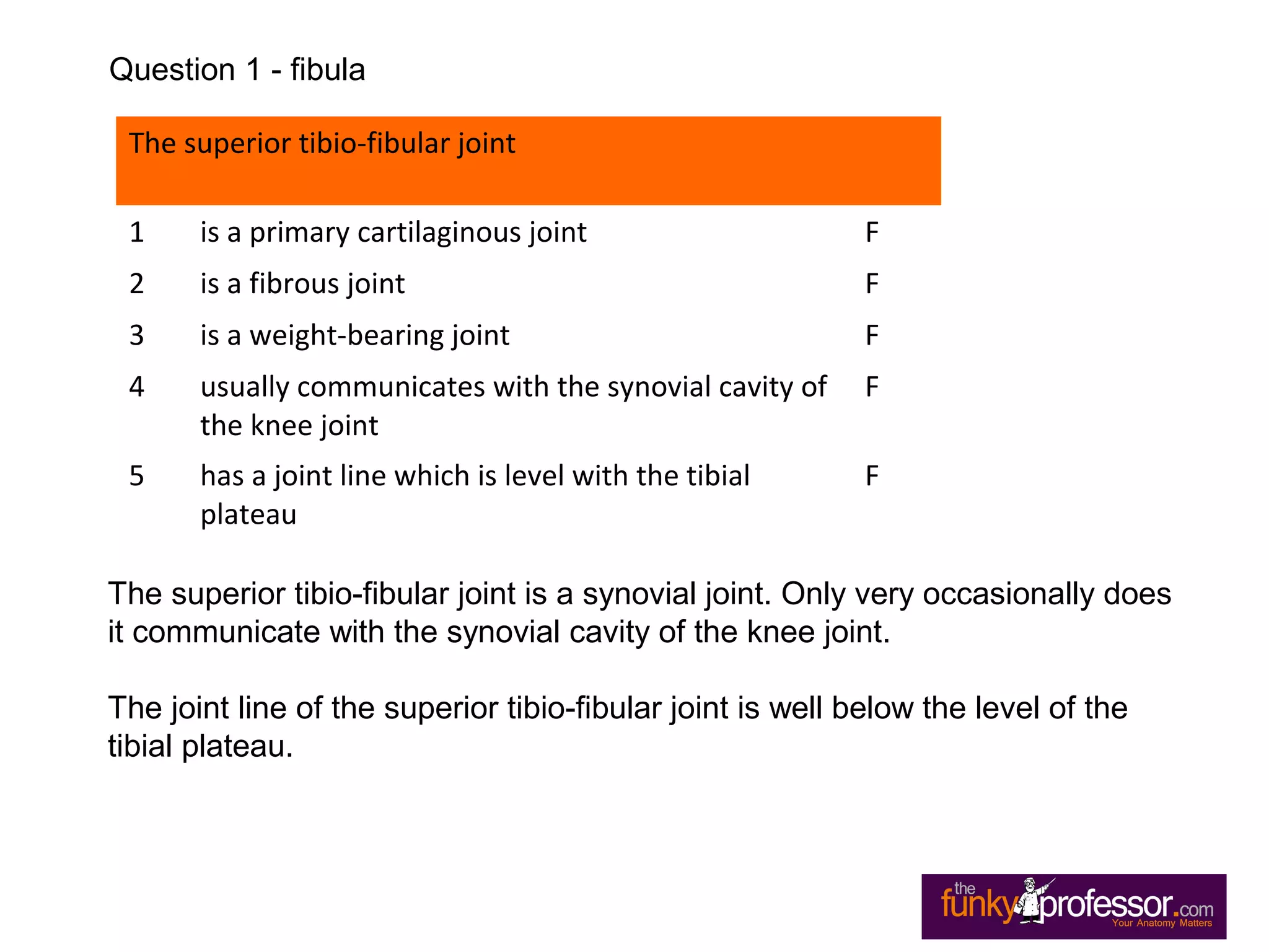
Questions
Fibula
The superior tibio-fibular joint
1 is a primary cartilaginous joint
2 is a fibrous joint
3 is a weight-bearing joint
4 usually communicates with the synovial cavity of
the knee joint
5 has a joint line which is level with the tibial
plateau
Question 1 - fibula
The superior tibio-fibular joint
1 is a primary cartilaginous joint F
2 is a fibrous joint F
3 is a weight-bearing joint F
4 usually communicates with the synovial cavity of
the knee joint
F
5 has a joint line which is level with the tibial
plateau
F
The superior tibio-fibular joint is a synovial joint. Only very occasionally does
it communicate with the synovial cavity of the knee joint.
The joint line of the superior tibio-fibular joint is well below the level of the
tibial plateau.
Question 1 - fibula
Concerning the fibula
1 the peroneus longus muscle arises form the lower two-thirds of the
lateral surface of the fibular shaft
2 the tip of the fibular malleolus normally does not project as distally as the
tip of the medial malleolus
3 all three bands of the lateral collateral ligament of the ankle joint are
attached to the lower end of the fibula
4 Both the superior and inferior tibio-fibular joints are synovial articulations
5 the tendon that grooves the posterior aspect of the lateral (fibular)
malleolus is that of flexor hallucis longus
Question 2 - fibula
Concerning the fibula
1 the peroneus longus muscle arises form the lower two-thirds of the
lateral surface of the fibular shaft
F
2 the tip of the fibular malleolus normally does not project as distally as the
tip of the medial malleolus
F
3 all three bands of the lateral collateral ligament of the ankle joint are
attached to the lower end of the fibula
T
4 Both the superior and inferior tibio-fibular joints are synovial articulations F
5 the tendon that grooves the posterior aspect of the lateral (fibular)
malleolus is that of flexor hallucis longus
F
Peroneus longus arises from the upper 2/3rds
of the lateral surface of the
fibula, and peroneus brevis takes origin from the lower 2/3rds
.
Normally the tip of the lateral malleolus is 1.5 to 2cm’s distal to the tip of the
medial malleolus.
The superior tibio-fibular joint is a synovial joint, but the inferior tibio-fibular
joint is a fibrous joint.
It is the peroneus brevis tendon that grooves the back of the lateral
malleolus.
Question 2 - fibula
Complete transection of the common peroneal (fibular) nerve
at the level of the fibular neck will result in:
1 foot-drop deformity
2 loss of inversion of foot
3 loss of cutaneous sensation over medial aspect
of leg
4 loss of cutaneous sensation over antero-lateral
aspect of leg
5 loss of eversion of foot
Question 3 - fibula
Complete transection of the common peroneal (fibular) nerve
at the level of the fibular neck will result in:
1 foot-drop deformity T
2 loss of inversion of foot F
3 loss of cutaneous sensation over medial aspect
of leg
F
4 loss of cutaneous sensation over antero-lateral
aspect of leg
T
5 loss of eversion of foot T
Tibialis posterior is quite a powerful invertor, and is not innervated by the
common peroneal nerve.
Cutaneous innervation over the medial aspect of the leg is by the saphenous
nerve, which is not a branch of the common peroneal nerve.
All evertors (ie peroneus brevis, longus and tertius) are innervated by the
common peroneal nerve.
Question 3 - fibula
The following muscles attach to the fibula
1 Extensor digitorum longus
2 Extensor hallucis longus
3 Flexor hallucis longus
4 Gastrocnemius
5 Peroneus longus
Question 4 - fibula
The following muscles attach to the fibula
1 Extensor digitorum longus T
2 Extensor hallucis longus T
3 Flexor hallucis longus T
4 Gastrocnemius F
5 Peroneus longus T
Gastronemius attaches to the distal femur and to the calcaneus.
Question 4 - fibula
With regard to the fibula
1 The lateral surface of the shaft lies between the
anterior and posterior borders
2 The posterior border of the shaft is continuous
with the medial malleolus
3 The flexor muscles arise from the lateral surface
4 The flexor muscles arise from the posterior
surface
5 It articulates with the calcaneus
Question 5 - fibula
With regard to the fibula
1 The lateral surface of the shaft lies between the
anterior and posterior borders
T
2 The posterior border of the shaft is continuous
with the medial malleolus
F
3 The flexor muscles arise from the lateral surface F
4 The flexor muscles arise from the posterior
surface
T
5 It articulates with the calcaneus F
The posterior border of the fibula is continuous with the lateral malleolus.
The medial malleolus is part of the distal tibia.
The peronei (longus and brevis) arise from the lateral surface.
The distal fibula articulates with the tibia and the talus.
Question 5 - fibula

Exam Questions Fibula

  • 1.
  • 2.
    The superior tibio-fibularjoint 1 is a primary cartilaginous joint 2 is a fibrous joint 3 is a weight-bearing joint 4 usually communicates with the synovial cavity of the knee joint 5 has a joint line which is level with the tibial plateau Question 1 - fibula
  • 3.
    The superior tibio-fibularjoint 1 is a primary cartilaginous joint F 2 is a fibrous joint F 3 is a weight-bearing joint F 4 usually communicates with the synovial cavity of the knee joint F 5 has a joint line which is level with the tibial plateau F The superior tibio-fibular joint is a synovial joint. Only very occasionally does it communicate with the synovial cavity of the knee joint. The joint line of the superior tibio-fibular joint is well below the level of the tibial plateau. Question 1 - fibula
  • 4.
    Concerning the fibula 1the peroneus longus muscle arises form the lower two-thirds of the lateral surface of the fibular shaft 2 the tip of the fibular malleolus normally does not project as distally as the tip of the medial malleolus 3 all three bands of the lateral collateral ligament of the ankle joint are attached to the lower end of the fibula 4 Both the superior and inferior tibio-fibular joints are synovial articulations 5 the tendon that grooves the posterior aspect of the lateral (fibular) malleolus is that of flexor hallucis longus Question 2 - fibula
  • 5.
    Concerning the fibula 1the peroneus longus muscle arises form the lower two-thirds of the lateral surface of the fibular shaft F 2 the tip of the fibular malleolus normally does not project as distally as the tip of the medial malleolus F 3 all three bands of the lateral collateral ligament of the ankle joint are attached to the lower end of the fibula T 4 Both the superior and inferior tibio-fibular joints are synovial articulations F 5 the tendon that grooves the posterior aspect of the lateral (fibular) malleolus is that of flexor hallucis longus F Peroneus longus arises from the upper 2/3rds of the lateral surface of the fibula, and peroneus brevis takes origin from the lower 2/3rds . Normally the tip of the lateral malleolus is 1.5 to 2cm’s distal to the tip of the medial malleolus. The superior tibio-fibular joint is a synovial joint, but the inferior tibio-fibular joint is a fibrous joint. It is the peroneus brevis tendon that grooves the back of the lateral malleolus. Question 2 - fibula
  • 6.
    Complete transection ofthe common peroneal (fibular) nerve at the level of the fibular neck will result in: 1 foot-drop deformity 2 loss of inversion of foot 3 loss of cutaneous sensation over medial aspect of leg 4 loss of cutaneous sensation over antero-lateral aspect of leg 5 loss of eversion of foot Question 3 - fibula
  • 7.
    Complete transection ofthe common peroneal (fibular) nerve at the level of the fibular neck will result in: 1 foot-drop deformity T 2 loss of inversion of foot F 3 loss of cutaneous sensation over medial aspect of leg F 4 loss of cutaneous sensation over antero-lateral aspect of leg T 5 loss of eversion of foot T Tibialis posterior is quite a powerful invertor, and is not innervated by the common peroneal nerve. Cutaneous innervation over the medial aspect of the leg is by the saphenous nerve, which is not a branch of the common peroneal nerve. All evertors (ie peroneus brevis, longus and tertius) are innervated by the common peroneal nerve. Question 3 - fibula
  • 8.
    The following musclesattach to the fibula 1 Extensor digitorum longus 2 Extensor hallucis longus 3 Flexor hallucis longus 4 Gastrocnemius 5 Peroneus longus Question 4 - fibula
  • 9.
    The following musclesattach to the fibula 1 Extensor digitorum longus T 2 Extensor hallucis longus T 3 Flexor hallucis longus T 4 Gastrocnemius F 5 Peroneus longus T Gastronemius attaches to the distal femur and to the calcaneus. Question 4 - fibula
  • 10.
    With regard tothe fibula 1 The lateral surface of the shaft lies between the anterior and posterior borders 2 The posterior border of the shaft is continuous with the medial malleolus 3 The flexor muscles arise from the lateral surface 4 The flexor muscles arise from the posterior surface 5 It articulates with the calcaneus Question 5 - fibula
  • 11.
    With regard tothe fibula 1 The lateral surface of the shaft lies between the anterior and posterior borders T 2 The posterior border of the shaft is continuous with the medial malleolus F 3 The flexor muscles arise from the lateral surface F 4 The flexor muscles arise from the posterior surface T 5 It articulates with the calcaneus F The posterior border of the fibula is continuous with the lateral malleolus. The medial malleolus is part of the distal tibia. The peronei (longus and brevis) arise from the lateral surface. The distal fibula articulates with the tibia and the talus. Question 5 - fibula