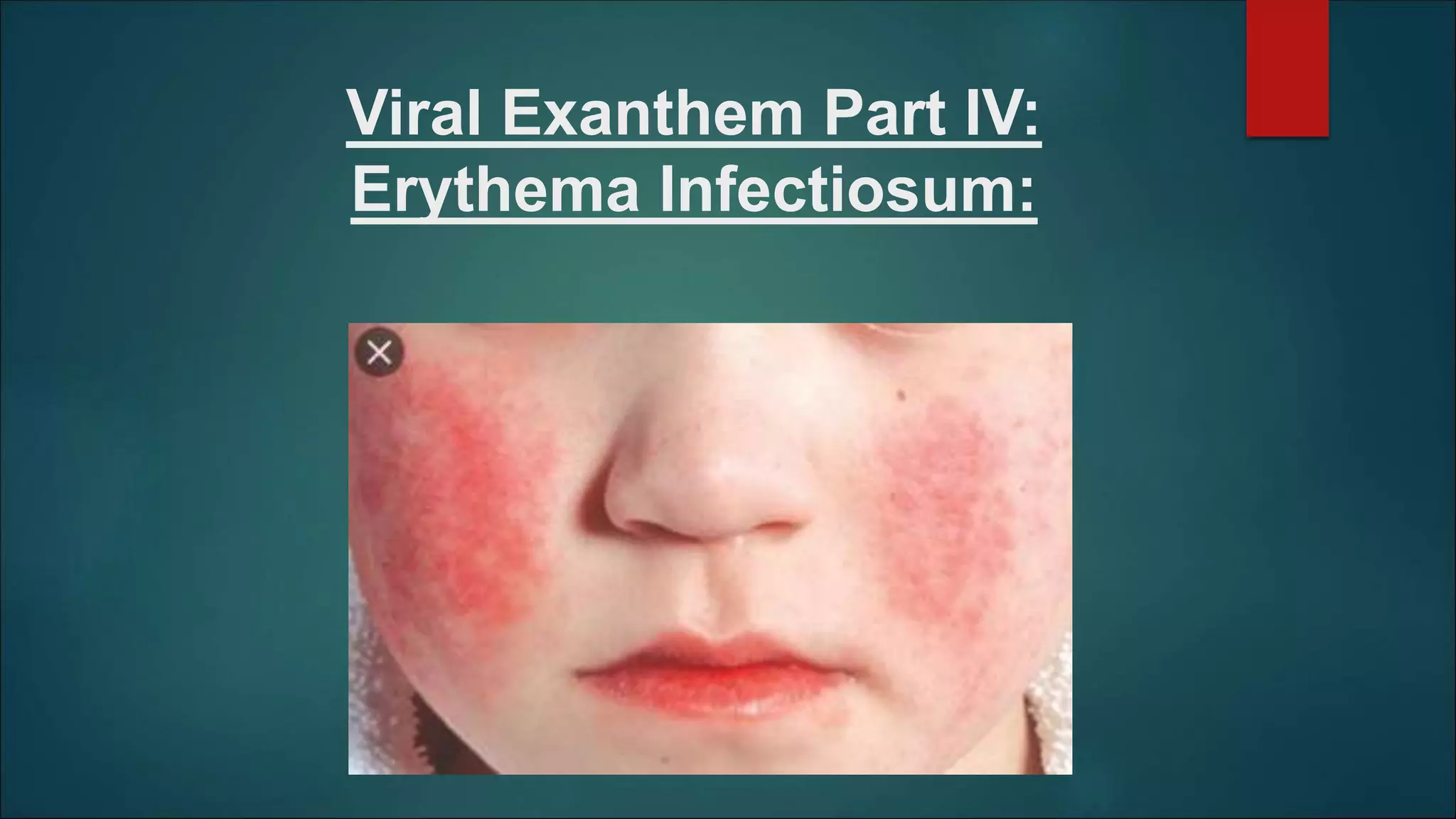
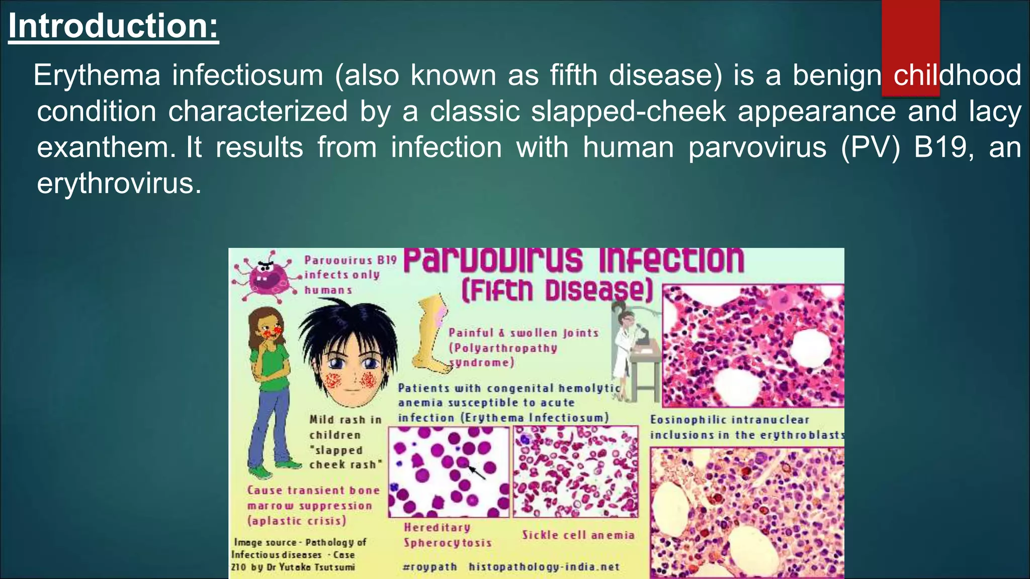
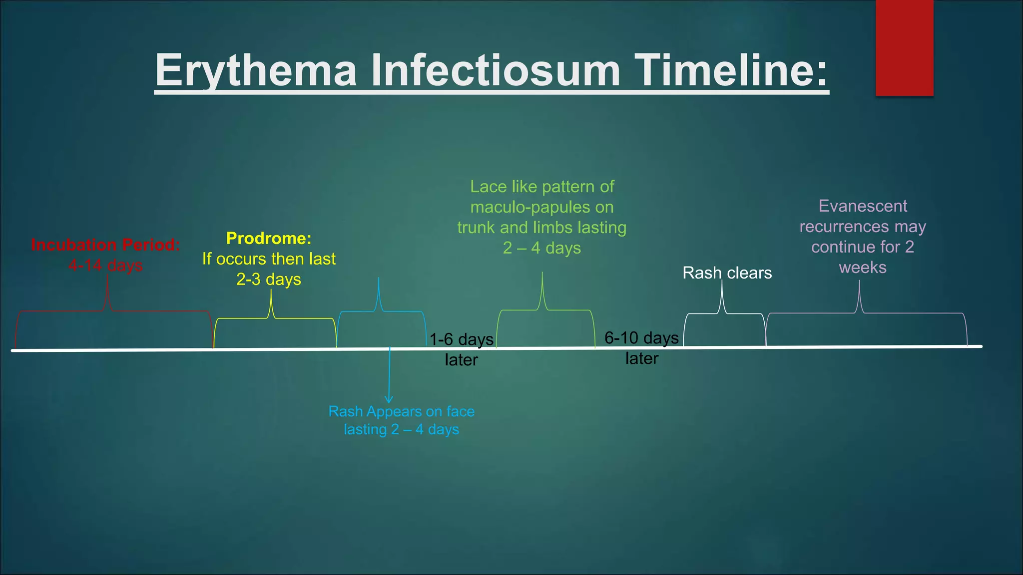
Viral Exanthem Part IV:
Erythema Infectiosum:
Introduction:
Erythema infectiosum (also known as fifth disease) is a benign childhood
condition characterized by a classic slapped-cheek appearance and lacy
exanthem. It results from infection with human parvovirus (PV) B19, an
erythrovirus.
Epidemiology
Incidence and prevalence:
 A childhood disease
 Infections is spread primarily via aerosolized respiratory droplets.
 Transmission also occurs through blood products and from mother to fetus.
Pathophysiology:
Causative organisms:
 Parvovirus B19 belongs to the genus Erythroparvovirus within the Parvoviridae family.
 Three genotypes are recognized. Most infections are due to type 1, but types 2 and 3 may be more
easily detectable in immunocompromised.
Clinical features in Children:
 Incubation Period: 4 to 14 days
 Usually no prodromal symptoms although there may be mild malaise, myalgia, sore throat, headache or
fever. Usually occurs 1 week after acquiring the infection and lasting 2 – 3 days.
 The infection progresses through the following stages:
Phase 1:
 The exanthem begins with the classic slapped-cheek appearance, which typically fades over 2-4
days.
 Rose‐red papules on the cheeks rapidly coalesce to form a hot turgid erythema, almost erysipeloid,
giving a ‘slapped cheek’ appearance.
 There is often perioral pallor.
Phase 2:
 This phase occurs 1-4 days later and is characterized by an
erythematous maculo-papular rash that fades in 2-4 days into
a classic lacelike reticular pattern.
Phase 3:
 Frequent clearing and recurrences for weeks or occasionally months may occur due to stimuli such as
exercise, irritation, stress, or overheating of the skin from sunlight or bathing in hot water
 Other Clinical Features:
 Palms and soles may be involved and acral lesions may be petechial.
 There may be dark‐red macules on the buccal and genital mucous membranes.
 The patient is no longer infectious when the rash appears.
This picture shows bilateral
erythema of the cheeks,
likened to ‘slapped
cheeks’, with circumoral
sparing.
Lacy/reticulated pattern is
seen, as seen on the inner
thigh of this patient.
 Clinical Features in Adults:
 In adults, polyarthralgia is often the predominant symptom of infection and systemic symptoms are
more marked.
 The following, in decreasing order of frequency, are the most commonly affected joint sites:
 Metacarpophalangeal and/or interphalangeal areas
 Knees
 Wrists
 Ankles
 Unlike rheumatoid arthritis, joint pain worsens over the day, and no joint destruction occurs. The synovial fluid is
acellular and devoid of viral particles
 When the exanthem does occur, the features of facial erythema are usually less marked than in
children.
Clinical variants:
 Rarely, lesions may be vesicular or pustular or
 The eruption may be petechial or purpuric in children, often generalized but
sometimes acral.
 Some conditions associated with B19 are listed as under:
Complications and co‐morbidities:
 In early stages:
 Leukocytosis
 Relative lymphopenia;
 Later:
 An eosinophilia of up to 36%.
 Lymphocytosis.
 Mild anaemia: Several days after the onset of symptoms, a clinically insignificant decline in
hemoglobin concentration is noted; the decreased level is maintained for 7 to 10 days, during which
time examination of bone marrow samples reveals a marked depletion of erythroid precursor cells.
 Aplastic anaemia in cases of immunosuppression
 Rare complications:
 Heamophagocytic syndrome and
 Severe liver damage
 Acute parvovirus infection in pregnancy can result in fetal death or hydrops fetalis
Disease course and prognosis:
 The eruption usually fades in 6–10 days,
 Evanescent recurrences on previously affected sites may continue for 2 weeks or
longer due to stimuli such as exercise, irritation, stress, or overheating of the skin from
sunlight or bathing in hot water.
 Arthropathy common in adults usually resolves in a few weeks, however in 10%
women joint symptoms can persist for more than 2 months.
 In some cases, the combination of parvovirus infection with rash and arthritis is
accompanied by other features diagnostic of connective tissue disease such as
systemic lupus erythematosus or rheumatoid arthritis.
Investigations:
 Confirmation of infection is usually made by serology.
 IgM antibodies against the capsid proteins VP1 and VP2 are detectable
during acute infection and for up to 2 months
 IgG antibodies are an indication of past infection.
 False negative results can occur.
 Parvovirus DNA can be detected by PCR in the early stages of disease in
serum and in later stages in tissues. This can be quicker and useful when
clinical suspicion is high but the immune test negative.
Management:
 It is a mild and self‐limiting infection, no specific treatment
is needed.
 In pregnant women, rapid diagnosis is essential.
 In immunosuppressed or haematological disease such as sickle cell disease, bone
marrow needs to be examined.
Erythema Infectiosum Timeline:
Prodrome:
If occurs then last
2-3 days
Rash Appears on face
lasting 2 – 4 days
Lace like pattern of
maculo-papules on
trunk and limbs lasting
2 – 4 days
Rash clears
Incubation Period:
4-14 days
Evanescent
recurrences may
continue for 2
weeks
6-10 days
later
1-6 days
later
Future Topics to be Discussed:
1)Infectious Mononucleosis
2)Adenovirus
3)Enterovirus
4)Cytomegalovirus
5)West Nile Fever
Erythema infectiosum made Very simple!!!!!!

Erythema infectiosum made Very simple!!!!!!

  • 1.
    Viral Exanthem PartIV: Erythema Infectiosum:
  • 2.
    Introduction: Erythema infectiosum (alsoknown as fifth disease) is a benign childhood condition characterized by a classic slapped-cheek appearance and lacy exanthem. It results from infection with human parvovirus (PV) B19, an erythrovirus.
  • 3.
    Epidemiology Incidence and prevalence: A childhood disease  Infections is spread primarily via aerosolized respiratory droplets.  Transmission also occurs through blood products and from mother to fetus.
  • 4.
    Pathophysiology: Causative organisms:  ParvovirusB19 belongs to the genus Erythroparvovirus within the Parvoviridae family.  Three genotypes are recognized. Most infections are due to type 1, but types 2 and 3 may be more easily detectable in immunocompromised.
  • 6.
    Clinical features inChildren:  Incubation Period: 4 to 14 days  Usually no prodromal symptoms although there may be mild malaise, myalgia, sore throat, headache or fever. Usually occurs 1 week after acquiring the infection and lasting 2 – 3 days.  The infection progresses through the following stages: Phase 1:  The exanthem begins with the classic slapped-cheek appearance, which typically fades over 2-4 days.  Rose‐red papules on the cheeks rapidly coalesce to form a hot turgid erythema, almost erysipeloid, giving a ‘slapped cheek’ appearance.  There is often perioral pallor.
  • 7.
    Phase 2:  Thisphase occurs 1-4 days later and is characterized by an erythematous maculo-papular rash that fades in 2-4 days into a classic lacelike reticular pattern. Phase 3:  Frequent clearing and recurrences for weeks or occasionally months may occur due to stimuli such as exercise, irritation, stress, or overheating of the skin from sunlight or bathing in hot water  Other Clinical Features:  Palms and soles may be involved and acral lesions may be petechial.  There may be dark‐red macules on the buccal and genital mucous membranes.  The patient is no longer infectious when the rash appears.
  • 8.
    This picture showsbilateral erythema of the cheeks, likened to ‘slapped cheeks’, with circumoral sparing. Lacy/reticulated pattern is seen, as seen on the inner thigh of this patient.
  • 9.
     Clinical Featuresin Adults:  In adults, polyarthralgia is often the predominant symptom of infection and systemic symptoms are more marked.  The following, in decreasing order of frequency, are the most commonly affected joint sites:  Metacarpophalangeal and/or interphalangeal areas  Knees  Wrists  Ankles  Unlike rheumatoid arthritis, joint pain worsens over the day, and no joint destruction occurs. The synovial fluid is acellular and devoid of viral particles  When the exanthem does occur, the features of facial erythema are usually less marked than in children.
  • 10.
    Clinical variants:  Rarely,lesions may be vesicular or pustular or  The eruption may be petechial or purpuric in children, often generalized but sometimes acral.  Some conditions associated with B19 are listed as under:
  • 11.
    Complications and co‐morbidities: In early stages:  Leukocytosis  Relative lymphopenia;  Later:  An eosinophilia of up to 36%.  Lymphocytosis.  Mild anaemia: Several days after the onset of symptoms, a clinically insignificant decline in hemoglobin concentration is noted; the decreased level is maintained for 7 to 10 days, during which time examination of bone marrow samples reveals a marked depletion of erythroid precursor cells.  Aplastic anaemia in cases of immunosuppression  Rare complications:  Heamophagocytic syndrome and  Severe liver damage  Acute parvovirus infection in pregnancy can result in fetal death or hydrops fetalis
  • 12.
    Disease course andprognosis:  The eruption usually fades in 6–10 days,  Evanescent recurrences on previously affected sites may continue for 2 weeks or longer due to stimuli such as exercise, irritation, stress, or overheating of the skin from sunlight or bathing in hot water.  Arthropathy common in adults usually resolves in a few weeks, however in 10% women joint symptoms can persist for more than 2 months.  In some cases, the combination of parvovirus infection with rash and arthritis is accompanied by other features diagnostic of connective tissue disease such as systemic lupus erythematosus or rheumatoid arthritis.
  • 13.
    Investigations:  Confirmation ofinfection is usually made by serology.  IgM antibodies against the capsid proteins VP1 and VP2 are detectable during acute infection and for up to 2 months  IgG antibodies are an indication of past infection.  False negative results can occur.  Parvovirus DNA can be detected by PCR in the early stages of disease in serum and in later stages in tissues. This can be quicker and useful when clinical suspicion is high but the immune test negative.
  • 14.
    Management:  It isa mild and self‐limiting infection, no specific treatment is needed.  In pregnant women, rapid diagnosis is essential.  In immunosuppressed or haematological disease such as sickle cell disease, bone marrow needs to be examined.
  • 15.
    Erythema Infectiosum Timeline: Prodrome: Ifoccurs then last 2-3 days Rash Appears on face lasting 2 – 4 days Lace like pattern of maculo-papules on trunk and limbs lasting 2 – 4 days Rash clears Incubation Period: 4-14 days Evanescent recurrences may continue for 2 weeks 6-10 days later 1-6 days later
  • 16.
    Future Topics tobe Discussed: 1)Infectious Mononucleosis 2)Adenovirus 3)Enterovirus 4)Cytomegalovirus 5)West Nile Fever