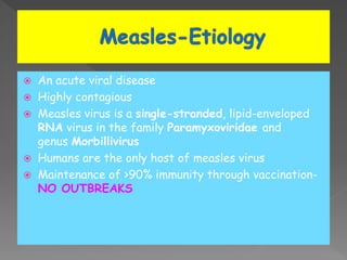
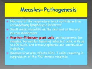
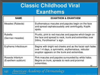
 An acute viral disease
 Highly contagious
 Measles virus is a single-stranded, lipid-enveloped
RNA virus in the family Paramyxoviridae and
genus Morbillivirus
 Humans are the only host of measles virus
 Maintenance of >90% immunity through vaccination-
NO OUTBREAKS
 Necrosis of the respiratory tract epithelium & an
accompanying lymphocytic infiltrate
 Small vessel vasculitis on the skin and on the oral
mucous membranes
 Warthin-Finkeldey giant cells: pathognomonic for
measles, formed by fusion of infected cells, with up
to 100 nuclei and intracytoplasmic and intranuclear
inclusions
 Measles virus also infects CD4+ T cells, resulting in
suppression of the Th1 immune response
 4 phases:
Incubation period
Prodromal illness
Exanthematous phase
Recovery
Incubation period: measles virus migrates to
regional lymph nodes primary viremia-
disseminates the virus to the
reticuloendothelial system secondary
viremia spreads virus to body surfaces
The prodromal illness begins after the
secondary viremia; associated with epithelial
necrosis, giant cell formation & virus shedding
With onset of the rash, antibody production
begins & viral replication & symptoms begin to
subside
 Through the respiratory tract or conjunctivae
 Following contact with large droplets or small-
droplet aerosols in which the virus is suspended
 Patients are infectious from 3-4 days before to up
to 4-6 days after the onset of rash
 High fever, an enanthem, cough, coryza,
conjunctivitis & a prominent exanthem
 Incubation period: 8-12 days
 Prodromal phase: mild fever, conjunctivitis with
photophobia, coryza, a prominent cough and KOPLIK’S
SPOTS
 Koplik spots: enanthem & the pathognomonic sign of
measles
 Appear 1 to 4 days prior to the onset of the rash
 Discrete red lesions with bluish-white spots in the
center on the inner aspects of the cheeks at the level
of the premolars
 Koplick’s spots: spread
to involve the lips,
hard palate & gingiva
 They also may occur
in conjunctival folds
 Temperature rises abruptly as rash appears & may
reach upto 40OC
 Measles rash: generalized, maculopapular,
erythematous, confluent
 The rash begins on the face around
the hairline & behind the ears
 It then spreads downward
to the neck, trunk, arms, legs
and feet over next 24-48 hours
 The rash fades over about 7 days in the same
progression as it evolved
 Leaves a fine, browny, branny desquamation of skin
 Severity of disease: related to the extent and
confluence of rash
 Rash: may be absent in immuno-compromised
children
 Hemorrhagic measles (black measles): bleeding from
mouth, nose or bowels
 Diarrhoea: more common in malnourished and small
children
 Severe cases: generalized lymphadenopathy
including cervical & mesenteric lymph nodes
 Mild splenomegaly
 Almost always based on clinical and epidemiologic
findings (history of contact)
 Fever of at least 3 days with at least one of three C
(cough, coryza, conjuctivitis)
 Decreased total white blood cell count, with relative
lymphocytosis
 IgM antibody in serum: appears 1-2 days after the
onset of the rash & remains detectable for about
1 mo
 Demonstration of a fourfold rise in IgG antibodies in
acute & convalescent specimens collected 2-4 wk
later
 Viral isolation from blood, urine or respiratory
secretions by culture or rt-PCR
 Rubella-rashes & fever are less striking
 Roseola infantum (exanthem subitum)- rash appear
as the fever disappears
 Echovirus
 Coxsachie
 Adenovirus
 Infectious mononucleosis
 Scarlet fever-diffuse fleshy papular rash with
“goose flesh” texture
 Meningococcemia-rashes are similar but NO
conjuctivitis & cough
 Kawasaki disease- no cough, elevations of
neutrophils and acute-phase reactants; the
characteristic thrombocytosis
 Drug fever
 Due to the pathogenic effects of the virus on the
respiratory tract and immune system
Risk factors for complications
 Children <5 years of age & adults >20 years of age
 Severe malnutrition
 Vitamin A deficiency
 Immuno-compromised persons
 Pneumonia- giant cell pneumonia (direct viral
infection) or super-imposed bacterial infection
(Streptococcus pneumoniae, Haemophilus
influenzae & Staphylococcus aureus)
 Croup, tracheitis or bronchiolitis
 Acute otitis media
 Sinusitis and mastoiditis
 Retropharyngeal abscess
 Activation of pulmonary tuberculosis
 Diarrhea & vomiting
 Appendicitis- obstruction of the appendiceal lumen
by lymphoid hyperplasia
 Febrile seizures
 Encephalitis- 1-3/1,000 cases of measles;
postinfectious, immunologically mediated process,
not due to a direct viral effect
 Measles encephalitis in immunocompromised
patients-from direct damage to the brain by the
virus
 Thrombocytopenia
 Myocarditis
 Bacteremia, cellulitis & toxic shock syndrome
 Measles during pregnancy-high maternal morbidity,
fetal wastage & stillbirths & congenital
malformations in 3% of live born infants
 Fatal degenerative disease of central nervous
system
 Chronic complication of measles
 Result from a persistent infection with an altered
measles virus that is harbored intracellularly in the
CNS for several years
 Usually after 7-10 year the virus apparently regains
virulence & attacks the cells in the CNS
 Change in personality, gradual onset of mental
deterioration and myoclonus
 Measles vaccination protects against SSPE
 SUPPORTIVE
 Maintenance of hydration, oxygenation and comfort
 Antipyretics-comfort and fever control
 Vitamin A supplementation-reduced morbidity and
mortality from measles
 Single dose of 200,000 IU orally for children
≥1 yr of age (100,000 IU for children 6 mo–1 yr
of age and 50,000 IU for infants <6 mo of age)
 Isolation- from 7 days after exposure to 4-6 days
after the onset of rash
 Vaccine or immunoglobulin- vaccine is effective in
prevention or modification of measles only if given
within 72 hr of exposure. Immune globulin may be
given up to 6 days after exposure to prevent or modify
infection.
 Immune globulin-for susceptible household contacts
younger than 6 months of age, pregnant women and
immunocompromised persons
 Immunization during an outbreak-immunize infant as
young as 6 months of age; additional dose at 12-15
months of age
 Rubella (German measles or 3-day measles)
 Mild exanthematous disease of infants and children
 Major clinical significance- fetal damage as part of
the congenital rubella syndrome
 Etiology: Rubella virus; RNA virus of genus Rubivirus
under family Togaviridae
 Humans are the only known host.
 Transmission-through oral droplet or transplacental
route
 Virus is shed in nasopharyngeal secretions 7 days
before exanthem & upto 7-8 days after its
disappearance
 Rubella susceptibility among women of child bearing
age in India- 4%-43%
 Infection virus replication in the respiratory
epithelium spreads to regional lymph nodes
viremia viral shedding from the nasopharynx
 Cellular and tissue damage in the infected fetus:
tissue necrosis, reduced cellular multiplication time,
chromosomal breaks & production of a protein
inhibitor causing mitotic arrests
 Most distinctive feature of congenital rubella:
chronicity
 Ongoing tissue damage and reactivation
 Risk factor for severe congenital defects: stage of
gestation at the time of infection
 Maternal infection during the 1st 8 wk of gestation:
most severe & widespread defects
 Risk for congenital defects: 90% for maternal
infection before 11 wk of gestation, 33% at 11-
12 wk, 11% at 13-14 wk & 24% at 15-16 wk
 After 16 wk of gestation: defects uncommon
POSTNATAL INFECTION
 Incubation period: 14-21 days
 Prodrome: low-grade fever, sore throat, red eyes
with or without eye pain, headache, malaise, anorexia
& lymphadenopathy (suboccipital, postauricular &
anterior cervical lymph nodes)
 Rash: begins on the face & neck as small, irregular
pink macules that coalesce and it spreads
centrifugally to involve the torso and extremities,
where it tends to occur as discrete macules
 Rash: fades from the face as it extends to the rest
of the body so that the whole body may not be
involved at any 1 time
 The duration of the rash is generally 3 days & it
resolves without desquamation
 About the time of onset of the rash, examination of
the oropharynx- reveal tiny, rose-colored lesions
(Forchheimer spots) or petechial hemorrhages on the
soft palate
 Subclinical infections are common (25-40%)
 Polyarthritis or arthralgia-common in adult females
 Lab findings: Leukopenia, neutropenia & mild
thrombocytopenia
 Mild form of measles
 Scarlet fever
 Roseola infantum
 Enteroviral infections
 Drug fever
 Infectoius mononucleosis
 Erythema infectiosum
 Supportive history of exposure or consistent clinical
findings
 Rubella specific IgM enzyme immunosorbent assay (4-
72 days)
 Fourfold rise in IgG in sequential sera
 Rubella virus culture from nasopharynx & blood by
tissue culture system or PCR
 WHO definition of PROBABLE infection: fever,
maculopapular rash, lymphadenopathy or
arthralgia/arthritis
 WHO definition of CONFORMED infection: probable
case with IgM positivity within 28 days of onset of rash
 Postinfectious thrombocytopenia
 Arthritis- classically involves the small joints of the
hands
 Encephalitis-a postinfectious syndrome following
acute rubella & a rare progressive panencephalitis
manifesting as a neurodegenerative disorder years
following rubella
 Guillain-Barré syndrome, peripheral neuritis
 Myocarditis
 Result of in utero fetal infection
 Classical CRS triad: cataract, sensorineural hearing
loss and congenital heart disease
Clinical manifestations:
 Intrauterine growth restriction, postnatal mental and
motor retardation
 Bilateral/unilateral cataract, salt-and-pepper
retinopathy, microphthalmia
 Nerve deafness
 Meningoencephalitis at birth
 Patent ductus arteriosus, pulmonary artery stenosis,
VSD & ASD, myocarditis
 Hepatitis
 Dermal erythropoiesis (blueberry muffin lesions)
 Thrombocytopenic purpura
 Anemia
 Hepatosplenomegaly
 Microcephaly
 Interstitial pneumonitis
 Delayed manifestations: Diabetes mellitus (20%),
thyroid dysfunction (5%)
 No specific treatment available for either acquired
rubella or CRS
 Supportive treatment- antipyretics and analgesics
 Intravenous immunoglobulin or corticosteroids-for
severe, nonremitting thrombocytopenia
 Hearing screening- important, early intervention
improve outcomes
Management of exposed pregnant women
 Rubella antibody status is tested immediately
result positive mother is immune no further
action
 Rubella antibody status negative repeat samples
after 1-2 weeks negative 1st specimen & positive
test result in either the 2nd or 3rd specimen
seroconversion suggesting recent infection
termination of pregnancy
Management of congenital rubella syndrome
 Children with CRS may excrete the virus in
respiratory secretions up to 1 yr of age
 Isolation & contact precautions maintained unless
repeated cultures of urine and pharyngeal secretions
have negative results
 Isolation at home my be required for 1 year
 Care of CRS infants require multidisciplinary team
 Prognosis poor
 PREVENTION by IMMUNIZATION
 Varicella is an acute febrile rash illness
 Caused by VZV which is a neurotropic human α-
herpesvirus
 Secondary attack rate: 90%
 Transmission: by airborne spread or through direct
contact with skin lesions
 Varicella results from inoculation of the virus onto the
mucosa of the upper respiratory tract and tonsillar
lymphoid tissue
Incubation period (10-21 days): replication
in the local lymphoid tissue primary
viremia-disseminates the virus to the
reticuloendothelial system secondary
viremia spreads virus to body surfaces
leading to widespread cutaneous lesions
During the late incubation period-VZV
transported back to the mucosa of the upper
respiratory tract & oropharynx, permitting
spread to susceptible contacts 1-2 days
before the appearance of rash
Host immune responses limit viral replication
and facilitate recovery from infection
Immunocompromised child-continued viral
replication -disseminated infection
 Transportation of virus in a retrograde manner
through sensory axons to the dorsal root ganglia
throughout the spinal cord establishment of
virus latent infection in the neurons
subsequent reactivation
herpes zoster, a vesicular rash that usually is
dermatomal in distribution
 Prodromal symptoms: fever (moderate), malaise,
anorexia, headache and occasionally mild abdominal
pain, 24-48 hours before the rash appears
 These symptoms resolve within 2-4 days after the
onset of the rash
 Varicella rash often appear first on the scalp, face, or
trunk
 The initial exanthem consists of intensely pruritic
erythematous macules that evolve through the papular
stage to form clear, fluid-filled vesicles
 Clouding & umbilication of the lesions begin in 24-
48 hr
 While the initial lesions are crusting, new crops form
on the trunk & then the extremities
 The simultaneous presence of lesions in various
stages of evolution is characteristic of varicella
 The distribution of the rash is predominantly central
or centripetal
Pearl on a rose patel
 The average number of varicella lesions is about 300
(10-1500)
 Hypopigmentation or hyperpigmentation of lesion sites
persists for days to weeks in some children
 Severe scarring is unusual unless the lesions were
secondarily infected
Vesicular rashes caused by
 Herpes simplex virus
 Enterovirus
 Rickettsial pox
 S. aureus
 Drug reactions
 Contact dermatitis
 Insect bites
 CLINICAL
 Leukopenia during the 1st 72 hours after onset of
rash; followed by a relative & absolute lymphocytosis
 Elevated hepatic enzymes
 Specific diagnosis of VZV infection: needed in
immunocompromised children
 Mild thrombocytopenia, petechiae (common); purpura,
hemorrhagic vesicles, hematuria & gastrointestinal
bleeding (rare)
 Cerebellar ataxia, encephalitis, Guillian-Barre
syndrome, transverse myelitis
 Pneumonia
 Nephritis, nephrotic syndrome, hemolytic-uremic
syndrome
 Arthritis
 Myocarditis, pericarditis
 Pancreatitis
 Orchitis
 Secondary bacterial infections of the skin (group A
streptococci & S. aureus): impetigo, cellulitis,
lymphadenitis & subcutaneous abscesses; varicella
gangrenosa- more invasive skin infections
 In infants born to women who have varicella before
20 wk of gestation
Characterized by
 Cicatricial skin scarring in a zoster-like distribution,
limb hypoplasia
 Neurologic abnormalities: microcephaly, cortical
atrophy, seizures & mental retardation
 Eye abnormalities: chorioretinitis, microphthalmia &
cataracts
 Renal abnormalities: hydroureter & hydronephrosis
 Autonomic nervous system abnormalities: neurogenic
bladder, swallowing dysfunction & aspiration pneumonia
 If a baby is born <4 days after onset of maternal
varicella or upto 2 days before the onset: high risk
for severe varicella & a high mortality rate
 Supportive treatment for fever & itching
Indications for acyclovir in children:
 Malignancies
 BMT
 Chmotherapy or high dose steroid treatment
 HIV infection
 Severe varicella
 Chronic skin disease
 Long term salicylate therapy
 Chlidren >12 years
Treatment should be initiated within 24 hr of the
onset of rash
 Foscarnet is the only drug for the treatment of
acyclovir-resistant VZV infections (in children infected
with HIV)
Viral exanthems

Viral exanthems

  • 2.
     An acuteviral disease  Highly contagious  Measles virus is a single-stranded, lipid-enveloped RNA virus in the family Paramyxoviridae and genus Morbillivirus  Humans are the only host of measles virus  Maintenance of >90% immunity through vaccination- NO OUTBREAKS
  • 3.
     Necrosis ofthe respiratory tract epithelium & an accompanying lymphocytic infiltrate  Small vessel vasculitis on the skin and on the oral mucous membranes  Warthin-Finkeldey giant cells: pathognomonic for measles, formed by fusion of infected cells, with up to 100 nuclei and intracytoplasmic and intranuclear inclusions  Measles virus also infects CD4+ T cells, resulting in suppression of the Th1 immune response
  • 4.
     4 phases: Incubationperiod Prodromal illness Exanthematous phase Recovery
  • 5.
    Incubation period: measlesvirus migrates to regional lymph nodes primary viremia- disseminates the virus to the reticuloendothelial system secondary viremia spreads virus to body surfaces The prodromal illness begins after the secondary viremia; associated with epithelial necrosis, giant cell formation & virus shedding With onset of the rash, antibody production begins & viral replication & symptoms begin to subside
  • 6.
     Through therespiratory tract or conjunctivae  Following contact with large droplets or small- droplet aerosols in which the virus is suspended  Patients are infectious from 3-4 days before to up to 4-6 days after the onset of rash
  • 7.
     High fever,an enanthem, cough, coryza, conjunctivitis & a prominent exanthem  Incubation period: 8-12 days  Prodromal phase: mild fever, conjunctivitis with photophobia, coryza, a prominent cough and KOPLIK’S SPOTS  Koplik spots: enanthem & the pathognomonic sign of measles  Appear 1 to 4 days prior to the onset of the rash  Discrete red lesions with bluish-white spots in the center on the inner aspects of the cheeks at the level of the premolars
  • 8.
     Koplick’s spots:spread to involve the lips, hard palate & gingiva  They also may occur in conjunctival folds
  • 9.
     Temperature risesabruptly as rash appears & may reach upto 40OC  Measles rash: generalized, maculopapular, erythematous, confluent  The rash begins on the face around the hairline & behind the ears  It then spreads downward to the neck, trunk, arms, legs and feet over next 24-48 hours
  • 10.
     The rashfades over about 7 days in the same progression as it evolved  Leaves a fine, browny, branny desquamation of skin  Severity of disease: related to the extent and confluence of rash  Rash: may be absent in immuno-compromised children  Hemorrhagic measles (black measles): bleeding from mouth, nose or bowels
  • 11.
     Diarrhoea: morecommon in malnourished and small children  Severe cases: generalized lymphadenopathy including cervical & mesenteric lymph nodes  Mild splenomegaly
  • 12.
     Almost alwaysbased on clinical and epidemiologic findings (history of contact)  Fever of at least 3 days with at least one of three C (cough, coryza, conjuctivitis)  Decreased total white blood cell count, with relative lymphocytosis
  • 13.
     IgM antibodyin serum: appears 1-2 days after the onset of the rash & remains detectable for about 1 mo  Demonstration of a fourfold rise in IgG antibodies in acute & convalescent specimens collected 2-4 wk later  Viral isolation from blood, urine or respiratory secretions by culture or rt-PCR
  • 14.
     Rubella-rashes &fever are less striking  Roseola infantum (exanthem subitum)- rash appear as the fever disappears  Echovirus  Coxsachie  Adenovirus  Infectious mononucleosis  Scarlet fever-diffuse fleshy papular rash with “goose flesh” texture
  • 15.
     Meningococcemia-rashes aresimilar but NO conjuctivitis & cough  Kawasaki disease- no cough, elevations of neutrophils and acute-phase reactants; the characteristic thrombocytosis  Drug fever
  • 16.
     Due tothe pathogenic effects of the virus on the respiratory tract and immune system Risk factors for complications  Children <5 years of age & adults >20 years of age  Severe malnutrition  Vitamin A deficiency  Immuno-compromised persons
  • 17.
     Pneumonia- giantcell pneumonia (direct viral infection) or super-imposed bacterial infection (Streptococcus pneumoniae, Haemophilus influenzae & Staphylococcus aureus)  Croup, tracheitis or bronchiolitis  Acute otitis media  Sinusitis and mastoiditis  Retropharyngeal abscess  Activation of pulmonary tuberculosis
  • 18.
     Diarrhea &vomiting  Appendicitis- obstruction of the appendiceal lumen by lymphoid hyperplasia  Febrile seizures  Encephalitis- 1-3/1,000 cases of measles; postinfectious, immunologically mediated process, not due to a direct viral effect
  • 19.
     Measles encephalitisin immunocompromised patients-from direct damage to the brain by the virus  Thrombocytopenia  Myocarditis  Bacteremia, cellulitis & toxic shock syndrome  Measles during pregnancy-high maternal morbidity, fetal wastage & stillbirths & congenital malformations in 3% of live born infants
  • 20.
     Fatal degenerativedisease of central nervous system  Chronic complication of measles  Result from a persistent infection with an altered measles virus that is harbored intracellularly in the CNS for several years  Usually after 7-10 year the virus apparently regains virulence & attacks the cells in the CNS  Change in personality, gradual onset of mental deterioration and myoclonus  Measles vaccination protects against SSPE
  • 21.
     SUPPORTIVE  Maintenanceof hydration, oxygenation and comfort  Antipyretics-comfort and fever control  Vitamin A supplementation-reduced morbidity and mortality from measles  Single dose of 200,000 IU orally for children ≥1 yr of age (100,000 IU for children 6 mo–1 yr of age and 50,000 IU for infants <6 mo of age)
  • 22.
     Isolation- from7 days after exposure to 4-6 days after the onset of rash  Vaccine or immunoglobulin- vaccine is effective in prevention or modification of measles only if given within 72 hr of exposure. Immune globulin may be given up to 6 days after exposure to prevent or modify infection.  Immune globulin-for susceptible household contacts younger than 6 months of age, pregnant women and immunocompromised persons  Immunization during an outbreak-immunize infant as young as 6 months of age; additional dose at 12-15 months of age
  • 23.
     Rubella (Germanmeasles or 3-day measles)  Mild exanthematous disease of infants and children  Major clinical significance- fetal damage as part of the congenital rubella syndrome  Etiology: Rubella virus; RNA virus of genus Rubivirus under family Togaviridae  Humans are the only known host.
  • 24.
     Transmission-through oraldroplet or transplacental route  Virus is shed in nasopharyngeal secretions 7 days before exanthem & upto 7-8 days after its disappearance  Rubella susceptibility among women of child bearing age in India- 4%-43%
  • 25.
     Infection virusreplication in the respiratory epithelium spreads to regional lymph nodes viremia viral shedding from the nasopharynx  Cellular and tissue damage in the infected fetus: tissue necrosis, reduced cellular multiplication time, chromosomal breaks & production of a protein inhibitor causing mitotic arrests  Most distinctive feature of congenital rubella: chronicity  Ongoing tissue damage and reactivation
  • 26.
     Risk factorfor severe congenital defects: stage of gestation at the time of infection  Maternal infection during the 1st 8 wk of gestation: most severe & widespread defects  Risk for congenital defects: 90% for maternal infection before 11 wk of gestation, 33% at 11- 12 wk, 11% at 13-14 wk & 24% at 15-16 wk  After 16 wk of gestation: defects uncommon
  • 27.
    POSTNATAL INFECTION  Incubationperiod: 14-21 days  Prodrome: low-grade fever, sore throat, red eyes with or without eye pain, headache, malaise, anorexia & lymphadenopathy (suboccipital, postauricular & anterior cervical lymph nodes)  Rash: begins on the face & neck as small, irregular pink macules that coalesce and it spreads centrifugally to involve the torso and extremities, where it tends to occur as discrete macules
  • 28.
     Rash: fadesfrom the face as it extends to the rest of the body so that the whole body may not be involved at any 1 time  The duration of the rash is generally 3 days & it resolves without desquamation
  • 29.
     About thetime of onset of the rash, examination of the oropharynx- reveal tiny, rose-colored lesions (Forchheimer spots) or petechial hemorrhages on the soft palate  Subclinical infections are common (25-40%)  Polyarthritis or arthralgia-common in adult females  Lab findings: Leukopenia, neutropenia & mild thrombocytopenia
  • 30.
     Mild formof measles  Scarlet fever  Roseola infantum  Enteroviral infections  Drug fever  Infectoius mononucleosis  Erythema infectiosum
  • 31.
     Supportive historyof exposure or consistent clinical findings  Rubella specific IgM enzyme immunosorbent assay (4- 72 days)  Fourfold rise in IgG in sequential sera  Rubella virus culture from nasopharynx & blood by tissue culture system or PCR  WHO definition of PROBABLE infection: fever, maculopapular rash, lymphadenopathy or arthralgia/arthritis  WHO definition of CONFORMED infection: probable case with IgM positivity within 28 days of onset of rash
  • 32.
     Postinfectious thrombocytopenia Arthritis- classically involves the small joints of the hands  Encephalitis-a postinfectious syndrome following acute rubella & a rare progressive panencephalitis manifesting as a neurodegenerative disorder years following rubella  Guillain-Barré syndrome, peripheral neuritis  Myocarditis
  • 33.
     Result ofin utero fetal infection  Classical CRS triad: cataract, sensorineural hearing loss and congenital heart disease Clinical manifestations:  Intrauterine growth restriction, postnatal mental and motor retardation  Bilateral/unilateral cataract, salt-and-pepper retinopathy, microphthalmia  Nerve deafness  Meningoencephalitis at birth
  • 34.
     Patent ductusarteriosus, pulmonary artery stenosis, VSD & ASD, myocarditis  Hepatitis  Dermal erythropoiesis (blueberry muffin lesions)  Thrombocytopenic purpura  Anemia  Hepatosplenomegaly  Microcephaly  Interstitial pneumonitis  Delayed manifestations: Diabetes mellitus (20%), thyroid dysfunction (5%)
  • 35.
     No specifictreatment available for either acquired rubella or CRS  Supportive treatment- antipyretics and analgesics  Intravenous immunoglobulin or corticosteroids-for severe, nonremitting thrombocytopenia  Hearing screening- important, early intervention improve outcomes
  • 36.
    Management of exposedpregnant women  Rubella antibody status is tested immediately result positive mother is immune no further action  Rubella antibody status negative repeat samples after 1-2 weeks negative 1st specimen & positive test result in either the 2nd or 3rd specimen seroconversion suggesting recent infection termination of pregnancy
  • 37.
    Management of congenitalrubella syndrome  Children with CRS may excrete the virus in respiratory secretions up to 1 yr of age  Isolation & contact precautions maintained unless repeated cultures of urine and pharyngeal secretions have negative results  Isolation at home my be required for 1 year  Care of CRS infants require multidisciplinary team  Prognosis poor  PREVENTION by IMMUNIZATION
  • 38.
     Varicella isan acute febrile rash illness  Caused by VZV which is a neurotropic human α- herpesvirus  Secondary attack rate: 90%  Transmission: by airborne spread or through direct contact with skin lesions  Varicella results from inoculation of the virus onto the mucosa of the upper respiratory tract and tonsillar lymphoid tissue
  • 39.
    Incubation period (10-21days): replication in the local lymphoid tissue primary viremia-disseminates the virus to the reticuloendothelial system secondary viremia spreads virus to body surfaces leading to widespread cutaneous lesions During the late incubation period-VZV transported back to the mucosa of the upper respiratory tract & oropharynx, permitting spread to susceptible contacts 1-2 days before the appearance of rash Host immune responses limit viral replication and facilitate recovery from infection Immunocompromised child-continued viral replication -disseminated infection
  • 40.
     Transportation ofvirus in a retrograde manner through sensory axons to the dorsal root ganglia throughout the spinal cord establishment of virus latent infection in the neurons subsequent reactivation herpes zoster, a vesicular rash that usually is dermatomal in distribution
  • 41.
     Prodromal symptoms:fever (moderate), malaise, anorexia, headache and occasionally mild abdominal pain, 24-48 hours before the rash appears  These symptoms resolve within 2-4 days after the onset of the rash  Varicella rash often appear first on the scalp, face, or trunk  The initial exanthem consists of intensely pruritic erythematous macules that evolve through the papular stage to form clear, fluid-filled vesicles  Clouding & umbilication of the lesions begin in 24- 48 hr
  • 42.
     While theinitial lesions are crusting, new crops form on the trunk & then the extremities  The simultaneous presence of lesions in various stages of evolution is characteristic of varicella  The distribution of the rash is predominantly central or centripetal Pearl on a rose patel
  • 43.
     The averagenumber of varicella lesions is about 300 (10-1500)  Hypopigmentation or hyperpigmentation of lesion sites persists for days to weeks in some children  Severe scarring is unusual unless the lesions were secondarily infected
  • 44.
    Vesicular rashes causedby  Herpes simplex virus  Enterovirus  Rickettsial pox  S. aureus  Drug reactions  Contact dermatitis  Insect bites
  • 45.
     CLINICAL  Leukopeniaduring the 1st 72 hours after onset of rash; followed by a relative & absolute lymphocytosis  Elevated hepatic enzymes  Specific diagnosis of VZV infection: needed in immunocompromised children
  • 46.
     Mild thrombocytopenia,petechiae (common); purpura, hemorrhagic vesicles, hematuria & gastrointestinal bleeding (rare)  Cerebellar ataxia, encephalitis, Guillian-Barre syndrome, transverse myelitis  Pneumonia  Nephritis, nephrotic syndrome, hemolytic-uremic syndrome  Arthritis  Myocarditis, pericarditis  Pancreatitis
  • 47.
     Orchitis  Secondarybacterial infections of the skin (group A streptococci & S. aureus): impetigo, cellulitis, lymphadenitis & subcutaneous abscesses; varicella gangrenosa- more invasive skin infections
  • 48.
     In infantsborn to women who have varicella before 20 wk of gestation Characterized by  Cicatricial skin scarring in a zoster-like distribution, limb hypoplasia  Neurologic abnormalities: microcephaly, cortical atrophy, seizures & mental retardation  Eye abnormalities: chorioretinitis, microphthalmia & cataracts  Renal abnormalities: hydroureter & hydronephrosis  Autonomic nervous system abnormalities: neurogenic bladder, swallowing dysfunction & aspiration pneumonia
  • 49.
     If ababy is born <4 days after onset of maternal varicella or upto 2 days before the onset: high risk for severe varicella & a high mortality rate
  • 50.
     Supportive treatmentfor fever & itching Indications for acyclovir in children:  Malignancies  BMT  Chmotherapy or high dose steroid treatment  HIV infection  Severe varicella  Chronic skin disease  Long term salicylate therapy  Chlidren >12 years Treatment should be initiated within 24 hr of the onset of rash
  • 51.
     Foscarnet isthe only drug for the treatment of acyclovir-resistant VZV infections (in children infected with HIV)