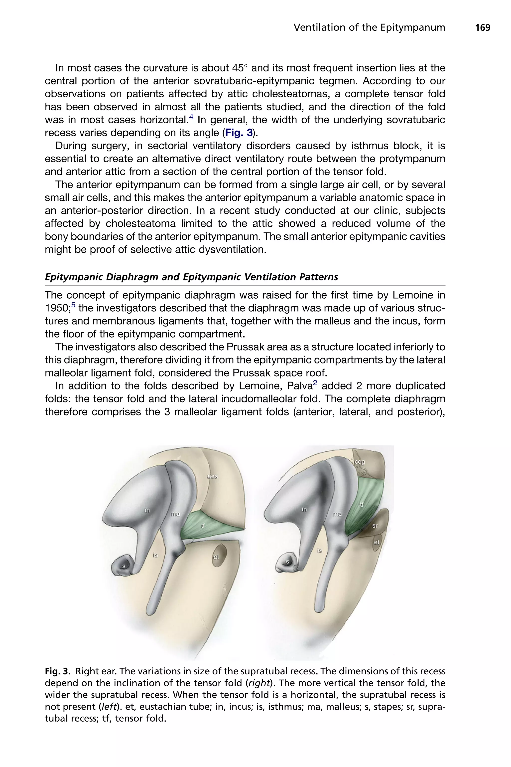
This document discusses the anatomy and ventilation patterns of the epitympanum. It describes:
1) The epitympanum is divided into anterior and posterior compartments. The superior attic communicates with the mesotympanum via the isthmus and is ventilated through the isthmus. The inferior lateral attic and Prussak space are ventilated by the mesotympanum.
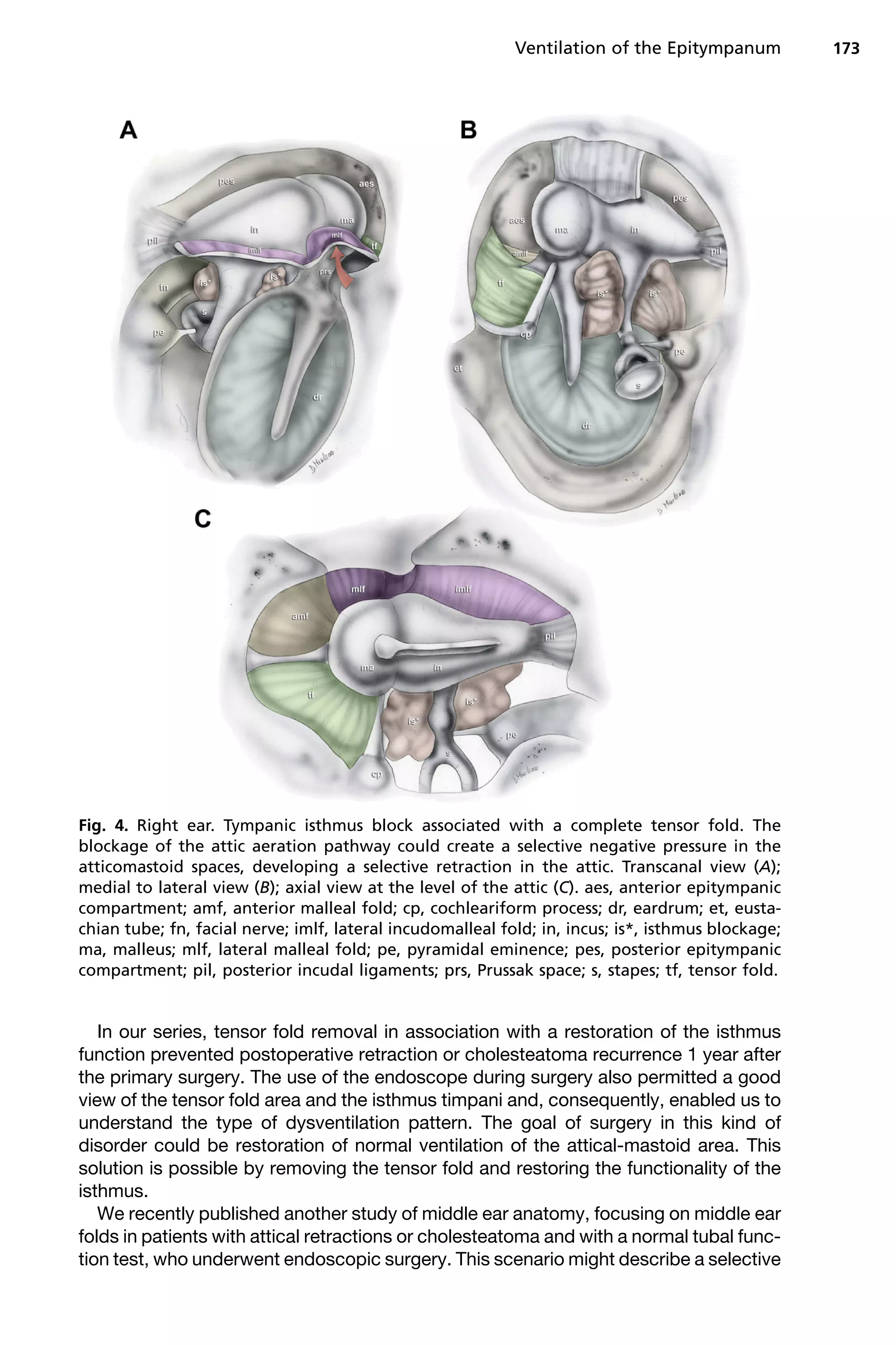
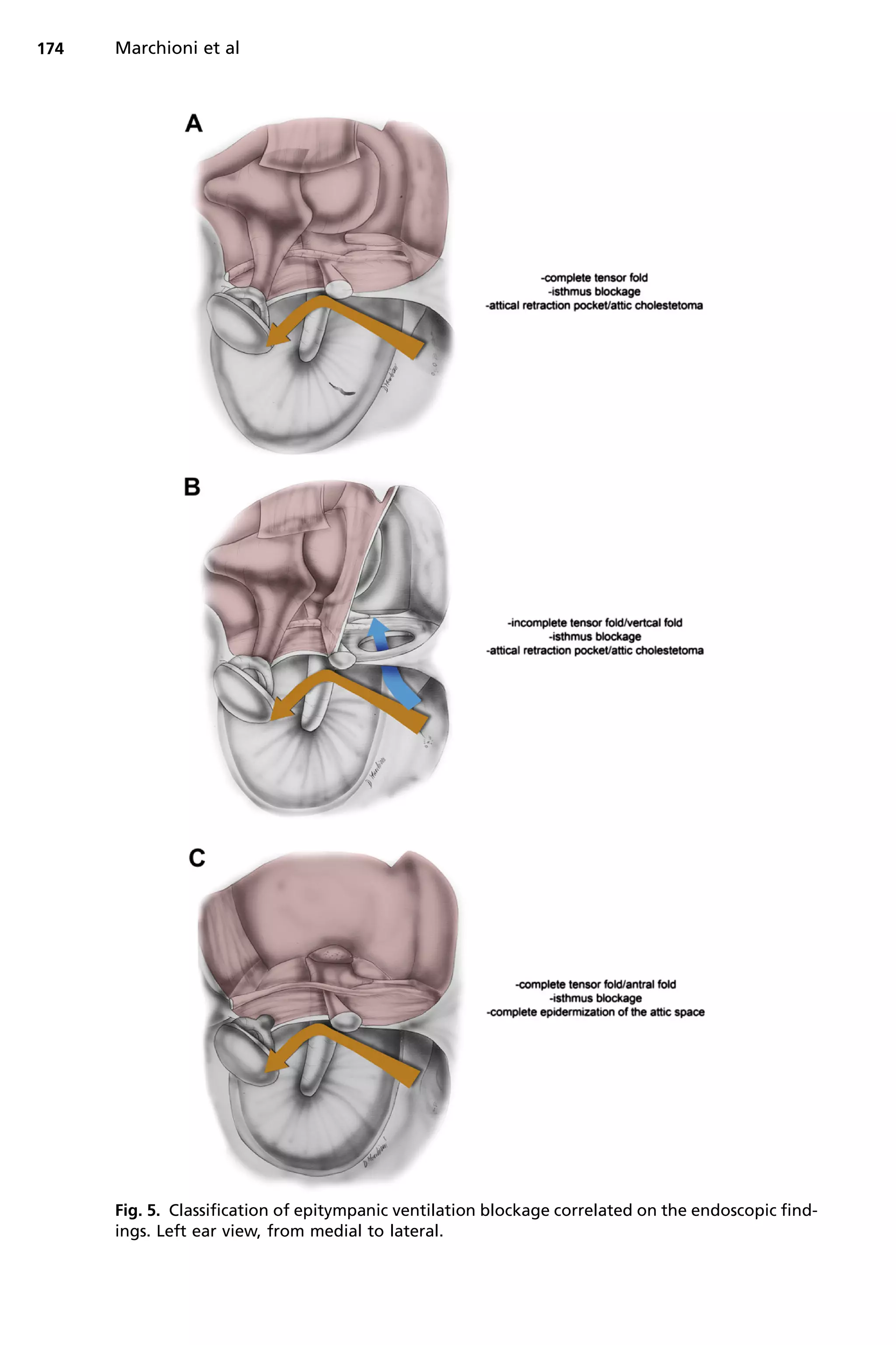
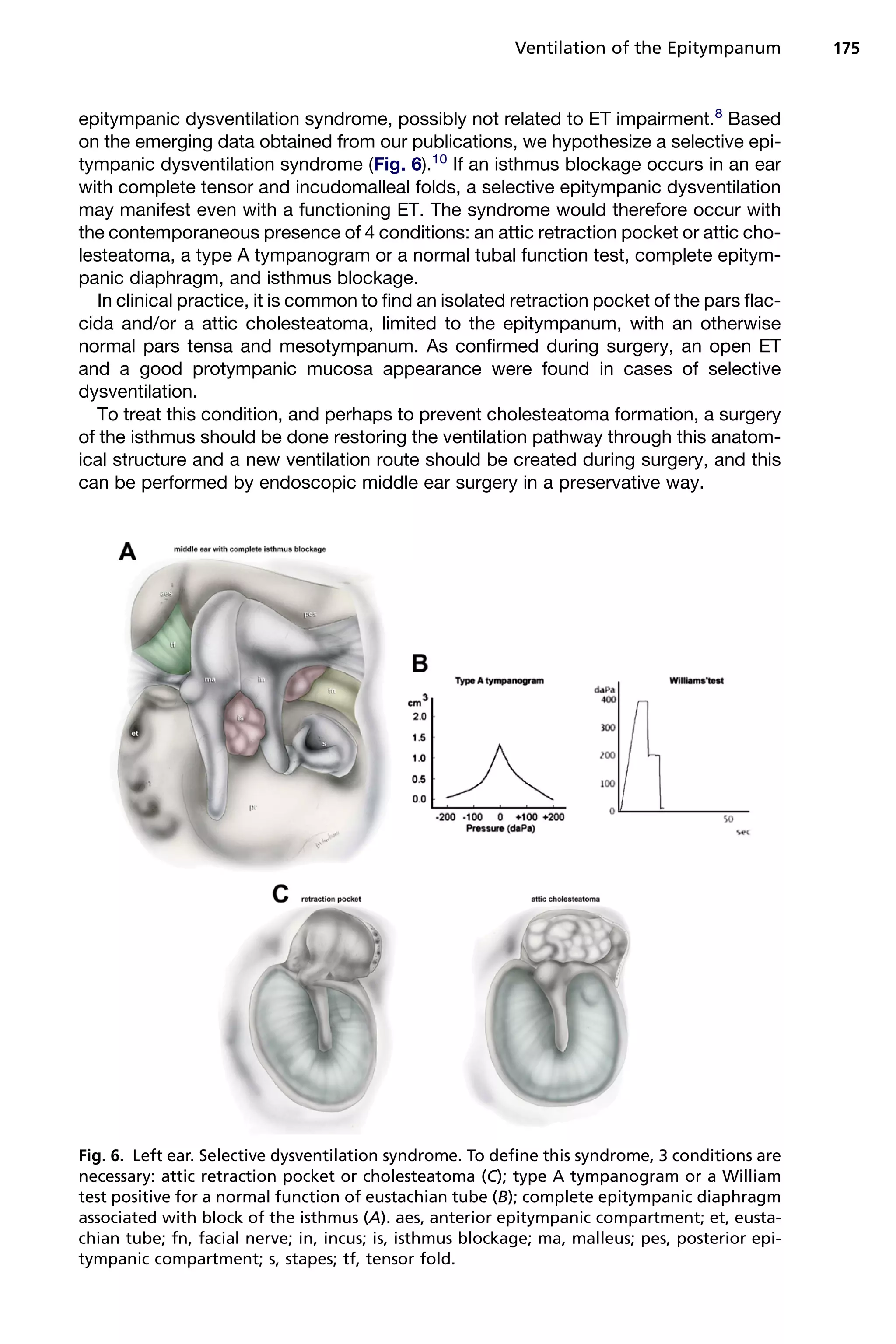
2) The tensor fold plays a role in ventilation, and can be complete or incomplete. A complete tensor fold with isthmus blockage can lead to inadequate ventilation and attic retraction.
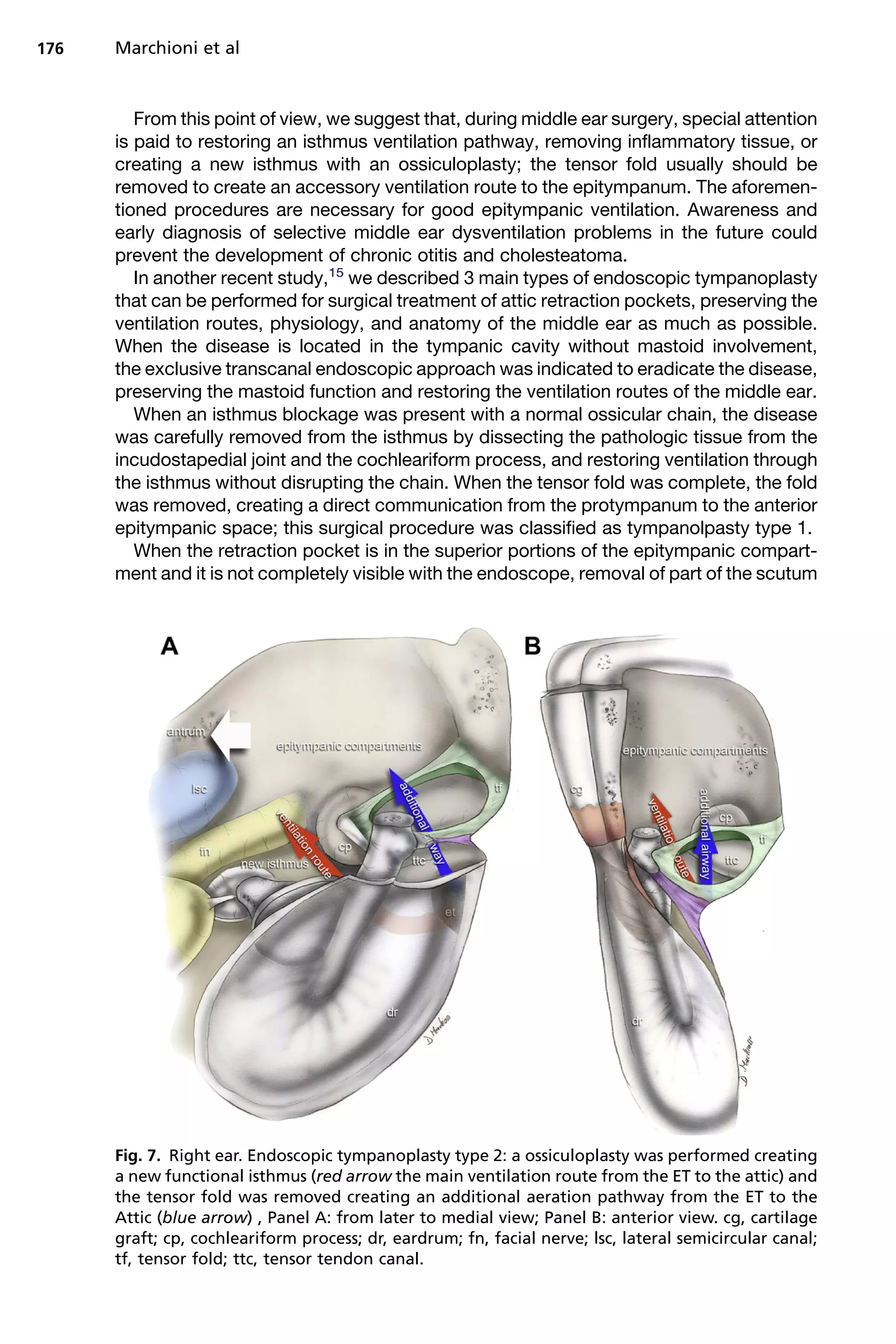
3) Endoscopic surgery aims to restore ventilation by enlarging the isthmus and
Spaces of middle ear and their surgical importanceDr Soumya Singh
one of the imp topics in ENT that should be understood very thoroughly if u want to pursue as an otologist.I tried to simplify the topic with simple diagrams and models for better understanding .
otosclerosis....
stapedectomy vs stapedotomy
complication of otosclerotic surgery
management of otosclerotic surgery complications
techniques
latest trends
Sinus tymapni shape and depth can influence surgical approach in cholesteatoma surgery. In the case of a shallower ST, an exclusive endoscopic exploration is chosen; while in the case of a deeper ST, a retrofacial approach is usually preferred.
Perilymph Fistula can be difficult to diagnose as a standalone condition. Post-trauma symptoms such as dizziness, headache, etc. can be linked to other conditions like a traumatic brain injury with a concussion.
Cavity obliteration is a procedure done at the end of Mastoidectomy to get a cavity-less mastoid cavity thus solving the problem of discharging post-operative cavity.
surgical anatomy of nose is a humble attempt to make the anatomy of nose simpler and easy for medical students and fellow physicians. at the end of the presentation the students will be able to identify all the structures.
Ct anatomy of paranasal sinuses( PNS) pk.pdf pptDr pradeep Kumar
This presentation includes cross sectional anatomy like axial,saggital and coronal images of paranasal sinuses and most important variation of paranasal sinus.This help alot. Must read topic for radiology resident. Thanks
I have tried my level best to complete this one. Basics & subjective details as much possible, are included here with understandable diagrams, CT-scans & charts. Clinical associations with possible anatomical structures are also touched . Frequent questions based on the topic discussed, will be there at the middle & end of presentation.
If you find it helpful then please like it & if any query regarding this ppt or upcoming ppts then mail me
drsuraj1997@gmail.com
Development of the middle ear is not covered in this presentation. If you are interested then please mail me. I will try to upload it as a separate one.
ARTIFICIAL INTELLIGENCE IN HEALTHCARE.pdfAnujkumaranit
Artificial intelligence (AI) refers to the simulation of human intelligence processes by machines, especially computer systems. It encompasses tasks such as learning, reasoning, problem-solving, perception, and language understanding. AI technologies are revolutionizing various fields, from healthcare to finance, by enabling machines to perform tasks that typically require human intelligence.
Couples presenting to the infertility clinic- Do they really have infertility...Sujoy Dasgupta
Dr Sujoy Dasgupta presented the study on "Couples presenting to the infertility clinic- Do they really have infertility? – The unexplored stories of non-consummation" in the 13th Congress of the Asia Pacific Initiative on Reproduction (ASPIRE 2024) at Manila on 24 May, 2024.
New Directions in Targeted Therapeutic Approaches for Older Adults With Mantl...i3 Health
i3 Health is pleased to make the speaker slides from this activity available for use as a non-accredited self-study or teaching resource.
This slide deck presented by Dr. Kami Maddocks, Professor-Clinical in the Division of Hematology and
Associate Division Director for Ambulatory Operations
The Ohio State University Comprehensive Cancer Center, will provide insight into new directions in targeted therapeutic approaches for older adults with mantle cell lymphoma.
STATEMENT OF NEED
Mantle cell lymphoma (MCL) is a rare, aggressive B-cell non-Hodgkin lymphoma (NHL) accounting for 5% to 7% of all lymphomas. Its prognosis ranges from indolent disease that does not require treatment for years to very aggressive disease, which is associated with poor survival (Silkenstedt et al, 2021). Typically, MCL is diagnosed at advanced stage and in older patients who cannot tolerate intensive therapy (NCCN, 2022). Although recent advances have slightly increased remission rates, recurrence and relapse remain very common, leading to a median overall survival between 3 and 6 years (LLS, 2021). Though there are several effective options, progress is still needed towards establishing an accepted frontline approach for MCL (Castellino et al, 2022). Treatment selection and management of MCL are complicated by the heterogeneity of prognosis, advanced age and comorbidities of patients, and lack of an established standard approach for treatment, making it vital that clinicians be familiar with the latest research and advances in this area. In this activity chaired by Michael Wang, MD, Professor in the Department of Lymphoma & Myeloma at MD Anderson Cancer Center, expert faculty will discuss prognostic factors informing treatment, the promising results of recent trials in new therapeutic approaches, and the implications of treatment resistance in therapeutic selection for MCL.
Target Audience
Hematology/oncology fellows, attending faculty, and other health care professionals involved in the treatment of patients with mantle cell lymphoma (MCL).
Learning Objectives
1.) Identify clinical and biological prognostic factors that can guide treatment decision making for older adults with MCL
2.) Evaluate emerging data on targeted therapeutic approaches for treatment-naive and relapsed/refractory MCL and their applicability to older adults
3.) Assess mechanisms of resistance to targeted therapies for MCL and their implications for treatment selection
MANAGEMENT OF ATRIOVENTRICULAR CONDUCTION BLOCK.pdfJim Jacob Roy
Cardiac conduction defects can occur due to various causes.
Atrioventricular conduction blocks ( AV blocks ) are classified into 3 types.
This document describes the acute management of AV block.
- Video recording of this lecture in English language: https://youtu.be/lK81BzxMqdo
- Video recording of this lecture in Arabic language: https://youtu.be/Ve4P0COk9OI
- Link to download the book free: https://nephrotube.blogspot.com/p/nephrotube-nephrology-books.html
- Link to NephroTube website: www.NephroTube.com
- Link to NephroTube social media accounts: https://nephrotube.blogspot.com/p/join-nephrotube-on-social-media.html
Title: Sense of Taste
Presenter: Dr. Faiza, Assistant Professor of Physiology
Qualifications:
MBBS (Best Graduate, AIMC Lahore)
FCPS Physiology
ICMT, CHPE, DHPE (STMU)
MPH (GC University, Faisalabad)
MBA (Virtual University of Pakistan)
Learning Objectives:
Describe the structure and function of taste buds.
Describe the relationship between the taste threshold and taste index of common substances.
Explain the chemical basis and signal transduction of taste perception for each type of primary taste sensation.
Recognize different abnormalities of taste perception and their causes.
Key Topics:
Significance of Taste Sensation:
Differentiation between pleasant and harmful food
Influence on behavior
Selection of food based on metabolic needs
Receptors of Taste:
Taste buds on the tongue
Influence of sense of smell, texture of food, and pain stimulation (e.g., by pepper)
Primary and Secondary Taste Sensations:
Primary taste sensations: Sweet, Sour, Salty, Bitter, Umami
Chemical basis and signal transduction mechanisms for each taste
Taste Threshold and Index:
Taste threshold values for Sweet (sucrose), Salty (NaCl), Sour (HCl), and Bitter (Quinine)
Taste index relationship: Inversely proportional to taste threshold
Taste Blindness:
Inability to taste certain substances, particularly thiourea compounds
Example: Phenylthiocarbamide
Structure and Function of Taste Buds:
Composition: Epithelial cells, Sustentacular/Supporting cells, Taste cells, Basal cells
Features: Taste pores, Taste hairs/microvilli, and Taste nerve fibers
Location of Taste Buds:
Found in papillae of the tongue (Fungiform, Circumvallate, Foliate)
Also present on the palate, tonsillar pillars, epiglottis, and proximal esophagus
Mechanism of Taste Stimulation:
Interaction of taste substances with receptors on microvilli
Signal transduction pathways for Umami, Sweet, Bitter, Sour, and Salty tastes
Taste Sensitivity and Adaptation:
Decrease in sensitivity with age
Rapid adaptation of taste sensation
Role of Saliva in Taste:
Dissolution of tastants to reach receptors
Washing away the stimulus
Taste Preferences and Aversions:
Mechanisms behind taste preference and aversion
Influence of receptors and neural pathways
Impact of Sensory Nerve Damage:
Degeneration of taste buds if the sensory nerve fiber is cut
Abnormalities of Taste Detection:
Conditions: Ageusia, Hypogeusia, Dysgeusia (parageusia)
Causes: Nerve damage, neurological disorders, infections, poor oral hygiene, adverse drug effects, deficiencies, aging, tobacco use, altered neurotransmitter levels
Neurotransmitters and Taste Threshold:
Effects of serotonin (5-HT) and norepinephrine (NE) on taste sensitivity
Supertasters:
25% of the population with heightened sensitivity to taste, especially bitterness
Increased number of fungiform papillae
Anti ulcer drugs and their Advance pharmacology ||
Anti-ulcer drugs are medications used to prevent and treat ulcers in the stomach and upper part of the small intestine (duodenal ulcers). These ulcers are often caused by an imbalance between stomach acid and the mucosal lining, which protects the stomach lining.
||Scope: Overview of various classes of anti-ulcer drugs, their mechanisms of action, indications, side effects, and clinical considerations.
HOT NEW PRODUCT! BIG SALES FAST SHIPPING NOW FROM CHINA!! EU KU DB BK substit...GL Anaacs
Contact us if you are interested:
Email / Skype : kefaya1771@gmail.com
Threema: PXHY5PDH
New BATCH Ku !!! MUCH IN DEMAND FAST SALE EVERY BATCH HAPPY GOOD EFFECT BIG BATCH !
Contact me on Threema or skype to start big business!!
Hot-sale products:
NEW HOT EUTYLONE WHITE CRYSTAL!!
5cl-adba precursor (semi finished )
5cl-adba raw materials
ADBB precursor (semi finished )
ADBB raw materials
APVP powder
5fadb/4f-adb
Jwh018 / Jwh210
Eutylone crystal
Protonitazene (hydrochloride) CAS: 119276-01-6
Flubrotizolam CAS: 57801-95-3
Metonitazene CAS: 14680-51-4
Payment terms: Western Union,MoneyGram,Bitcoin or USDT.
Deliver Time: Usually 7-15days
Shipping method: FedEx, TNT, DHL,UPS etc.Our deliveries are 100% safe, fast, reliable and discreet.
Samples will be sent for your evaluation!If you are interested in, please contact me, let's talk details.
We specializes in exporting high quality Research chemical, medical intermediate, Pharmaceutical chemicals and so on. Products are exported to USA, Canada, France, Korea, Japan,Russia, Southeast Asia and other countries.
The prostate is an exocrine gland of the male mammalian reproductive system
It is a walnut-sized gland that forms part of the male reproductive system and is located in front of the rectum and just below the urinary bladder
Function is to store and secrete a clear, slightly alkaline fluid that constitutes 10-30% of the volume of the seminal fluid that along with the spermatozoa, constitutes semen
A healthy human prostate measures (4cm-vertical, by 3cm-horizontal, 2cm ant-post ).
It surrounds the urethra just below the urinary bladder. It has anterior, median, posterior and two lateral lobes
It’s work is regulated by androgens which are responsible for male sex characteristics
Generalised disease of the prostate due to hormonal derangement which leads to non malignant enlargement of the gland (increase in the number of epithelial cells and stromal tissue)to cause compression of the urethra leading to symptoms (LUTS
Flu Vaccine Alert in Bangalore Karnatakaaddon Scans
As flu season approaches, health officials in Bangalore, Karnataka, are urging residents to get their flu vaccinations. The seasonal flu, while common, can lead to severe health complications, particularly for vulnerable populations such as young children, the elderly, and those with underlying health conditions.
Dr. Vidisha Kumari, a leading epidemiologist in Bangalore, emphasizes the importance of getting vaccinated. "The flu vaccine is our best defense against the influenza virus. It not only protects individuals but also helps prevent the spread of the virus in our communities," he says.
This year, the flu season is expected to coincide with a potential increase in other respiratory illnesses. The Karnataka Health Department has launched an awareness campaign highlighting the significance of flu vaccinations. They have set up multiple vaccination centers across Bangalore, making it convenient for residents to receive their shots.
To encourage widespread vaccination, the government is also collaborating with local schools, workplaces, and community centers to facilitate vaccination drives. Special attention is being given to ensuring that the vaccine is accessible to all, including marginalized communities who may have limited access to healthcare.
Residents are reminded that the flu vaccine is safe and effective. Common side effects are mild and may include soreness at the injection site, mild fever, or muscle aches. These side effects are generally short-lived and far less severe than the flu itself.
Healthcare providers are also stressing the importance of continuing COVID-19 precautions. Wearing masks, practicing good hand hygiene, and maintaining social distancing are still crucial, especially in crowded places.
Protect yourself and your loved ones by getting vaccinated. Together, we can help keep Bangalore healthy and safe this flu season. For more information on vaccination centers and schedules, residents can visit the Karnataka Health Department’s official website or follow their social media pages.
Stay informed, stay safe, and get your flu shot today!
TEST BANK for Operations Management, 14th Edition by William J. Stevenson, Ve...kevinkariuki227
TEST BANK for Operations Management, 14th Edition by William J. Stevenson, Verified Chapters 1 - 19, Complete Newest Version.pdf
Prix Galien International 2024 Forum ProgramLevi Shapiro
June 20, 2024, Prix Galien International and Jerusalem Ethics Forum in ROME. Detailed agenda including panels:
- ADVANCES IN CARDIOLOGY: A NEW PARADIGM IS COMING
- WOMEN’S HEALTH: FERTILITY PRESERVATION
- WHAT’S NEW IN THE TREATMENT OF INFECTIOUS,
ONCOLOGICAL AND INFLAMMATORY SKIN DISEASES?
- ARTIFICIAL INTELLIGENCE AND ETHICS
- GENE THERAPY
- BEYOND BORDERS: GLOBAL INITIATIVES FOR DEMOCRATIZING LIFE SCIENCE TECHNOLOGIES AND PROMOTING ACCESS TO HEALTHCARE
- ETHICAL CHALLENGES IN LIFE SCIENCES
- Prix Galien International Awards Ceremony
1. Endoscopic Anatomy and
Ventilation of the Epitympanum
Daniele Marchioni, MD*, Alessia Piccinini, MD,
Matteo Alicandri-Ciufelli, MD, Livio Presutti, MD
INTRODUCTION
Aeration of the tympanic cavity and mastoid cells and anatomic pathways for middle
ear ventilation have been studied since the end of the nineteenth century, starting with
the work of Prussak1
in 1867. More recently, Palva and Johnsson2
were the first to
describe middle ear anatomy focusing on ventilation patterns and their implications
for middle ear disease.
The eustachian tube (ET) plays a crucial role in maintaining middle ear aeration and
atmospheric pressure. Inflammatory middle ear chronic disease is usually related to
Disclosures: All of the authors have read and approved the manuscript. None of the authors
have any financial relationships to disclose.
Conflict of interest: None.
Otolaryngology, Head and Neck Surgery Department, Policlinic of Modena, University Hospital
of Modena, Via del Pozzo 71, Modena 41100, Italy
* Corresponding author.
E-mail address: marchionidaniele@yahoo.it
KEYWORDS
Endoscopic ear surgery Epitympanum Middle ear ventilation Surgical anatomy
Prussak space
KEY POINTS
The superior attic (upper unit) is in communication with the mesotympanum through the
underlying tympanic isthmus and posteriorly it is open to the aditus ad antrum.
The inferior lateral attic and the Prussack space are lower than the epitympanic dia-
phragm, and it is ventilated by the mesotympanum.
An isthmus blockage associated with a complete tensor fold leads to inadequate ventila-
tion of the mastoid cells and this scenario could be at the basis of the attic retraction
pocket development.
During surgery, in sectorial disventilatory disorders caused by isthmus block, it is essential
to restore the ventilation pathway through the isthmus and to create an alternative direct
ventilatory route between the protympanum and anterior attic from a section of the central
portion of the tensor fold.
Endoscopic middle ear surgery may help in understanding the physiopathology of the
middle ear, allowing the surgeon to explore middle ear anatomy, and thus all ventilation
pathways.
Otolaryngol Clin N Am 46 (2013) 165–178
http://dx.doi.org/10.1016/j.otc.2012.10.002 oto.theclinics.com
0030-6665/13/$ – see front matter Ó 2013 Elsevier Inc. All rights reserved.
2. ET dysfunction caused by poor tympanic ventilation. This condition is also related to
hearing impairment and poor postoperative outcomes.3
Although middle ear aeration
is related to ET function, other anatomic factors may play important roles in ventilation
of these spaces and, in particular, in the pathophysiology of selective epitympanic
retraction.
In recent years, the use of endoscope with varied angulations has allowed the
surgeon to explore all of the hidden areas that are often not visualized using a micro-
scope.4–7
Endoscopes have also improved knowledge of the complex fold anatomy
and functional interventions in middle ear inflammatory disorders during middle ear
surgery, particularly in the case of selective dysventilation.8
This article discusses the anatomy of the epitympanum and the ventilation patterns
and pathophysiology of epitympanic retraction.
The Epitympanic Compartments (Anterior and Posterior Epitympanum) and the
Concept of Upper and Lower Units
The epitympanum is divided into 2 compartments: a large posterior compartment and
a smaller anterior compartment. The demarcation between the anterior and posterior
epitympanum depends on the anatomic variations of important structures such as the
cog and the tensor fold. In most people, the demarcation between the anterior and
posterior epitympanum is represented by the transverse ridge or cog. The cog is
a bony septum that detaches from the tegmen tympani cranially, leading vertically
toward the cochleariform process in front of the malleus head.
Much of the posterior epitympanic volume is occupied by the body and short
process of the incus together with the head of the malleus. The lateral portion of the
posterior epitympanum is narrow and is divided by the lateral incudomalleolar fold
in 2 further portions, the superior and inferior lateral attic, positioned separately one
above the other.
The incudomalleolar fold originates at the posterior extremity of the short process of
incus and the lateral portion of the posterior incudal fold, continuing anteriorly between
the body of the incus, the head of the malleus and the lateral aspect of the attic. At this
level, the fold bends inferiorly, joining the posterior malleolar ligament fold and the
lateral malleolar ligament fold, with which it forms the medial and superior aspect of
the Prussak space (Fig. 1).
The inferior lateral attic is bounded superiorly by the lateral incudo-malleolar fold.
This anatomic area is therefore in a lower position than the epitympanic diaphragm
in communication with the underlying mesotympanum. Ventilation of the inferior lateral
attic is provided by the mesotympanic region. In a more cranial position than the infe-
rior lateral attic lies the superior lateral attic, whose floor or inferior limit is represented
by the incudomalleolar fold. Together with the medial attic, this anatomic area is called
the superior attic or upper unit.
The superior attic is in communication with the mesotympanum through the under-
lying tympanic isthmus, and posteriorly it is opened to the aditus ad antrum. Its upper
limit is the tegmen tympani, the lower limit is the second (intratympanic) portion of the
facial nerve, and laterally it is bounded by the bony lateral wall of the atticus. The supe-
rior attic is therefore ventilated through the isthmus (Fig. 2).
The lower unit is formed by the reduced space represented by the Prussak space,
which is separated in its anatomy and ventilation from the upper unit by its vault, rep-
resented by the lateral malleolar ligament fold. This inferior epitympanic portion is
ventilated in most cases from the posterior pocket through mesotympanum (see
Fig. 1 panel D).
Marchioni et al166
3. Fig. 1. The malleolar ligament folds and membranous folds representing the complete epi-
tympanic diaphragm, and the 2 major middle ear ventilation pathways of the epitympanic
compartments (blue arrow) and Prussak space (orange arrow). (A) Anterior view. (B) Poste-
rior view. (C) Axial view according to Palva. (D) Lateral view of Prussak space. AES, anterior
epitympanic space; amlf, anterior malleal ligamental fold; as, anterior spine; bin, body of
the incus; cp, cochleariform process; ct, corda tympani; et, eustachian tube; fn, facial nerve;
hma, head of the malleus; imlf, lateral incudomalleal fold; in, incus; ma, malleus; mlf, lateral
malleal ligamental fold; PES, posterior epitympanic space; pil, lateral and medial posterior
incudal ligaments; plm, posterior malleal ligamental fold; prs, Prussak space; ps, posterior
spine; s, stapes; sr, supratubal recess; tf, tensor fold. The orange arrow indicates the major
ventilation pathway through the posterior pocket.
Ventilation of the Epitympanum 167
4. The 2 ventilatory trajectories of the epitympanic units are therefore separated from
each other; this pathophysiologic concept is important in transcanalar endoscopic
surgery, because surgical treatment is based on the restoration of ventilation and
on the unification of the upper unit with the lower unit, through the creation of a large
tympanic isthmus and an accessory route through the tensor fold. The surgical solu-
tion must ensure the ventilation of all parts of the epitympanum.
The anterior epitympanum is delimited anteriorly by the root of the zygomatic arch
(a thick bony plate that separates it from pericarotic cells), superiorly by the tegmen
tympani (which separates it from the meninges), laterally by the tympanic bone and
chorda tympani, and medially by a bony wall that separates it from the geniculate
fossa, which contains the homonymous ganglion. Its inferior limit is represented by
the tensor fold, which, if complete, separates it from the underlying sovratubaric
recess. The tensor fold presents a variable anatomy: according to Palva and col-
leagues,9
the tensor fold is incomplete in only 25% of cases, allowing an alternative
ventilation route directed from the sovratubaric recess toward the attic (Fig. 7). It
extends laterally from the semicanal of the tensor tympani muscle to the lateral aspect
of the protympanum, posteriorly adhering to the cochleariform process and to the
tensor tympani tendon, and extends anteriorly to the root of the zygomatic bone to
provide the epitympanic floor.
If it inserts on the transverse crest, its direction is almost vertical, whereas, if inserts
on the tubaric tegmen, its direction is horizontal.
Fig. 2. Right ear. The posterior isthmus after posterior atticotomy maintaining the integrity
of the ossicular chain (the red arrow represents the main ventilation route from the ET to
the antrum through the isthmus). aes, anterior epitympanic compartment; cp, cochleariform
process; fn, facial nerve; in, incus; lsc, lateral semicircular canal; ma, malleus; pe, pyramidal
eminence; pes, posterior epitympanic compartment; s, stapes; tf, tensor fold; ttc, tensor
tympani canal.
Marchioni et al168
5. In most cases the curvature is about 45
and its most frequent insertion lies at the
central portion of the anterior sovratubaric-epitympanic tegmen. According to our
observations on patients affected by attic cholesteatomas, a complete tensor fold
has been observed in almost all the patients studied, and the direction of the fold
was in most cases horizontal.4
In general, the width of the underlying sovratubaric
recess varies depending on its angle (Fig. 3).
During surgery, in sectorial ventilatory disorders caused by isthmus block, it is
essential to create an alternative direct ventilatory route between the protympanum
and anterior attic from a section of the central portion of the tensor fold.
The anterior epitympanum can be formed from a single large air cell, or by several
small air cells, and this makes the anterior epitympanum a variable anatomic space in
an anterior-posterior direction. In a recent study conducted at our clinic, subjects
affected by cholesteatoma limited to the attic showed a reduced volume of the
bony boundaries of the anterior epitympanum. The small anterior epitympanic cavities
might be proof of selective attic dysventilation.
Epitympanic Diaphragm and Epitympanic Ventilation Patterns
The concept of epitympanic diaphragm was raised for the first time by Lemoine in
1950;5
the investigators described that the diaphragm was made up of various struc-
tures and membranous ligaments that, together with the malleus and the incus, form
the floor of the epitympanic compartment.
The investigators also described the Prussak area as a structure located inferiorly to
this diaphragm, therefore dividing it from the epitympanic compartments by the lateral
malleolar ligament fold, considered the Prussak space roof.
In addition to the folds described by Lemoine, Palva2
added 2 more duplicated
folds: the tensor fold and the lateral incudomalleolar fold. The complete diaphragm
therefore comprises the 3 malleolar ligament folds (anterior, lateral, and posterior),
Fig. 3. Right ear. The variations in size of the supratubal recess. The dimensions of this recess
depend on the inclination of the tensor fold (right). The more vertical the tensor fold, the
wider the supratubal recess. When the tensor fold is a horizontal, the supratubal recess is
not present (left). et, eustachian tube; in, incus; is, isthmus; ma, malleus; s, stapes; sr, supra-
tubal recess; tf, tensor fold.
Ventilation of the Epitympanum 169
6. the posterior incudal fold, and the 2 duplicated membranous folds (tensor fold and
the lateral incudomalleolar fold) associated with the incus and the malleus.
Palva classified Proctor’s anterior tympanic isthmus as a single entity that is always
present, and defined it simply as the tympanic isthmus, whereas the ventilation route
posterior to the incus, an irregular feature named by Proctor6
the posterior isthmus,
provides inconsistent ventilation from the fossa incudis.
The tympanic isthmus described by Palva represents a wide ventilation route for the
epitympanum, excluding the Prussak space.
This structure extends anteriorly from the tensor fold to the pyramidal process
(inferiorly and posteriorly) and to the medial portion of the posterior incus ligament
(superiorly and posteriorly). Its medial limit is the attic bony wall and the lateral
limit is the body of the incus, the incus short process, and the head of the
malleus.
The space bounded by these structures is called the middle attic, which becomes
the mesotympanum inferior to the body incus.
The anterior portion of the tympanic isthmus, superior to the level of the tensor
tympani tendon, represents a wide communication with the anterior epitympanum.
In healthy ears, the tympanic isthmus is an open structure with no fold.
Although Palva noticed a wide opening just behind the incus short process in 25%
of Proctor posterior tympanic isthmus, in most cases this potential posterior ventila-
tion route was blocked by the posterior incus ligament fold.
Given this, all the compartments leading to the epitympanic diaphragm receive air
through the only ventilation way that is always present, the tympanic isthmus route,
located between the medial aspect of the posterior incus ligament and the tensor fold.
However, Palva2,9
noticed that membranous folds that formed the epitympanic dia-
phragm could have structural defects resulting in incomplete folds.
In this way, additional ventilation patterns arise for the structures above the epitym-
panic diaphragm. Most of the defects were of the tensor fold (29% of cases), followed
by the lateral incudomalleolar fold in its anterior portion (15% of cases).
The Prussak Space
The medial and inferior aspects of the Prussak space are formed respectively by the
neck and the short process of the malleus.
The superior limit is the fold of the lateral malleolar ligament, which also represents
the floor of the lateral malleolar space; this ligament inserts laterally on the medial wall
of the scutum. The lateral malleolar ligament fold is integral in most case, because
according to Palva9
only 19% of subjects showed a defect in the anterior portion of
the fold, whereas, in some rare cases (7% of the subjects examined by Palva), the
defect was in the posterior portion of the ligament, the latter involving the lateral mal-
leolar ligament fold, creating a communication between the upper epitympanic portion
(upper unit) and the lower (lower unit), resulting in a communication between the Prus-
sak space and the lateral malleolar space and creating a change in the classic epitym-
panic diaphragm.
Theanterior aspect ofthePrussak area is boundedby a thin, membranous fold among
the tympanic membrane and the anterior malleolar ligament fold, which inserts laterally
on the tympanic membrane and medially on the neck and long process of the malleus.
In some cases, this fold is absent, causing a further anterior ventilation trajectory to
the Prussak space.
The lateral aspect is represented by the Sharpnell membrane.
The posterior wall is represented by a large posterior pocket (the posterior
pocket of von Tro¨ ltsch), which is the main route of ventilation. This posterior pocket
Marchioni et al170
7. is bounded laterally by the pars tensa and pars flaccida of the tympanic
membrane, and medially by the posterior malleolar ligament fold, which originates
from the posterior portion of the malleus neck and the upper third of the malleus
handle and inserts posteriorly in the posterior tympanic spine (Fig. 1; Panel D).
This posterior pocket develops in a posterior-inferior direction and opens at the
most cranial portion of mesotympanum, so, in most people, ventilation of the Prus-
sak space occurs through the communication with the mesotympanum, the only
ventilation route that is separated from the epitympanic upper unit. This ventilation
route of the inferior epitympanic compartment through the posterior pocket of von
Tro¨ ltsch is rough and narrow, especially compared with the ventilation route
through tympanic isthmus, which aerates the upper epitympanic compartment
and is wider. For these reasons, the possibility of anatomic reduction of the
passage until the closing of the posterior pocket is plausible, especially the pres-
ence of thick and viscous secretions within the Prussak space that could cause
a chronic sectorial dysventilation associated with a retraction of the Sharpnell
membrane and its adhesion with the malleus neck.
For these reasons, although the Prussak space is anatomically inseparable from the
epitympanum, in terms of ventilation and drainage, it represents an independent unit.
This space may have a block and/or an obliteration without any involvement of the
compartments above the epitympanic diaphragm, like the anterior and posterior epi-
tympanum, the aditus, and mastoid cells.
Palva dissected subjects on ventilation tubes for epitympanic retraction, showed
that, despite the surgical treatment, an attical dysventilation was still present.2
Given these phenomena, Palva assumed that behind the genesis of attical cholestea-
toma there was a progressive closure of the ventilation route of the inferior epitympanic
unit (lower unit) initially derived from mucous tissue inflammation in the posterior pocket
and Prussack space, then from the granulation tissue formation that progressively
causes a total block to the passage of air from this route. These events could lead to
retraction of the Sharpnell membrane toward the malleus neck. The positioning of
a ventilation tube causes an improvement in mesotympanic and hypotympanic ventila-
tion, but does not address the blockage in the posterior pocket, and the process of
retraction would be irreversible. It is still debated whether these phenomena are suffi-
cient to cause an attical cholesteatoma.10
DISCUSSION OF MIDDLE EAR ANATOMY
Intraoperative evaluation of middle ear anatomy during endoscopic surgery for inflam-
matory disorders allows the visualization of anatomic blockages of the middle ear
ventilation patterns.
Many other investigators have described the anatomy and development of
tympanic compartments and folds because this knowledge is crucial in the under-
standing and treatment of middle ear disease.
More recently, tympanic isthmus and middle ear ventilation patterns have been
described in several articles.
Palva and Johnsson2,9
studied the anatomy of the tensor fold during temporal bone
dissection. They observed that, in most patients, the tensor fold was a complete fold
separating the epitympanic compartment from the protympanum. In these patients,
the isthmus was the only aeration pathway; however, in rare cases, it is possible to
observe an incomplete tensor fold; in these cases, the anterior epitympanic space
received aeration directly from the protympanum through the communication in the
tensor fold area.
Ventilation of the Epitympanum 171
8. Although exploration of the tensor fold region during middle ear surgery for chronic
disease has already been established in the international literature, it is not easy to
reach this region in otomicroscopy.
Several approaches have been described in the international literature, but we
suggest an endoscopic approach to the tensor fold in patients with attic disease,
which could be exclusive or combined with the traditional microscopic approach.11
In our previous study12
focused on epitympanic size in patients affected by a limited
attic cholesteatoma, we observed that the anterior epitympanic recess (AER) in an
affected ear is smaller than in an unaffected ear. We hypothesized that the presence
of a tympanic isthmus blockage associated with a complete tensor fold could exclude
the AER from the posterior epitympanic space and from the protympanum. The
blockage of the tympanic isthmus could create a selective negative pressure in the
atticomastoid spaces; this chronic lack of aeration could provoke a hypodevelopment
of the AER with a reduction of pressure level and, consequently, an attic retraction and
cholesteatoma sac development (Fig. 4). This process is also possible in patients with
a normally functioning ET.
Middle Ear Blockage
An isthmus blockage caused by chronic inflammatory disease in association with
a complete tensor fold leads to inadequate ventilation of the mastoid cells and epi-
tympanic recess. Middle ear pressure seems related not only to a functioning ET
but also to transmucosal gas exchange through the mastoid mucosa. The mucosal
gas exchange is related to the degree of mastoid pneumatization.13
Because of
these 2 gas pressure regulation systems, even if the ET is functioning, an isthmus
blockage could impair ventilation of the mastoid cells, causing sclerotization of
the mastoid. It is not clear whether chronic middle ear disease leads to inadequate
mastoid pneumatization or, conversely, a sclerotic mastoid leads to chronic middle
ear disease.14
In a recent study,4
we described the kinds of anatomic blockage of the middle ear
ventilation trajectories that may be identified during endoscopic surgery to understand
whether those alterations could be associated with anomalous mastoid pneumatiza-
tion, a classic sign of middle ear dysventilation problems.
In this study, the anatomic structure that separates epitympanic space from the
mesotympanum (tympanic isthmus and tensor fold area) was studied. No previous
studies have been performed on the surgical approach for patients affected by
a middle ear chronic disease with blockage of the isthmus. Intraoperative evaluation
of middle ear anatomy during endoscopic surgery allowed us to clearly visualize the
presence of anatomic blockage of the middle ear ventilation trajectories. We classified
these anatomic blockage patterns into 3 types (Fig. 5):
Type A: blockage of the isthmus associated with a complete tensor fold (most of
these patients presented a selective attic retraction pocket or attic cholesteatoma
without pathologic tissue in the mesotympanic spaces)
Type B: blockage of the isthmus associated with an attical vertical blockage
(consisting of a fold or granulation tissue involving the incudomalleal fold) sepa-
rating the anterior epitympanic space from the posterior epitympanic space with
or without a complete tensor fold, in these subjects a selective retraction pocket
into the posterior attic was found
Type C: a complete epidermization of the attic space causing a blockage of the
isthmus and a complete antral blockage excluding the mesotympanic space
from the epitympanic and mastoid spaces
Marchioni et al172
9. In our series, tensor fold removal in association with a restoration of the isthmus
function prevented postoperative retraction or cholesteatoma recurrence 1 year after
the primary surgery. The use of the endoscope during surgery also permitted a good
view of the tensor fold area and the isthmus timpani and, consequently, enabled us to
understand the type of dysventilation pattern. The goal of surgery in this kind of
disorder could be restoration of normal ventilation of the attical-mastoid area. This
solution is possible by removing the tensor fold and restoring the functionality of the
isthmus.
We recently published another study of middle ear anatomy, focusing on middle ear
folds in patients with attical retractions or cholesteatoma and with a normal tubal func-
tion test, who underwent endoscopic surgery. This scenario might describe a selective
Fig. 4. Right ear. Tympanic isthmus block associated with a complete tensor fold. The
blockage of the attic aeration pathway could create a selective negative pressure in the
atticomastoid spaces, developing a selective retraction in the attic. Transcanal view (A);
medial to lateral view (B); axial view at the level of the attic (C). aes, anterior epitympanic
compartment; amf, anterior malleal fold; cp, cochleariform process; dr, eardrum; et, eusta-
chian tube; fn, facial nerve; imlf, lateral incudomalleal fold; in, incus; is*, isthmus blockage;
ma, malleus; mlf, lateral malleal fold; pe, pyramidal eminence; pes, posterior epitympanic
compartment; pil, posterior incudal ligaments; prs, Prussak space; s, stapes; tf, tensor fold.
Ventilation of the Epitympanum 173
10. Fig. 5. Classification of epitympanic ventilation blockage correlated on the endoscopic find-
ings. Left ear view, from medial to lateral.
Marchioni et al174
11. epitympanic dysventilation syndrome, possibly not related to ET impairment.8
Based
on the emerging data obtained from our publications, we hypothesize a selective epi-
tympanic dysventilation syndrome (Fig. 6).10
If an isthmus blockage occurs in an ear
with complete tensor and incudomalleal folds, a selective epitympanic dysventilation
may manifest even with a functioning ET. The syndrome would therefore occur with
the contemporaneous presence of 4 conditions: an attic retraction pocket or attic cho-
lesteatoma, a type A tympanogram or a normal tubal function test, complete epitym-
panic diaphragm, and isthmus blockage.
In clinical practice, it is common to find an isolated retraction pocket of the pars flac-
cida and/or a attic cholesteatoma, limited to the epitympanum, with an otherwise
normal pars tensa and mesotympanum. As confirmed during surgery, an open ET
and a good protympanic mucosa appearance were found in cases of selective
dysventilation.
To treat this condition, and perhaps to prevent cholesteatoma formation, a surgery
of the isthmus should be done restoring the ventilation pathway through this anatom-
ical structure and a new ventilation route should be created during surgery, and this
can be performed by endoscopic middle ear surgery in a preservative way.
Fig. 6. Left ear. Selective dysventilation syndrome. To define this syndrome, 3 conditions are
necessary: attic retraction pocket or cholesteatoma (C); type A tympanogram or a William
test positive for a normal function of eustachian tube (B); complete epitympanic diaphragm
associated with block of the isthmus (A). aes, anterior epitympanic compartment; et, eusta-
chian tube; fn, facial nerve; in, incus; is, isthmus blockage; ma, malleus; pes, posterior epi-
tympanic compartment; s, stapes; tf, tensor fold.
Ventilation of the Epitympanum 175
12. From this point of view, we suggest that, during middle ear surgery, special attention
is paid to restoring an isthmus ventilation pathway, removing inflammatory tissue, or
creating a new isthmus with an ossiculoplasty; the tensor fold usually should be
removed to create an accessory ventilation route to the epitympanum. The aforemen-
tioned procedures are necessary for good epitympanic ventilation. Awareness and
early diagnosis of selective middle ear dysventilation problems in the future could
prevent the development of chronic otitis and cholesteatoma.
In another recent study,15
we described 3 main types of endoscopic tympanoplasty
that can be performed for surgical treatment of attic retraction pockets, preserving the
ventilation routes, physiology, and anatomy of the middle ear as much as possible.
When the disease is located in the tympanic cavity without mastoid involvement,
the exclusive transcanal endoscopic approach was indicated to eradicate the disease,
preserving the mastoid function and restoring the ventilation routes of the middle ear.
When an isthmus blockage was present with a normal ossicular chain, the disease
was carefully removed from the isthmus by dissecting the pathologic tissue from the
incudostapedial joint and the cochleariform process, and restoring ventilation through
the isthmus without disrupting the chain. When the tensor fold was complete, the fold
was removed, creating a direct communication from the protympanum to the anterior
epitympanic space; this surgical procedure was classified as tympanolpasty type 1.
When the retraction pocket is in the superior portions of the epitympanic compart-
ment and it is not completely visible with the endoscope, removal of part of the scutum
Fig. 7. Right ear. Endoscopic tympanoplasty type 2: a ossiculoplasty was performed creating
a new functional isthmus (red arrow the main ventilation route from the ET to the attic) and
the tensor fold was removed creating an additional aeration pathway from the ET to the
Attic (blue arrow) , Panel A: from later to medial view; Panel B: anterior view. cg, cartilage
graft; cp, cochleariform process; dr, eardrum; fn, facial nerve; lsc, lateral semicircular canal;
tf, tensor fold; ttc, tensor tendon canal.
Marchioni et al176
13. is necessary and a wide atticotomy should be performed. A tragal graft has been used
to reconstruct small defects, whereas a segment of mastoid cortical bone has been
used to reconstruct larger scutum defects.
In patients with an attical aeration pattern with erosion of the ossicular chain or in
subjects in whom erosion of the incus is present with disruption of the incudostapedial
joint associated with blockage of the isthmus an Endoscopic tympanoplasty type 2 is
attempted, new isthmus was created with a lower ossiculoplasty and the head of the
malleus was cut, creating a wide and well-ventilated epitympanic compartment. When
an isthmus blockage was present with erosion of the long process of the incus, a lower
ossiculoplasty was performed with a remodeled incus placed on the stapes. In this
way, it was possible to create a new wide isthmus, the tensor fold was removed,
creating a direct communication between the protympanum and the anterior epitym-
panic space and permitting an additional aeration pathway (Fig. 7).
In some cases, a complete epidermization of the attical and the antrum region was
present, and it was not possible to restore good ventilation in the epitympanic
compartments because of the high risk of residual cholesteatoma. What might be
termed an endoscopic open technique was performed (Tympanoplasty type 3),
excluding the epitympanic compartment from the tympanic cavity by temporalis
fascia interposition; the tympanic cavity was excluded from the epitympanum by
a complete lateral attic bony wall removal and with interposition of the temporalis
fascia in the antrum. This approach allowed us to create ventilation of the middle
ear, excluding the mastoid and the epitympanum from the tympanic cavity. In this
way, tympanoplasty tensor fold resection was not required because fascia was placed
over the tensor fold.
SUMMARY
The physiopathology of middle ear disease requires proper understanding, and endo-
scopic middle ear surgery may help provide this, allowing the surgeon to explore all
ventilation pathways without radically changing middle ear anatomy. In this way, the
surgical approach must be the focused. The restoration of an adequate ventilation
route between the mesotympanum and epitympanum, and, in our experience, surgical
treatment of attic retraction or cholesteatoma limited to the tympanic cavity, can be
achieved exclusively by the endoscopic approach.
REFERENCES
1. Prussak A. Zur Anatomie des menschlichen Trommelfells. Arch Ohrenheilkd
1867;3:255–78.
2. Palva T, Johnsson L. Epitympanic compartment surgical considerations: reevalu-
ation. Am J Otol 1995;16:505–13.
3. Lin AC, Messner AH. Pediatric tympanoplasty: factors affecting success. Curr
Opin Otolaryngol Head Neck Surg 2008;16:64–8.
4. Marchioni D, Mattioli F, Alicandri-Ciufelli M, et al. Endoscopic evaluation of middle
ear ventilation route blockage. Am J Otolaryngol 2010;31(6):453–66.
5. Lemoine J. The role of interattico-tympanic diaphragm in the pathogenesis of
otitis in infants, Nourrisson 1950;38(1):1–64.
6. Proctor B. The development of the middle ear spaces and their surgical signifi-
cance. J Laryngol Otol 1964;78:631–48.
7. Marchioni D, Alicandri-Ciufelli M, Grammatica A, et al. Lateral endoscopic
approach to epitympanic diaphragm and Prussak’s space: a dissection study.
Surg Radiol Anat 2010;32(9):843–52.
Ventilation of the Epitympanum 177
14. 8. Marchioni D, Alicandri-Ciufelli M, Molteni G, et al. Selective epitympanic dysven-
tilation syndrome. Laryngoscope 2010;120(5):1028–33.
9. Palva T, Ramsay H, Bohling T. Tensor fold and anterior epitympanum. Am J Otol
1997;18:307–16.
10. Marchioni D, Grammatica A, Alicandri-Ciufelli M. The contribution of selective
dysventilation to attical middle ear pathology. Med Hypotheses 2011;77(1):
116–20.
11. Marchioni D, Mattioli F, Alicandri-Ciufelli M, et al. Endoscopic approach to tensor
fold in patients with attic cholesteatoma. Acta Otolaryngol 2009;129(9):946–54.
12. Marchioni D, Mattioli F, Cobelli M, et al. CT morphological evaluation of anterior epi-
tympanic recess in patients with attic cholesteatoma. Eur Arch Otorhinolaryngol
2008;12:12–3.
13. Tanabe M, Takahashi H, Honjo I, et al. Gas exchange function of middle ear in
patients with otitis media with effusion. Eur Arch Otorhinolaryngol 1997;254:
453–5.
14. Gorur K, Ozcan C, Talas DU. The computed tomographical and tympanometrical
evaluation of mastoid pneumatization and attic blockage in patients with chronic
otitis media with effusion. Int J Pediatr Otorhinolaryngol 2006;70:481–5.
15. Marchioni D, Alicandri-Ciufelli M, Molteni G, et al. Endoscopic tympanoplasty in
patients with attic retraction pockets. Laryngoscope 2010;120(9):1847–55.
Marchioni et al178