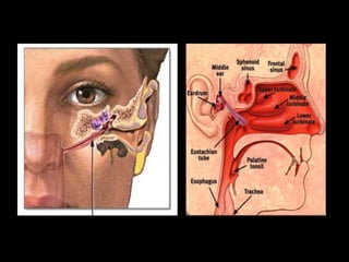
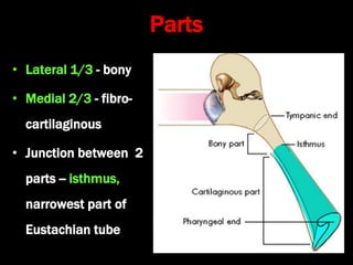
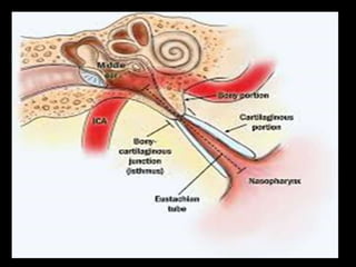
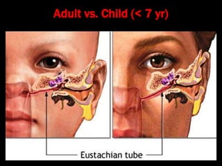
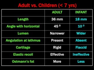
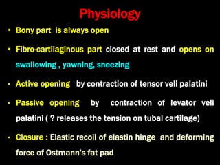
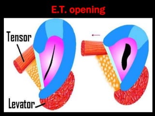
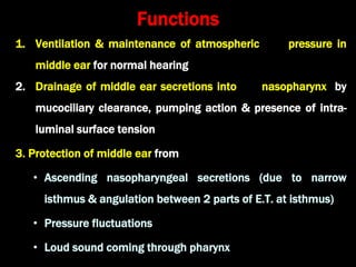
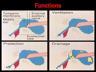
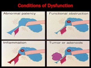
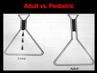
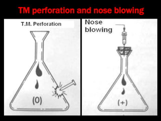
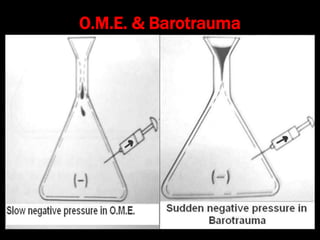
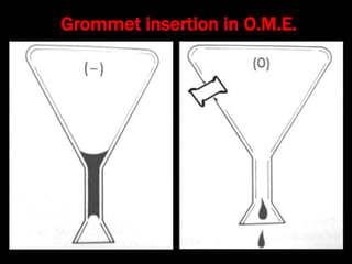
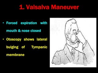
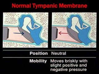
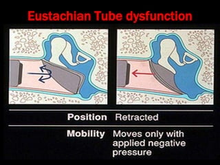
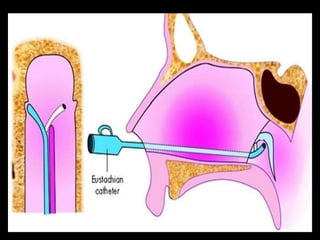
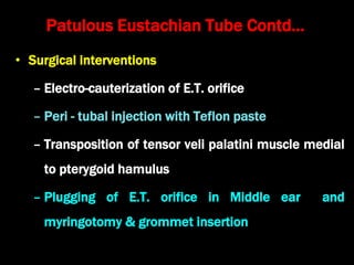
Anatomy and Physiology
of the Eustachian Tube
Dr. Krishna Koirala
• Links the pharynx to the middle ear
• Eustachius (1562) : Pharyngotympanic tube
• Antonio Valsalva : Eustachian tube
• Develops from tubotympanic recess which is derived from
endoderm of 1st pharyngeal pouch
• 36 mm long in adults
• Directed anteriorly, inferiorly and medially from anterior wall of
middle ear forming angle of 450 with horizontal and sagittal
planes
• Enters the nasopharynx 1.25 cm behind posterior end of
inferior turbinate
Parts
• Lateral 1/3 - bony
• Medial 2/3 - fibro-
cartilaginous
• Junction between 2
parts -- isthmus,
narrowest part of
Eustachian tube
Anatomy of medial 2/3rd
• Cartilage plate
– Lies postero-medially
– Consists of medial and
lateral laminae separated
by elastin hinge
• Fibrous tissue and
Ostmann’s fat pad lie infero-
laterally
Muscles
1. Tensor veli palatini or dilator tubae
2. Levator veli palatini
3. Salpingopharyngeus
4. Tensor tympani
Nerve supply
1. Sphenopalatine ganglion
2. Mandibular nerve
3. Tympanic plexus
• Lining epithelium
− Respiratory epithelium
• Arterial supply
– Ascending pharyngeal & middle meningeal
arteries
• Venous drainage
− Pharyngeal & pterygoid venous plexus
• Lymphatic drainage
− Retropharyngeal node
Endoscopic Anatomy
• Medial end forms tubal
elevation / torus tubaris
• Lymphoid collection over
torus is called Gerlach’s tubal
tonsil
• Postero-superior to torus is
fossa of Rosenmüller
Adult vs. Child (< 7 yr)
Adult vs. Children (< 7 yrs)
ADULT INFANT
Length 36 mm 18 mm
Angle with horizontal 45 0 10 0
Lumen Narrower Wider
Angulation at isthmus Present Absent
Cartilage Rigid Flaccid
Elastic recoil Effective Ineffective
Ostmann’s fat More Less
Physiology
• Bony part is always open
• Fibro-cartilaginous part closed at rest and opens on
swallowing , yawning, sneezing
• Active opening by contraction of tensor veli palatini
• Passive opening by contraction of levator veli
palatini ( ? releases the tension on tubal cartilage)
• Closure : Elastic recoil of elastin hinge and deforming
force of Ostmann’s fat pad
E.T. opening
Functions
1. Ventilation & maintenance of atmospheric pressure in
middle ear for normal hearing
2. Drainage of middle ear secretions into nasopharynx by
mucociliary clearance, pumping action & presence of intra-
luminal surface tension
3. Protection of middle ear from
• Ascending nasopharyngeal secretions (due to narrow
isthmus & angulation between 2 parts of E.T. at isthmus)
• Pressure fluctuations
• Loud sound coming through pharynx
Functions
Conditions of Dysfunction
Bluestone’s Flask
Model
Adult vs. Pediatric
TM perforation and nose blowing
O.M.E. & Barotrauma
Grommet insertion in O.M.E.
Tests for E.T. function
1. Valsalva Maneuver
• Forced expiration with
mouth & nose closed
• Otoscopy shows lateral
bulging of Tympanic
membrane
2. Frenzel Maneuver
• Hands free Valsalva
• Compression of nasopharyngeal
air by muscles of tongue
• Otoscopy shows lateral bulging
of tympanic membrane
3. Toynbee Maneuver
• More physiological
• Swallowing with mouth &
nose closed
• Otoscopy shows retraction
of tympanic membrane
• Air pressure is alternately increased & decreased
within external auditory canal
• Mobility of tympanic membrane is observed
• Normal mobility indicates good patency of
Eustachian tube
4. Pneumatic otoscopy & Siegelization
Siegelization
Pneumatic Otoscope
Normal Tympanic Membrane
Eustachian Tube dysfunction
Early otitis media with effusion
Late otitis media with effusion
Acute suppurative otitis media
Ear drum perforation
5. Politzerization
• Rubber tube attached to a Politzer bag put into one
nostril and both nostrils are pinched
• Patient asked to swallow or repeat “k”
• Politzer bag is squeezed simultaneously
• Otoscopy shows lateral bulging of ear drum in patent
Eustachian tube
6. E.T. catheterization
• E.T. catheter passed along nasal floor till it touches
posterior wall of nasopharynx
• Catheter rotated 90° medially & pulled forward till it
impinges on posterior nasal septum
• Catheter rotated 180° laterally, & its tip inserted into
opening of E.T.
• Politzer bag attached to outer end of catheter
• Air pushed into E.T. catheter by squeezing Politzer
bag
• Examiner hears by Toynbee auscultation tube put in
pt's ear
• Blowing sound  normal E.T. patency
• Bubbling sound  middle ear fluid
• Whistling sound  partial E.T. obstruction
• No sound  complete obstruction of E.T.
Eustachian tube catheter
7. Tymapanometry
• Type C = E.T. dysfunction
• Type B = fluid in middle ear
• 200 mm H2
O pressure is created in patient’s external
auditory canal
• Patient asked to swallow 10 times
• Residual pressure in patient’s external auditory canal
after 10th swallow is noted
• Test repeated with -ve 200 mm H2
O pressure created
in patient’s external auditory canal
8. William’s pressure equalization test
William’s Test
Residual Pressure Result
Up to + 50 mm H2
O normal E.T. function
+ 51 to + 100 mm H2
O mild dysfunction
+ 101 to + 199 mm H2
O moderate dysfunction
+ 200 mm H2
O severe dysfunction
9. Sono-tubometry
• Sound made in pt’s nasal cavity & detected with
stethoscope in patient’s external auditory canal
• Loud sound = patent Eustachian tube
10. Eustachian tube Salpingogram
• Dye instilled through E.T. catheter & X-ray taken
11. C.T. scan & M.R.I. of skull
12. Trans-nasal E.T. video-endoscopy
13. Test for E.T. patency in T.M. perforation
• Saccharine crystal / antibiotic ear drop /
methylene blue placed in middle ear via ear drum
perforation
• Sweet taste / bitter taste / blue staining of
secretions indicates patent Eustachian tube
Patulous Eustachian Tube
• Aural fullness, humming tinnitus, autophony, hearing own
breath sounds (tympanophonia)
• Symptoms resolve in supine position, in forward bending with
head between knees, in U.R.T.I. and aggravated by
mastication
• Otoscopy: T.M. moves during breathing
• Associated conditions: radiation therapy, hormonal therapy,
nasal decongestants, 3rd trimester pregnancy, stress, sudden
weight loss, multiple sclerosis
• Treatment: Reassurance, weight gain, oral potassium iodide
Patulous Eustachian Tube Contd…
• Surgical interventions
– Electro-cauterization of E.T. orifice
– Peri - tubal injection with Teflon paste
– Transposition of tensor veli palatini muscle medial
to pterygoid hamulus
– Plugging of E.T. orifice in Middle ear and
myringotomy & grommet insertion

3. Eustachian tube

  • 1.
    Anatomy and Physiology ofthe Eustachian Tube Dr. Krishna Koirala
  • 2.
    • Links thepharynx to the middle ear • Eustachius (1562) : Pharyngotympanic tube • Antonio Valsalva : Eustachian tube • Develops from tubotympanic recess which is derived from endoderm of 1st pharyngeal pouch • 36 mm long in adults • Directed anteriorly, inferiorly and medially from anterior wall of middle ear forming angle of 450 with horizontal and sagittal planes • Enters the nasopharynx 1.25 cm behind posterior end of inferior turbinate
  • 4.
    Parts • Lateral 1/3- bony • Medial 2/3 - fibro- cartilaginous • Junction between 2 parts -- isthmus, narrowest part of Eustachian tube
  • 6.
    Anatomy of medial2/3rd • Cartilage plate – Lies postero-medially – Consists of medial and lateral laminae separated by elastin hinge • Fibrous tissue and Ostmann’s fat pad lie infero- laterally
  • 7.
    Muscles 1. Tensor velipalatini or dilator tubae 2. Levator veli palatini 3. Salpingopharyngeus 4. Tensor tympani Nerve supply 1. Sphenopalatine ganglion 2. Mandibular nerve 3. Tympanic plexus
  • 9.
    • Lining epithelium −Respiratory epithelium • Arterial supply – Ascending pharyngeal & middle meningeal arteries • Venous drainage − Pharyngeal & pterygoid venous plexus • Lymphatic drainage − Retropharyngeal node
  • 10.
    Endoscopic Anatomy • Medialend forms tubal elevation / torus tubaris • Lymphoid collection over torus is called Gerlach’s tubal tonsil • Postero-superior to torus is fossa of Rosenmüller
  • 11.
  • 12.
    Adult vs. Children(< 7 yrs) ADULT INFANT Length 36 mm 18 mm Angle with horizontal 45 0 10 0 Lumen Narrower Wider Angulation at isthmus Present Absent Cartilage Rigid Flaccid Elastic recoil Effective Ineffective Ostmann’s fat More Less
  • 13.
    Physiology • Bony partis always open • Fibro-cartilaginous part closed at rest and opens on swallowing , yawning, sneezing • Active opening by contraction of tensor veli palatini • Passive opening by contraction of levator veli palatini ( ? releases the tension on tubal cartilage) • Closure : Elastic recoil of elastin hinge and deforming force of Ostmann’s fat pad
  • 14.
  • 15.
    Functions 1. Ventilation &maintenance of atmospheric pressure in middle ear for normal hearing 2. Drainage of middle ear secretions into nasopharynx by mucociliary clearance, pumping action & presence of intra- luminal surface tension 3. Protection of middle ear from • Ascending nasopharyngeal secretions (due to narrow isthmus & angulation between 2 parts of E.T. at isthmus) • Pressure fluctuations • Loud sound coming through pharynx
  • 16.
  • 17.
  • 18.
  • 19.
  • 20.
    TM perforation andnose blowing
  • 21.
  • 22.
  • 23.
  • 24.
    1. Valsalva Maneuver •Forced expiration with mouth & nose closed • Otoscopy shows lateral bulging of Tympanic membrane
  • 26.
    2. Frenzel Maneuver •Hands free Valsalva • Compression of nasopharyngeal air by muscles of tongue • Otoscopy shows lateral bulging of tympanic membrane
  • 27.
    3. Toynbee Maneuver •More physiological • Swallowing with mouth & nose closed • Otoscopy shows retraction of tympanic membrane
  • 28.
    • Air pressureis alternately increased & decreased within external auditory canal • Mobility of tympanic membrane is observed • Normal mobility indicates good patency of Eustachian tube 4. Pneumatic otoscopy & Siegelization
  • 29.
  • 30.
  • 31.
  • 32.
  • 33.
    Early otitis mediawith effusion
  • 34.
    Late otitis mediawith effusion
  • 35.
  • 36.
  • 37.
    5. Politzerization • Rubbertube attached to a Politzer bag put into one nostril and both nostrils are pinched • Patient asked to swallow or repeat “k” • Politzer bag is squeezed simultaneously • Otoscopy shows lateral bulging of ear drum in patent Eustachian tube
  • 40.
    6. E.T. catheterization •E.T. catheter passed along nasal floor till it touches posterior wall of nasopharynx • Catheter rotated 90° medially & pulled forward till it impinges on posterior nasal septum • Catheter rotated 180° laterally, & its tip inserted into opening of E.T. • Politzer bag attached to outer end of catheter
  • 41.
    • Air pushedinto E.T. catheter by squeezing Politzer bag • Examiner hears by Toynbee auscultation tube put in pt's ear • Blowing sound  normal E.T. patency • Bubbling sound  middle ear fluid • Whistling sound  partial E.T. obstruction • No sound  complete obstruction of E.T.
  • 42.
  • 44.
    7. Tymapanometry • TypeC = E.T. dysfunction • Type B = fluid in middle ear
  • 45.
    • 200 mmH2 O pressure is created in patient’s external auditory canal • Patient asked to swallow 10 times • Residual pressure in patient’s external auditory canal after 10th swallow is noted • Test repeated with -ve 200 mm H2 O pressure created in patient’s external auditory canal 8. William’s pressure equalization test
  • 46.
    William’s Test Residual PressureResult Up to + 50 mm H2 O normal E.T. function + 51 to + 100 mm H2 O mild dysfunction + 101 to + 199 mm H2 O moderate dysfunction + 200 mm H2 O severe dysfunction
  • 47.
    9. Sono-tubometry • Soundmade in pt’s nasal cavity & detected with stethoscope in patient’s external auditory canal • Loud sound = patent Eustachian tube 10. Eustachian tube Salpingogram • Dye instilled through E.T. catheter & X-ray taken 11. C.T. scan & M.R.I. of skull
  • 48.
    12. Trans-nasal E.T.video-endoscopy 13. Test for E.T. patency in T.M. perforation • Saccharine crystal / antibiotic ear drop / methylene blue placed in middle ear via ear drum perforation • Sweet taste / bitter taste / blue staining of secretions indicates patent Eustachian tube
  • 49.
    Patulous Eustachian Tube •Aural fullness, humming tinnitus, autophony, hearing own breath sounds (tympanophonia) • Symptoms resolve in supine position, in forward bending with head between knees, in U.R.T.I. and aggravated by mastication • Otoscopy: T.M. moves during breathing • Associated conditions: radiation therapy, hormonal therapy, nasal decongestants, 3rd trimester pregnancy, stress, sudden weight loss, multiple sclerosis • Treatment: Reassurance, weight gain, oral potassium iodide
  • 50.
    Patulous Eustachian TubeContd… • Surgical interventions – Electro-cauterization of E.T. orifice – Peri - tubal injection with Teflon paste – Transposition of tensor veli palatini muscle medial to pterygoid hamulus – Plugging of E.T. orifice in Middle ear and myringotomy & grommet insertion