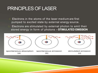
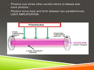
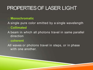
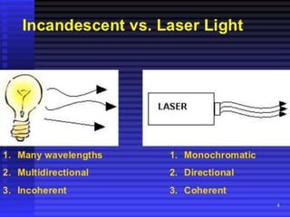
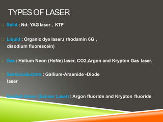
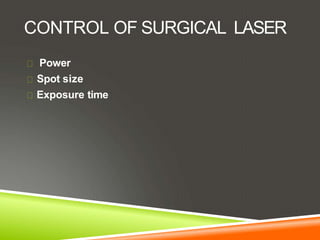
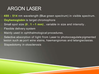
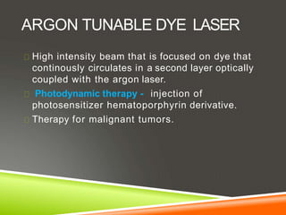
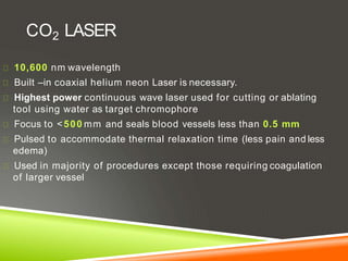
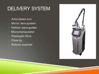
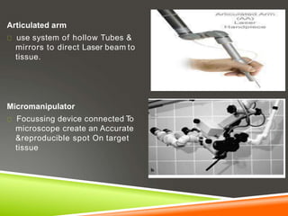
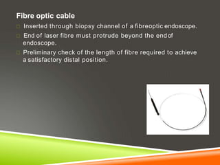
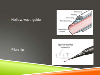
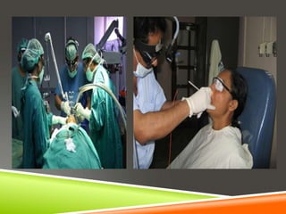
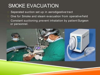
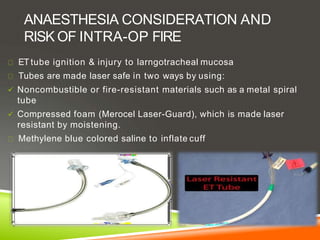
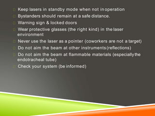
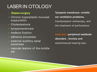
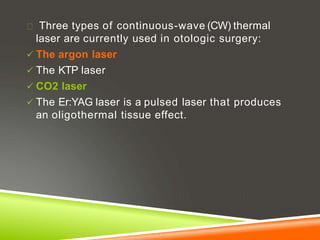
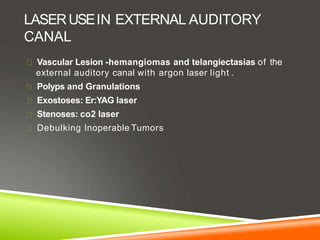
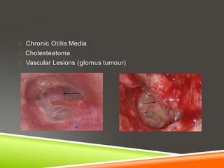
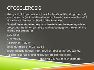
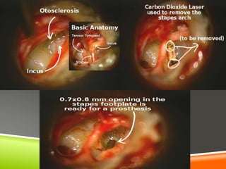
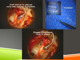
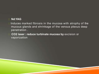
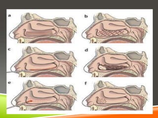
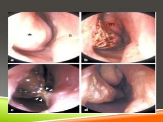
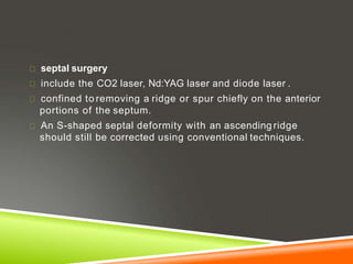
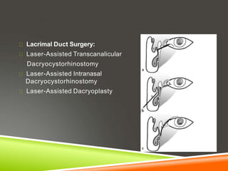
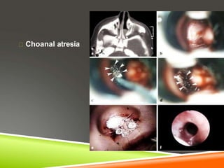
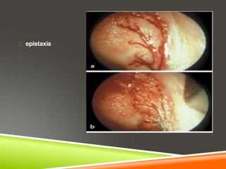
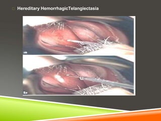
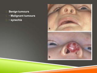
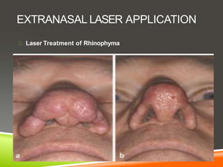
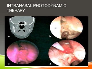
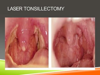
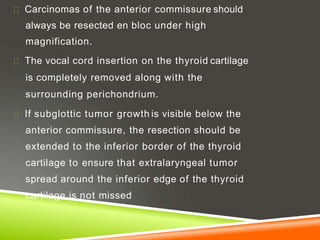
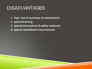
LASER IN OTOLARYNGOLOGY
DR PRASANNA DATTA
MS(ENT)
INTRODUCTION
LASER-light amplification by stimulated emission
of radiation
Albert einstein Theodore Maiman C.Kumar N.Patel
STRONGAND JAKO
PRINCIPLES OFLASER
Electrons in the atoms of the laser medium are first
pumped to excited state by external energy source.
Electrons are stimulated by external photon to emit their
stored energy in form of photons – STIMULATED EMISSION
Photons now strike other excited atoms to release even
more photons .
Photons move back and forth between two parallel mirrors
LIGHT AMPLIFICATION.
PROPERTIESOF LASER LIGHT
Monochromatic
A single pure color emitted by a single wavelength
Collimated
A beam in which all photons travel in same parallel
direction
coherent
All waves or photons travel in steps, or in phase
with one another.
TYPESOF LASER
Solid : Nd: YAG laser , KTP
Liquid : Organic dye laser.( rhodamin 6G ,
disodium fluorescein)
Gas : Helium Neon (HeNe) laser, CO2,Argon and Krypton Gas laser.
Semiconductors : Gallium-Arsenide -Diode
laser
Excited dimer (Eximer Laser) : Argon fluoride and Krypton fluoride
PATTERNS OF LASER OUTPUT
Continuous : continuously pumped ,emits light continuously
Pulse: laser energy delivered with each peak over an extremely short
period of a few nanoseconds with rest period (allows time for tissues
to cool down)
Q-switched: Allows a high build-up of energy within the tube which is
then released over a very short duration of a few nanoseconds
Cavity dumped Lasers- produces slightlyshorter
pulse of light
Mode locked lasers-produces pulses of light as
short as few pico seconds
CONTROL OF SURGICAL LASER
Power
Spot size
Exposure time
TISSUE EFFECT
Absorption
Scattering
Reflection
Transmision
BASIC LASERTISSUE INTERACTION
Photothermal
Photomechanical
Photochemical
Photoablative
When laser radiation strikes a tissue, the temperature begins to rise
100 C – 45 0C : Conformation change of proteins
500 C : Reduction of enzyme activity
60o - 99°C : Coagulation begins
100°C and above : Vaporization starts
400 -500°C : Char starts to burn
ARGON LASER
488 - 514 nm wavelength (Blue green spectrum) in visible spectrum.
Oxyhemoglobin is target chromophore
Small spot size (0 . 1 – 1 mm) , variable in size and intensity .
Flexible delivery system
Mainly used in ophthalmological procedures.
Selective absorption of light from Laser to photocoagulate pigmented
lesion such as port wine stains, haemangiomas and telangiectasias.
Stapedotomy in otosclerosis
ARGON TUNABLE DYE LASER
High intensity beam that is focused on dye that
continously circulates in a second layer optically
coupled with the argon laser.
Photodynamic therapy - injection of
photosensitizer hematoporphyrin derivative.
Therapy for malignant tumors.
Limitations –
Also absorbed by epidermal and dermal tissues
due to melanin
Continuous mode of operation-Higher fibrosis
prevalence of postoperative pigmentary alteration
CO2 LASER
10,600 nm wavelength
Built –in coaxial helium neon Laser is necessary.
Highest power continuous wave laser used for cutting or ablating
tool using water as target chromophore
Focus to <500 mm and seals blood vessels less than 0.5 mm
Pulsed to accommodate thermal relaxation time (less pain and less
edema)
Used in majority of procedures except those requiring coagulation
of larger vessel
Comparatively a poor hemostat (not being
effective in controlling bleeding from vessels
greater than 0.5 mm in diameter)
Not transmissible through the common optical
fibre
Its use on the cords has the advantage of
producing minimal scarring therefore glottic
competency is rarely jeopardized
USES OF CO2 LASER
Laser stapedotomy
Recurrent respiratory papillomatosis
In paediatric patients surgery for web , subglottic
stenosis, capillary hemangiomas.
Laser cordotomy , arytenoidectomy.
Malignant & benign laryngeal tumours.
Transoral robotic surgery.
ND YAG: NEODYMIUM-DOPED
YTTRIUM ALUMINIUM GARNET
1064 nm wavelength with Helium-Neon (He-Ne) beam
Solid state laser with fiberoptic carrier
Deeper penetration (up to 4 mm)
Radiant energy transmitted through clear fluid used in eye and
water filled cavity urinary bladder.Ideal laser for ablation,
coagulation and hemostasis in vascular malformations
USESOF ND-YAG IN ENT
Ablation of obstructing tracheo bronchial lesion,
oesophageal lesions.
Removal of malignant tumors in oral cavity.
Obstruction of tracheobronchial tree –
complication hemorrhage- ND-YAG laser deep
penetration
Limitations
Greater scatter than CO2
Deep thermal injury
Risk for transmural injury
KTP LASER- POTASSIUM TITANYL
PHOSPHATE
532 nm wavelength with Oxyhemoglobinas primary
chromophore
 • Continuous wave (CW) mode to cut tissue
 • Pulsed mode for vascular lesions.
 • Q-Switched mode for red/orange tattoo pigment Delivery
 • Insulated fiber, fiber handpiece, scanner, or microscope
for CW/pulsed mode
 • Articulating arm for Q-Switched mode
USES
Tosillectomy
Pigment dermal lesion
Revision stapedotomy
Limitation – unintended thermal injury
585-NM PULSEDDYELASER
Used in larynx, absorption peak 577nm
Targeted chromophore is oxyhemoglobin
Papilloma ,vascular polyps, varices and vocal fold polyps
Unlike CO2 laser ablation effects pulsed dye laser causes
involution of lesion through disruption of vascular supply .
Reduced risk of collateral thermal injury
DELIVERY SYSTEM
Articulated arm
Mirror lens system
Hollow wave guides
Micromanipulator
Fibreoptic fibre
Fibre tip
Robotic scanner
Articulated arm
use system of hollow Tubes &
mirrors to direct Laser beam to
tissue.
Micromanipulator
Focussing device connected To
microscope create an Accurate
&reproducible spot On target
tissue
Fibre optic cable
Inserted through biopsy channel of a fibreoptic endoscope.
End of laser fibre must protrude beyond the endof
endoscope.
Preliminary check of the length of fibre required to achieve
a satisfactory distal position.
Hollow wave guide
Fibre tip
Class 1 lasers pose no safety hazard (e.g., aCD
player).
Class 2 lasers emit only wavelengths in the visible
range of the spectrum and are not hazardous even
when shined directly into the eye(helium-neon laser
pointers).
Class 3a lasers are hazardous to the eye .
Class 3b and 4 laser, looking directly into the beam
close to its emergence from the applicator can
injure the eye regardless of the lens systems used.
Medical lasers are in classes 3b and4
Education-
1 .Appropriate credential certifying mechanism required for
physician,and nurses.
2 .Develop education policies for surgeon anaesthesiologist and
nurses
3 .Periodic review of all laser related complications
EYEPROTECTION AND SKIN
PROTECTION
Lasers absorbed by water (e.g., CO2) damage the anterior
portions of the eye (cornea, lens)
Wavelengths in the visible and NIR range (e.g., argon and
Nd:YAG lasers) pass through the optical media of the eye
and damage the retina.
Wavelenght specific protected eye glass with side
protectors
Double layer of saline moistened eye pad
Saline saturated surgical towel completely drape
SMOKE EVACUATION
Seperated suction set up in aerodigestive tract
One for Smoke and steam evacuation from operative field
Constant suctioning prevent inhalation by patient Surgeon
or personnel.
ANAESTHESIA CONSIDERATION AND
RISK OF INTRA-OP FIRE
ET tube ignition & injury to larngotracheal mucosa
Tubes are made laser safe in two ways by using:
 Noncombustible or fire-resistant materials such as a metal spiral
tube
 Compressed foam (Merocel Laser-Guard), which is made laser
resistant by moistening.
Methylene blue colored saline to inflate cuff
1. An endotracheal tube should be kept out of the operating field if at
all possible
2. If this cannot be done, a laser-resistant tube should be used
3. If a laser-resistant tube cannot be used, the surgeon should be able to
identify the tube the operative field at any time.
4. In this case the part of the tube closest to the surgical site can be
protected by covering it with wet neurosurgical cotton.
5. Wrapping the tube with aluminum foil can give a false sense of
security and is not advised.
IF LASERWILL FALL ON ET TUBE……
50ml bulb syringe and basin of saline should be available
Stop ventilation immediately
Withdraw tube and flush saline
Re establish airway immediately
Bronchoscopy to assess degree of injury
I/V steroids
Remain intubated
Repeat bronchoscopy
Keep lasers in standby mode when not in operation
Bystanders should remain at a safe distance.
Warning sign & locked doors
Wear protective glasses (the right kind) in the laser
environment
Never use the laser as a pointer (coworkers are not a target)
Do not aim the beam at other instruments(reflections)
Do not aim the beam at flammable materials (especiallythe
endotracheal tube)
Check your system (be informed)
LASERIN OTOLOGY
Stapes surgery
 Chronic hyperplastic mucosal
suppuration
 Cholesteatoma
 tympanosclerosis
 malleus fixation
 adhesive processes
 external auditory canal
exostoses
 vascular lesions of the middle
ear
Tympanic membrane -middle
ear ventilation problems,
transtympanic endoscopy, and
the treatment of perforations
Inner ear- peripheral vestibular
disorders , tinnitus and
sensorineural hearing loss .
Three types of continuous-wave (CW) thermal
laser are currently used in otologic surgery:
 The argon laser
 The KTP laser
 CO2 laser
 The Er:YAG laser is a pulsed laser that produces
an oligothermal tissue effect.
LASERUSEIN EXTERNAL AUDITORY
CANAL
Vascular Lesion -hemangiomas and telangiectasias of the
external auditory canal with argon laser light .
Polyps and Granulations
Exostoses: Er:YAG laser
Stenoses: co2 laser
Debulking Inoperable Tumors
LASERUSEON TYMPANIC MEMBRANE
 laser myringotomy :
 CO2 and Er:YAG lasers
opening of at least 2 mm should be created with the CO2 laser
 Secretory Otitis Media
 Acute Eustachian Tube Dysfunction
 Barotrauma
 Acute Otitis Media With Vestibulocochlear Complications
 Transtympanic Endoscopy
LASER MYRINGOTOMY
Tympanic Membrane Perforations and Atrophic Scars
Graft Fixation for Tympanic Membrane Defects
epidermoid Cysts of the Tympanic Membrane
USEOF LASERIN MIDDLE EAR
Medialization of the Malleus
CO2 laser for dividing scar tissue and
exposing the malleus.
Resecting the distal third of the malleus
handle
Malleus Fixation
Vaporization of the malleus neck or
sclerotic foci around the malleus head
with the laser can mobilize the chain
Tympanosclerosis:
On the tympanic
membrane and on the
ossicular chain and its
surroundings causing
fixation and obliteration
of the window niches can
be removed
Chronic Otitis Media
Cholesteatoma
Vascular Lesions (glomus tumour)
OTOSCLEROSIS
Using a drill to perforate a thick footplate obliterating the oval
window niche (as in obliterative otosclerosis) can cause harmful
vibrations to be transmitted to the inner ear.
Goal of laser stapedotomy is to create a precise opening while
protecting the inner ear and avoiding damage to the remaining
middle ear structures
CO2 laser
 C/W mode.
 A power of 1–22 W
 pulse duration of 0.03–0.05 s
 power density ranges from 4000 W/cm2 to 80,000W/cm2.
 A single laser applicationpoduces precise footplate
opening 0.5–0.7 mm in diameter .
Obliterative Otosclerosis-
 The CO2 laser can vaporize a fenestra in the stapes footplate,
regardless of its thickness or degree of fixation, without
mechanical trauma to the inner ear.
Overhanging Facial Nerve
 CO2 laser beam can be carefully applied tangentially at low
power (1–2 W), using short pulse lengths of 0.05 s, to remove
the bone.
 A conventional stapedotomy with a curved perforator.
 Redirect the CO2 laser beam with a mirror.
Overhanging Promontory:
Covering the footplate with saline solution or moist gelatin
sponge), the bony overhang can be ablated.
Inaccessible Footplate
 Due to an abnormal course of the facial nerve or a
vascular anomaly
 Fenestration of the promontory
Floating Footplate: Laser enables to create a fenestration of
the desired diameter even in a floating footplate
USEOF LASERIN INNER EAR
Cochleostomy
Laser cochleostomy inserting the electrode of acochlear
implant.
Effective for an ossified cochlea
Peripheral Vestibular Disorders:
Tinnitus and Sensorineural Hearing LossLow-level laser therapy
Acoustic Neuroma:
Availability of fiberoptic delivery; spot size focusable to 0.15 mm,
free passage of argon laser light through media such as
cerebrospinal fluid, and good hemostatic effect of the argon laser
wavelength.
pulsed holmium:YAG laser for the removal of cranial and spinal
meningiomas and neuromas
LASERIN RHINOLOGY: INTRANASAL
LASER APPLICATION
Turbinate Reduction
enlarged inferior nasal turbinates secondary to allergic or
vasomotor rhinitis
Thermal damage due to laser energy causes scarring of the
mucosal epithelium.
In the submucosa, reduces the swelling capacity and secretory
functions of the turbinate.
Laser surgery of hypertrophic inferior turbinates is appropriate
only if the obstruction is largely due to severe mucosal swelling
Nd:YAG
Induces marked fibrosis in the mucosa with atrophy of the
mucous glands and shrinkage of the venous plexus deep
penetration .
CO2 laser : reduce turbinate mucosa by excision or
vaporization
septal surgery
include the CO2 laser, Nd:YAG laser and diode laser .
confined to removing a ridge or spur chiefly on the anterior
portions of the septum.
An S-shaped septal deformity with an ascending ridge
should still be corrected using conventional techniques.
Paranasal sinus surgery:
The removal of polyps in patients who refuse conventional
surgery or are poor candidates for general anesthesia.
treatment of circumscribed recurrent polyposis following
prior intranasal surgery .
Lacrimal Duct Surgery:
Laser-Assisted Transcanalicular
Dacryocystorhinostomy
Laser-Assisted Intranasal
Dacryocystorhinostomy
Laser-Assisted Dacryoplasty
Choanal atresia
epistaxis
Hereditary HemorrhagicTelangiectasia
Benign tumours
Malignant tumours
synechia
EXTRANASAL LASER APPLICATION
Laser Treatment of Rhinophyma
INTRANASAL PHOTODYNAMIC
THERAPY
LASERUSEIN ORAL CAVITY AND
OROPHARYNX
The (CO2), (Nd:YAG), (KTP), and argon lasers are most commonly
used for soft-tissue surgery in the oral cavity and oropharynx.
occlusion of small transected vessels, providing hemostasis
bloodless field;
no-touch operating technique;
precise incisions
no need for sutures.
Hyperplasia of the Lingual Tonsil
Vascular Malformations
Other Benign Tumors: papillomas,fibromas, cysts, and
ranulas excision and vaporization.
Premalignant Lesions: leukoplakia and erythroplakia.
Labial and Lingual Frenoplasties
LASERFOR TREATMENT OF SNORING
AND SLEEP APNOEA
Laser-assisted uvulopalatoplasty (LAUP)
Appropriate for higher grades of OSA.
Contraindications for LAUPin primary snoring:
AHI greater than 20–30/h
BMI greater than 28 kg/m2
Midfacial deformities
Posterior airway space at the mandibular level smaller than
10mm
Severe concomitant medical disease
Severe neurologic or psychiatric comorbidity
LASER TONSILLECTOMY
Lasers in the Treatment of Salivary Gland Disease “optical
breakdown.”
When laser pulses of sufficiently high energy and short duration
are applied to tissue, they form a plasma that causes the sudden
volume expansion of fluids and generates a shockwave.
The laser fiber is advanced to the stone through anendoscope
The particles of the fragmented stone are either passed
spontaneously with the salivary flow or flushed out through the
working channel of the endoscope.
LASERFOR BENING CONDITIONS OF
LARYNX, HYPOPHARYNX AND TRACHEA
Laser Surgery of the Vocal Cords epithelial changes (vocal
nodules, leukoplakia, hyperkeratosis, acanthosis, dysplasia,
etc.),
Exudative changes in the Reinke space (vocal cord polyps,
Reinke edema).
Granulomas: (contact granuloma, intubation granuloma),
scarring, and subepithelial lesions (cysts)
Laser surgery can be done under general
endotracheal anesthesia and using jet ventilation
Postoperative monitoring in an intensive care unit (ICU)
following laser surgery for airway stenosis.
Contact granuloma
Vocal cord polyp
Subepithelial Vocal cord cyst
LASERSURGERYTO IMPROVE
SWALLOWING
Zenker diverticulum
Deficient or delayed
Relaxation of the
cricopharyngeus
muscle
LASERTREATMENT FOR AIRWAY
STENOSIS
Supraglottic Stenosis
Glottic Airway Stenosis
Bilateral Recurrent Nerve Paralysis
Arytenoidectomy
Cordectomy
Posterior cordectomy
Temporary lateral fixation
Subglottic and Tracheal Stenoses
Airway Stenosis Due to Malignant Disease
POSTERIOR CORDECTOMY OF B/L REC
LARYNGEAL PALSY-
MICROLARYNGOSCOPIC VIEW
LASERSURGERYOF BENIGN TUMOURS
OF LARYNX AND TRACHEA
RECURRENTLARYNGEALPAPILLOMATOSIS
SUBGLOTIC AND TRACHEAL STENOSIS
CO2 laser therapy is well accepted as the first approach to
the problem of benign stenosis of the upper airway.
Use of a rigid bronchoscope is necessary for the CO2 laser
bronchoscopy because the CO2 laser energy cannot be
transmitted through a flexible fibre.
COMPLICATION IN ENDOSCOPIC LASER
SURGERYIN LARYNX, HYPOPHARYNX
AND TRACHEA
Combustion of ventilation tube materials and anesthetic
gas mixtures during surgical laser use in the larynx .
Combustion of tube materials can be avoided by the use of laser-
safe tubes.
Ignition of anesthetic gas mixtures during procedures using jet
ventilation can be prevented by ventilating the patient with room
air (rather than pure oxygen) and by operating in intermittent
apnea.
On the whole, such incidents can be safely avoided by the selection
of suitable materials, operating methods, and analgesic
techniques.
LASERIN MALIGNANT LESIONS OF
UPPERAERODIGESTIVE TRACT
Diameter of the CO2 laser beam :
adjusted to produce either of two effects:
• Tissue ablation with a spot size of 1–4 mm or
• Tissue cutting with a spot size of 0.2–1 mm.
•The limits of the resection are defined by the tumor extent
visible under the operating microscope and can be adapted
to individual circumstances.
Carcinoma of the Oral Cavity
Pharyngeal Carcinoma
Laser Microsurgery of Glottic Carcinoma
T1 and T2a Glottic Carcinoma
Glottic Carcinoma with Involvement of the
Anterior Commissure
EARLY GLOTTIC CANCER
Carcinomas of the anterior commissure should
always be resected en bloc under high
magnification.
The vocal cord insertion on the thyroid cartilage
is completely removed along with the
surrounding perichondrium.
If subglottic tumor growth is visible below the
anterior commissure, the resection should be
extended to the inferior border of the thyroid
cartilage to ensure that extralaryngeal tumor
spread around the inferior edge of the thyroid
cartilage is not missed
DISADVANTAGES
1. high cost of purchase & maintenance
2. special training
3. special precautions & safety measures
4. special anaesthesia requirements
Thank you

Lasers in ENT

  • 1.
    LASER IN OTOLARYNGOLOGY DRPRASANNA DATTA MS(ENT)
  • 2.
    INTRODUCTION LASER-light amplification bystimulated emission of radiation
  • 3.
    Albert einstein TheodoreMaiman C.Kumar N.Patel
  • 4.
  • 5.
    PRINCIPLES OFLASER Electrons inthe atoms of the laser medium are first pumped to excited state by external energy source. Electrons are stimulated by external photon to emit their stored energy in form of photons – STIMULATED EMISSION
  • 6.
    Photons now strikeother excited atoms to release even more photons . Photons move back and forth between two parallel mirrors LIGHT AMPLIFICATION.
  • 7.
    PROPERTIESOF LASER LIGHT Monochromatic Asingle pure color emitted by a single wavelength Collimated A beam in which all photons travel in same parallel direction coherent All waves or photons travel in steps, or in phase with one another.
  • 9.
    TYPESOF LASER Solid :Nd: YAG laser , KTP Liquid : Organic dye laser.( rhodamin 6G , disodium fluorescein) Gas : Helium Neon (HeNe) laser, CO2,Argon and Krypton Gas laser. Semiconductors : Gallium-Arsenide -Diode laser Excited dimer (Eximer Laser) : Argon fluoride and Krypton fluoride
  • 10.
    PATTERNS OF LASEROUTPUT Continuous : continuously pumped ,emits light continuously Pulse: laser energy delivered with each peak over an extremely short period of a few nanoseconds with rest period (allows time for tissues to cool down) Q-switched: Allows a high build-up of energy within the tube which is then released over a very short duration of a few nanoseconds
  • 11.
    Cavity dumped Lasers-produces slightlyshorter pulse of light Mode locked lasers-produces pulses of light as short as few pico seconds
  • 12.
    CONTROL OF SURGICALLASER Power Spot size Exposure time
  • 13.
  • 14.
  • 15.
    When laser radiationstrikes a tissue, the temperature begins to rise 100 C – 45 0C : Conformation change of proteins 500 C : Reduction of enzyme activity 60o - 99°C : Coagulation begins 100°C and above : Vaporization starts 400 -500°C : Char starts to burn
  • 17.
    ARGON LASER 488 -514 nm wavelength (Blue green spectrum) in visible spectrum. Oxyhemoglobin is target chromophore Small spot size (0 . 1 – 1 mm) , variable in size and intensity . Flexible delivery system Mainly used in ophthalmological procedures. Selective absorption of light from Laser to photocoagulate pigmented lesion such as port wine stains, haemangiomas and telangiectasias. Stapedotomy in otosclerosis
  • 18.
    ARGON TUNABLE DYELASER High intensity beam that is focused on dye that continously circulates in a second layer optically coupled with the argon laser. Photodynamic therapy - injection of photosensitizer hematoporphyrin derivative. Therapy for malignant tumors.
  • 19.
    Limitations – Also absorbedby epidermal and dermal tissues due to melanin Continuous mode of operation-Higher fibrosis prevalence of postoperative pigmentary alteration
  • 20.
    CO2 LASER 10,600 nmwavelength Built –in coaxial helium neon Laser is necessary. Highest power continuous wave laser used for cutting or ablating tool using water as target chromophore Focus to <500 mm and seals blood vessels less than 0.5 mm Pulsed to accommodate thermal relaxation time (less pain and less edema) Used in majority of procedures except those requiring coagulation of larger vessel
  • 21.
    Comparatively a poorhemostat (not being effective in controlling bleeding from vessels greater than 0.5 mm in diameter) Not transmissible through the common optical fibre Its use on the cords has the advantage of producing minimal scarring therefore glottic competency is rarely jeopardized
  • 22.
    USES OF CO2LASER Laser stapedotomy Recurrent respiratory papillomatosis In paediatric patients surgery for web , subglottic stenosis, capillary hemangiomas. Laser cordotomy , arytenoidectomy. Malignant & benign laryngeal tumours. Transoral robotic surgery.
  • 23.
    ND YAG: NEODYMIUM-DOPED YTTRIUMALUMINIUM GARNET 1064 nm wavelength with Helium-Neon (He-Ne) beam Solid state laser with fiberoptic carrier Deeper penetration (up to 4 mm) Radiant energy transmitted through clear fluid used in eye and water filled cavity urinary bladder.Ideal laser for ablation, coagulation and hemostasis in vascular malformations
  • 24.
    USESOF ND-YAG INENT Ablation of obstructing tracheo bronchial lesion, oesophageal lesions. Removal of malignant tumors in oral cavity. Obstruction of tracheobronchial tree – complication hemorrhage- ND-YAG laser deep penetration
  • 25.
    Limitations Greater scatter thanCO2 Deep thermal injury Risk for transmural injury
  • 26.
    KTP LASER- POTASSIUMTITANYL PHOSPHATE 532 nm wavelength with Oxyhemoglobinas primary chromophore  • Continuous wave (CW) mode to cut tissue  • Pulsed mode for vascular lesions.  • Q-Switched mode for red/orange tattoo pigment Delivery  • Insulated fiber, fiber handpiece, scanner, or microscope for CW/pulsed mode  • Articulating arm for Q-Switched mode
  • 27.
    USES Tosillectomy Pigment dermal lesion Revisionstapedotomy Limitation – unintended thermal injury
  • 28.
    585-NM PULSEDDYELASER Used inlarynx, absorption peak 577nm Targeted chromophore is oxyhemoglobin Papilloma ,vascular polyps, varices and vocal fold polyps Unlike CO2 laser ablation effects pulsed dye laser causes involution of lesion through disruption of vascular supply . Reduced risk of collateral thermal injury
  • 29.
    DELIVERY SYSTEM Articulated arm Mirrorlens system Hollow wave guides Micromanipulator Fibreoptic fibre Fibre tip Robotic scanner
  • 30.
    Articulated arm use systemof hollow Tubes & mirrors to direct Laser beam to tissue. Micromanipulator Focussing device connected To microscope create an Accurate &reproducible spot On target tissue
  • 31.
    Fibre optic cable Insertedthrough biopsy channel of a fibreoptic endoscope. End of laser fibre must protrude beyond the endof endoscope. Preliminary check of the length of fibre required to achieve a satisfactory distal position.
  • 32.
  • 33.
    Class 1 laserspose no safety hazard (e.g., aCD player). Class 2 lasers emit only wavelengths in the visible range of the spectrum and are not hazardous even when shined directly into the eye(helium-neon laser pointers). Class 3a lasers are hazardous to the eye . Class 3b and 4 laser, looking directly into the beam close to its emergence from the applicator can injure the eye regardless of the lens systems used. Medical lasers are in classes 3b and4
  • 34.
    Education- 1 .Appropriate credentialcertifying mechanism required for physician,and nurses. 2 .Develop education policies for surgeon anaesthesiologist and nurses 3 .Periodic review of all laser related complications
  • 35.
    EYEPROTECTION AND SKIN PROTECTION Lasersabsorbed by water (e.g., CO2) damage the anterior portions of the eye (cornea, lens) Wavelengths in the visible and NIR range (e.g., argon and Nd:YAG lasers) pass through the optical media of the eye and damage the retina. Wavelenght specific protected eye glass with side protectors Double layer of saline moistened eye pad Saline saturated surgical towel completely drape
  • 37.
    SMOKE EVACUATION Seperated suctionset up in aerodigestive tract One for Smoke and steam evacuation from operative field Constant suctioning prevent inhalation by patient Surgeon or personnel.
  • 38.
    ANAESTHESIA CONSIDERATION AND RISKOF INTRA-OP FIRE ET tube ignition & injury to larngotracheal mucosa Tubes are made laser safe in two ways by using:  Noncombustible or fire-resistant materials such as a metal spiral tube  Compressed foam (Merocel Laser-Guard), which is made laser resistant by moistening. Methylene blue colored saline to inflate cuff
  • 39.
    1. An endotrachealtube should be kept out of the operating field if at all possible 2. If this cannot be done, a laser-resistant tube should be used 3. If a laser-resistant tube cannot be used, the surgeon should be able to identify the tube the operative field at any time. 4. In this case the part of the tube closest to the surgical site can be protected by covering it with wet neurosurgical cotton. 5. Wrapping the tube with aluminum foil can give a false sense of security and is not advised.
  • 40.
    IF LASERWILL FALLON ET TUBE…… 50ml bulb syringe and basin of saline should be available Stop ventilation immediately Withdraw tube and flush saline Re establish airway immediately Bronchoscopy to assess degree of injury I/V steroids Remain intubated Repeat bronchoscopy
  • 41.
    Keep lasers instandby mode when not in operation Bystanders should remain at a safe distance. Warning sign & locked doors Wear protective glasses (the right kind) in the laser environment Never use the laser as a pointer (coworkers are not a target) Do not aim the beam at other instruments(reflections) Do not aim the beam at flammable materials (especiallythe endotracheal tube) Check your system (be informed)
  • 42.
    LASERIN OTOLOGY Stapes surgery Chronic hyperplastic mucosal suppuration  Cholesteatoma  tympanosclerosis  malleus fixation  adhesive processes  external auditory canal exostoses  vascular lesions of the middle ear Tympanic membrane -middle ear ventilation problems, transtympanic endoscopy, and the treatment of perforations Inner ear- peripheral vestibular disorders , tinnitus and sensorineural hearing loss .
  • 43.
    Three types ofcontinuous-wave (CW) thermal laser are currently used in otologic surgery:  The argon laser  The KTP laser  CO2 laser  The Er:YAG laser is a pulsed laser that produces an oligothermal tissue effect.
  • 44.
    LASERUSEIN EXTERNAL AUDITORY CANAL VascularLesion -hemangiomas and telangiectasias of the external auditory canal with argon laser light . Polyps and Granulations Exostoses: Er:YAG laser Stenoses: co2 laser Debulking Inoperable Tumors
  • 45.
    LASERUSEON TYMPANIC MEMBRANE laser myringotomy :  CO2 and Er:YAG lasers opening of at least 2 mm should be created with the CO2 laser  Secretory Otitis Media  Acute Eustachian Tube Dysfunction  Barotrauma  Acute Otitis Media With Vestibulocochlear Complications  Transtympanic Endoscopy
  • 46.
  • 47.
    Tympanic Membrane Perforationsand Atrophic Scars Graft Fixation for Tympanic Membrane Defects epidermoid Cysts of the Tympanic Membrane
  • 48.
    USEOF LASERIN MIDDLEEAR Medialization of the Malleus CO2 laser for dividing scar tissue and exposing the malleus. Resecting the distal third of the malleus handle Malleus Fixation Vaporization of the malleus neck or sclerotic foci around the malleus head with the laser can mobilize the chain Tympanosclerosis: On the tympanic membrane and on the ossicular chain and its surroundings causing fixation and obliteration of the window niches can be removed
  • 49.
  • 50.
    OTOSCLEROSIS Using a drillto perforate a thick footplate obliterating the oval window niche (as in obliterative otosclerosis) can cause harmful vibrations to be transmitted to the inner ear. Goal of laser stapedotomy is to create a precise opening while protecting the inner ear and avoiding damage to the remaining middle ear structures CO2 laser  C/W mode.  A power of 1–22 W  pulse duration of 0.03–0.05 s  power density ranges from 4000 W/cm2 to 80,000W/cm2.  A single laser applicationpoduces precise footplate opening 0.5–0.7 mm in diameter .
  • 53.
    Obliterative Otosclerosis-  TheCO2 laser can vaporize a fenestra in the stapes footplate, regardless of its thickness or degree of fixation, without mechanical trauma to the inner ear. Overhanging Facial Nerve  CO2 laser beam can be carefully applied tangentially at low power (1–2 W), using short pulse lengths of 0.05 s, to remove the bone.  A conventional stapedotomy with a curved perforator.  Redirect the CO2 laser beam with a mirror.
  • 54.
    Overhanging Promontory: Covering thefootplate with saline solution or moist gelatin sponge), the bony overhang can be ablated. Inaccessible Footplate  Due to an abnormal course of the facial nerve or a vascular anomaly  Fenestration of the promontory Floating Footplate: Laser enables to create a fenestration of the desired diameter even in a floating footplate
  • 55.
    USEOF LASERIN INNEREAR Cochleostomy Laser cochleostomy inserting the electrode of acochlear implant. Effective for an ossified cochlea Peripheral Vestibular Disorders:
  • 56.
    Tinnitus and SensorineuralHearing LossLow-level laser therapy Acoustic Neuroma: Availability of fiberoptic delivery; spot size focusable to 0.15 mm, free passage of argon laser light through media such as cerebrospinal fluid, and good hemostatic effect of the argon laser wavelength. pulsed holmium:YAG laser for the removal of cranial and spinal meningiomas and neuromas
  • 57.
    LASERIN RHINOLOGY: INTRANASAL LASERAPPLICATION Turbinate Reduction enlarged inferior nasal turbinates secondary to allergic or vasomotor rhinitis Thermal damage due to laser energy causes scarring of the mucosal epithelium. In the submucosa, reduces the swelling capacity and secretory functions of the turbinate. Laser surgery of hypertrophic inferior turbinates is appropriate only if the obstruction is largely due to severe mucosal swelling
  • 58.
    Nd:YAG Induces marked fibrosisin the mucosa with atrophy of the mucous glands and shrinkage of the venous plexus deep penetration . CO2 laser : reduce turbinate mucosa by excision or vaporization
  • 61.
    septal surgery include theCO2 laser, Nd:YAG laser and diode laser . confined to removing a ridge or spur chiefly on the anterior portions of the septum. An S-shaped septal deformity with an ascending ridge should still be corrected using conventional techniques.
  • 62.
    Paranasal sinus surgery: Theremoval of polyps in patients who refuse conventional surgery or are poor candidates for general anesthesia. treatment of circumscribed recurrent polyposis following prior intranasal surgery .
  • 63.
    Lacrimal Duct Surgery: Laser-AssistedTranscanalicular Dacryocystorhinostomy Laser-Assisted Intranasal Dacryocystorhinostomy Laser-Assisted Dacryoplasty
  • 64.
  • 65.
  • 66.
  • 67.
  • 68.
    EXTRANASAL LASER APPLICATION LaserTreatment of Rhinophyma
  • 69.
  • 70.
    LASERUSEIN ORAL CAVITYAND OROPHARYNX The (CO2), (Nd:YAG), (KTP), and argon lasers are most commonly used for soft-tissue surgery in the oral cavity and oropharynx. occlusion of small transected vessels, providing hemostasis bloodless field; no-touch operating technique; precise incisions no need for sutures.
  • 71.
    Hyperplasia of theLingual Tonsil Vascular Malformations Other Benign Tumors: papillomas,fibromas, cysts, and ranulas excision and vaporization. Premalignant Lesions: leukoplakia and erythroplakia. Labial and Lingual Frenoplasties
  • 72.
    LASERFOR TREATMENT OFSNORING AND SLEEP APNOEA Laser-assisted uvulopalatoplasty (LAUP) Appropriate for higher grades of OSA. Contraindications for LAUPin primary snoring: AHI greater than 20–30/h BMI greater than 28 kg/m2 Midfacial deformities Posterior airway space at the mandibular level smaller than 10mm Severe concomitant medical disease Severe neurologic or psychiatric comorbidity
  • 74.
  • 75.
    Lasers in theTreatment of Salivary Gland Disease “optical breakdown.” When laser pulses of sufficiently high energy and short duration are applied to tissue, they form a plasma that causes the sudden volume expansion of fluids and generates a shockwave. The laser fiber is advanced to the stone through anendoscope The particles of the fragmented stone are either passed spontaneously with the salivary flow or flushed out through the working channel of the endoscope.
  • 76.
    LASERFOR BENING CONDITIONSOF LARYNX, HYPOPHARYNX AND TRACHEA Laser Surgery of the Vocal Cords epithelial changes (vocal nodules, leukoplakia, hyperkeratosis, acanthosis, dysplasia, etc.), Exudative changes in the Reinke space (vocal cord polyps, Reinke edema). Granulomas: (contact granuloma, intubation granuloma), scarring, and subepithelial lesions (cysts)
  • 77.
    Laser surgery canbe done under general endotracheal anesthesia and using jet ventilation Postoperative monitoring in an intensive care unit (ICU) following laser surgery for airway stenosis.
  • 78.
  • 79.
  • 80.
  • 81.
    LASERSURGERYTO IMPROVE SWALLOWING Zenker diverticulum Deficientor delayed Relaxation of the cricopharyngeus muscle
  • 82.
    LASERTREATMENT FOR AIRWAY STENOSIS SupraglotticStenosis Glottic Airway Stenosis Bilateral Recurrent Nerve Paralysis Arytenoidectomy Cordectomy Posterior cordectomy Temporary lateral fixation Subglottic and Tracheal Stenoses Airway Stenosis Due to Malignant Disease
  • 83.
    POSTERIOR CORDECTOMY OFB/L REC LARYNGEAL PALSY- MICROLARYNGOSCOPIC VIEW
  • 84.
    LASERSURGERYOF BENIGN TUMOURS OFLARYNX AND TRACHEA RECURRENTLARYNGEALPAPILLOMATOSIS
  • 85.
    SUBGLOTIC AND TRACHEALSTENOSIS CO2 laser therapy is well accepted as the first approach to the problem of benign stenosis of the upper airway. Use of a rigid bronchoscope is necessary for the CO2 laser bronchoscopy because the CO2 laser energy cannot be transmitted through a flexible fibre.
  • 86.
    COMPLICATION IN ENDOSCOPICLASER SURGERYIN LARYNX, HYPOPHARYNX AND TRACHEA Combustion of ventilation tube materials and anesthetic gas mixtures during surgical laser use in the larynx . Combustion of tube materials can be avoided by the use of laser- safe tubes. Ignition of anesthetic gas mixtures during procedures using jet ventilation can be prevented by ventilating the patient with room air (rather than pure oxygen) and by operating in intermittent apnea. On the whole, such incidents can be safely avoided by the selection of suitable materials, operating methods, and analgesic techniques.
  • 87.
    LASERIN MALIGNANT LESIONSOF UPPERAERODIGESTIVE TRACT Diameter of the CO2 laser beam : adjusted to produce either of two effects: • Tissue ablation with a spot size of 1–4 mm or • Tissue cutting with a spot size of 0.2–1 mm. •The limits of the resection are defined by the tumor extent visible under the operating microscope and can be adapted to individual circumstances.
  • 88.
    Carcinoma of theOral Cavity Pharyngeal Carcinoma Laser Microsurgery of Glottic Carcinoma T1 and T2a Glottic Carcinoma Glottic Carcinoma with Involvement of the Anterior Commissure
  • 89.
  • 90.
    Carcinomas of theanterior commissure should always be resected en bloc under high magnification. The vocal cord insertion on the thyroid cartilage is completely removed along with the surrounding perichondrium. If subglottic tumor growth is visible below the anterior commissure, the resection should be extended to the inferior border of the thyroid cartilage to ensure that extralaryngeal tumor spread around the inferior edge of the thyroid cartilage is not missed
  • 91.
    DISADVANTAGES 1. high costof purchase & maintenance 2. special training 3. special precautions & safety measures 4. special anaesthesia requirements
  • 92.