Cerebrospinal fluid rhinorrhoea
•Download as DOC, PDF•
2 likes•149 views

Cerebrospinal fluid rhinorrhoea is the leakage of cerebrospinal fluid from the subarachnoid space into the nasal cavity through a defect in the dura, bone, and mucosa. Common causes include head trauma, intranasal surgery, and skull base tumors. Diagnosis involves examining nasal fluid for beta-2 transferrin and imaging tests like CT and MRI to locate the leak site. Treatment is usually surgical to repair the defect, with an endoscopic approach being preferred over craniotomy in most cases. Success rates are high but raised intracranial pressure can cause repairs to fail and may require additional neurosurgical procedures.

Recommended
More Related Content
What's hot
What's hot (20)
Viewers also liked
Viewers also liked (14)
Similar to Cerebrospinal fluid rhinorrhoea
Similar to Cerebrospinal fluid rhinorrhoea (20)
More from Shekhar Krishna Debnath
More from Shekhar Krishna Debnath (20)
Cerebrospinal fluid rhinorrhoea
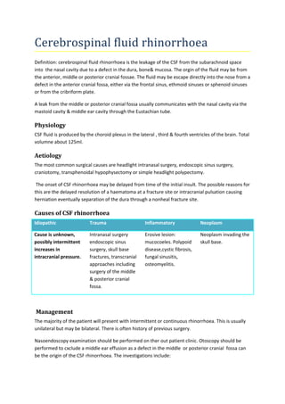
- 1. Cerebrospinal fluid rhinorrhoea Definition: cerebrospinal fluid rhinorrhoea is the leakage of the CSF from the subarachnoid space into the nasal cavity due to a defect in the dura, bone& mucosa. The orgin of the fluid may be from the anterior, middle or posterior cranial fossae. The fluid may be escape directly into the nose from a defect in the anterior cranial fossa, either via the frontal sinus, ethmoid sinuses or sphenoid sinuses or from the cribriform plate. A leak from the middle or posterior cranial fossa usually communicates with the nasal cavity via the mastoid cavity & middle ear cavity through the Eustachian tube. Physiology CSF fluid is produced by the choroid plexus in the lateral , third & fourth ventricles of the brain. Total volumne about 125ml. Aetiology The most common surgical causes are headlight intranasal surgery, endoscopic sinus surgery, craniotomy, transphenoidal hypophysectomy or simple headlight polypectomy. The onset of CSF rhinorrhoea may be delayed from time of the initial insult. The possible reasons for this are the delayed resolution of a haematoma at a fracture site or intracranial pulsation causing herniation eventually separation of the dura through a nonheal fracture site. Causes of CSF rhinorrhoea Idiopathic Trauma Inflammatory Neoplasm Cause is unknown, possibly intermittent increases in intracranial pressure. Intranasal surgery endoscopic sinus surgery, skull base fractures, transcranial approaches including surgery of the middle & posterior cranial fossa. Erosive lesion: mucocoeles. Polypoid disease,cystic fibrosis, fungal sinusitis, osteomyelitis. Neoplasm invading the skull base. Management The majority of the patient will present with intermittent or continuous rhinorrhoea. This is usually unilateral but may be bilateral. There is often history of previous surgery. Nasoendoscopy examination should be performed on ther out patient clinic. Otoscopy should be performed to cxclude a middle ear effusion as a defect in the middle or posterior cranial fossa can be the origin of the CSF rhinorrhoea. The investigations include:
- 2. 1.Laboratory investigation of rhinorrhoea fluid. 2. Imaging 3.Intrathecal dyes &markers. 1.Laboratory investigations Beta -2 transferrin is a protein involved in ferrous ion transport & it is also found in perilymph & aquous humour. Only few drops of CSF is required. The physician should aware that ceratin conditions abnormal transferring metabolism,& thus the beta-2 transferrin can appear in the blood, which could potentially lead to a false positive result. These are chronic liver disease, inborn errors of glycogen metabolism, neurophyschiatric disease & rectal carcinoma. 2. Imaging With the advent of high resolution CT , MRI & fluorescein lumber puncture, these modalities now form the mainstay of investigation. High resulation coronal scans (1-2 mm slices) can offer detection in up to 84% of cases. Axial views are helpful in detecting leaks from the posterior wall of frontal sinus & sphenoid sinus. 3. Intrathecal dyes & markers Fluorescein dye commonly use today. It can be exceedingly helpful either preoperatively in outpatient setting intraoperatively or both. Typically .25ml of 5% fluorescein is mixed with 10ml of CSF from a routine lumber puncture. The mixture is introduce via a polymedic pencil-point spinal needle & the patient is placed in the trendelenberg position for approximately for an hour. Then endoscopic examination is performed, fluorescein can be seen coming out from the defect. The use of blue filter on the endoscope light source can be increase the ease of detection. If at operation , fluorescein is not seen then the anaesthetist can temporarily raise the intracranial pressure by making the patient cough on their endotracheal tube tube as this will often cause the fluorescein to appear through the leak. Complication using a fluorescein lumber puncture have been described but with higher concentration than recommended here. They includes knee & ankle clonus, seizure, opisthonos & cranial nerve defect. None of the complications have been permanent & their occurance is extremely rare. Previous spinal surgery may prevent the use of fluorescein lumber puncture. Antibiotics There is debate as to whether antibiotic prophylactic should be prescribed in patients with known CSF rhinorrhoea. One meta analysis concluded that there is a significant reduction in the incidence of meningitis with prophylactic antibiotic therapy. Other go further & would not recommended prophylaxis in absence of infection as this can lead to a change in nasopharynx flora , potentially causing a partially treated or gram-negative meningitis.
- 3. Surgery Traditionally two approaches are available a craniotomy & an extradural external approach. With the development of endoscopic sinus surgery, this is now the the approach of choice with excellent success rate & minimal surgical morbidity (exception associated with malignancy). 1.Intracranial approach 2. Extracranial approach This remain the method of choice for assessing most leaks of the posterior wall of the frontal sinus that defy an endoscopic approach. Via an external ethmoidectomy for access to the cribriform plate & fovea ethmoidalis. Via transmastoid for defects in the tegmen& petrous temporal bone. Via transseptosphenoidal for access to the sphenoidal sinus . Via a coronal or eyebrow incision to the frontal sinus using an osteoplastic flap. In frontal & sphenoidal sinuses the mucosa can be removed, the decfect patched with fascia & the sinus can be can be obliterated by packing with fat. Nasal septum or turbinate can be used to support the graft. Extradural approach does have the advantage that it minimizes the incidence of intracranial complications. 3. Endoscopic surgery All techniques require accurate localization of of the leak intraoperatively. The edge of the defect are then freshened. The graft material is then placed into the defect as an underlay graft where possible. Majority of surgeons support their graft using nasal packing such as oxidized cellulose followed by a bismth iodoform paraffine paste pack (BIPP). The grafts are nasal mucosa flap or free nasal mucosal, which may be a composite graft incorporating turbinate bone ,conchal or septal scartilage, temporal fascia & fascia lata. Nearly all use antibiotic cover for the procedure. The patient should be advised not to blow their nose, to sneeze with their mouth open to avoid any abrupt increase in intracranial pressure. The supporting pack should be removed 7 to 10 days after surgery. Reason to failure Raised intracranial pressure is the most common reason for failure to repair a CSF leak. The raised intracranial pressure due to stenosis of sylvian duct or benign raised intracranial pressure mainly occur in obese young women with vague menstrual irregularities. A raised intracranial pressure is far more prevalent in patients with a spontaneous CSF leak. If imaging shows dilated cerebral ventricles then a neurosurgical procedure to lower the pressure such as shunt, should be performed at the time of repair of the leak.