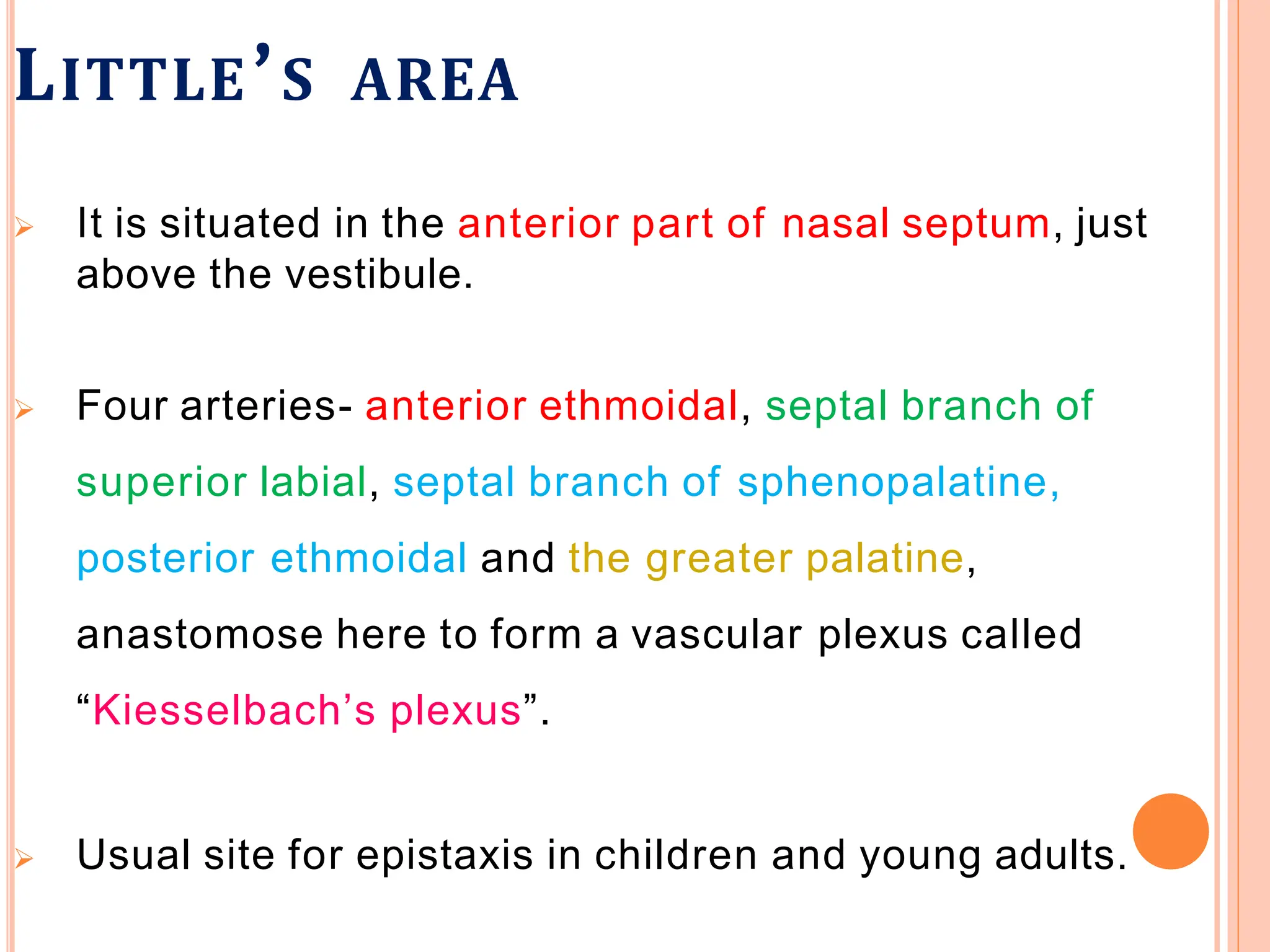
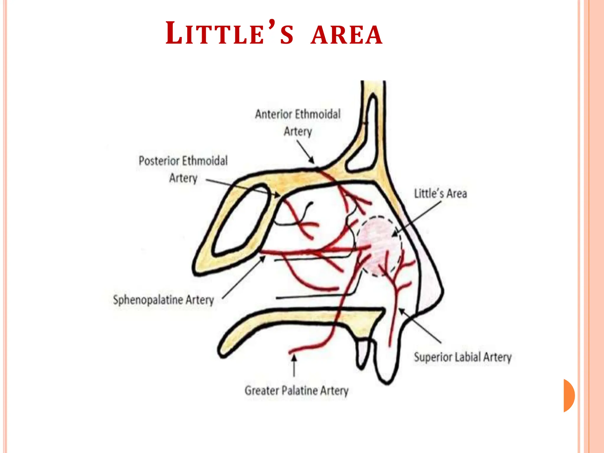
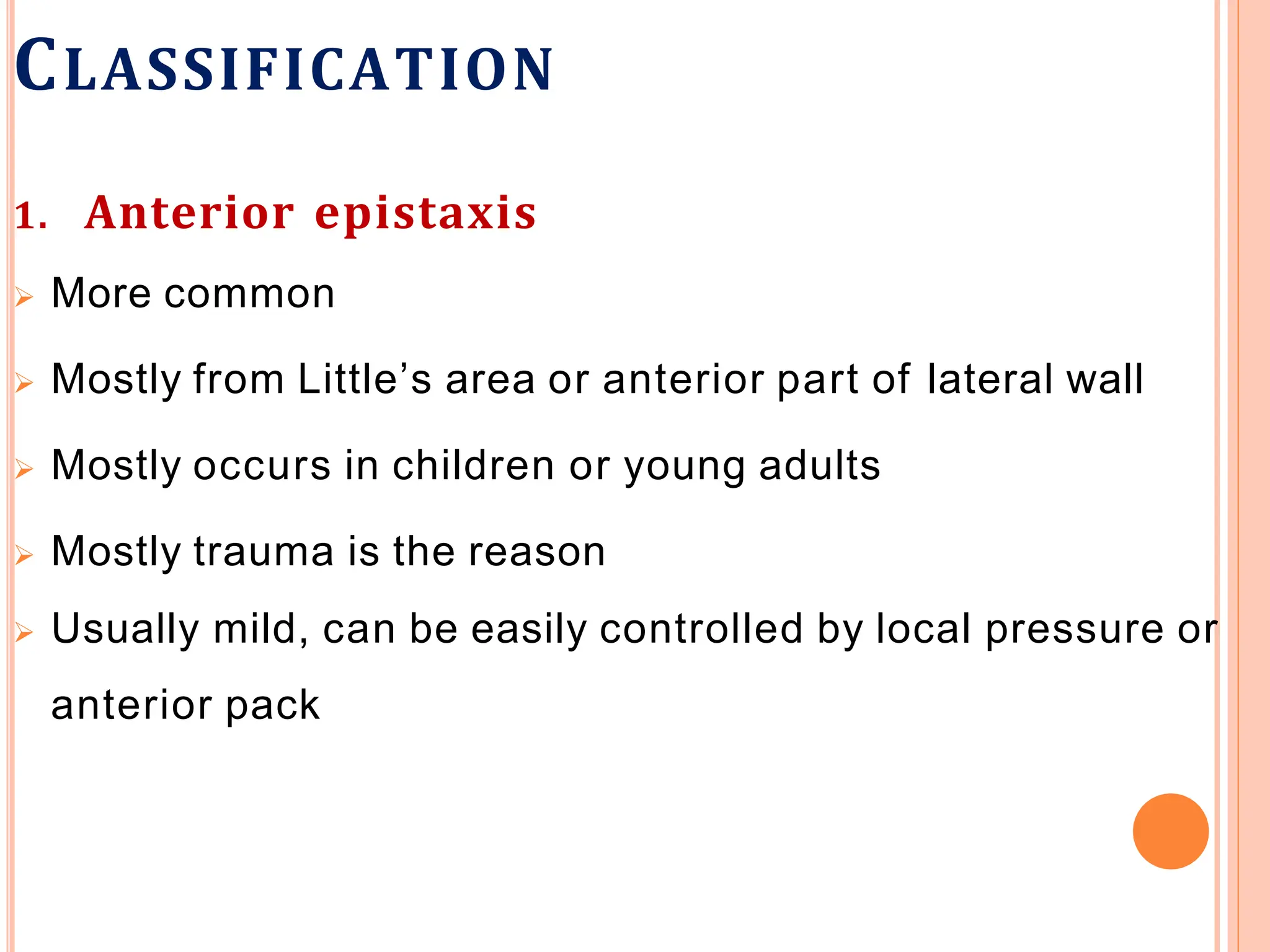
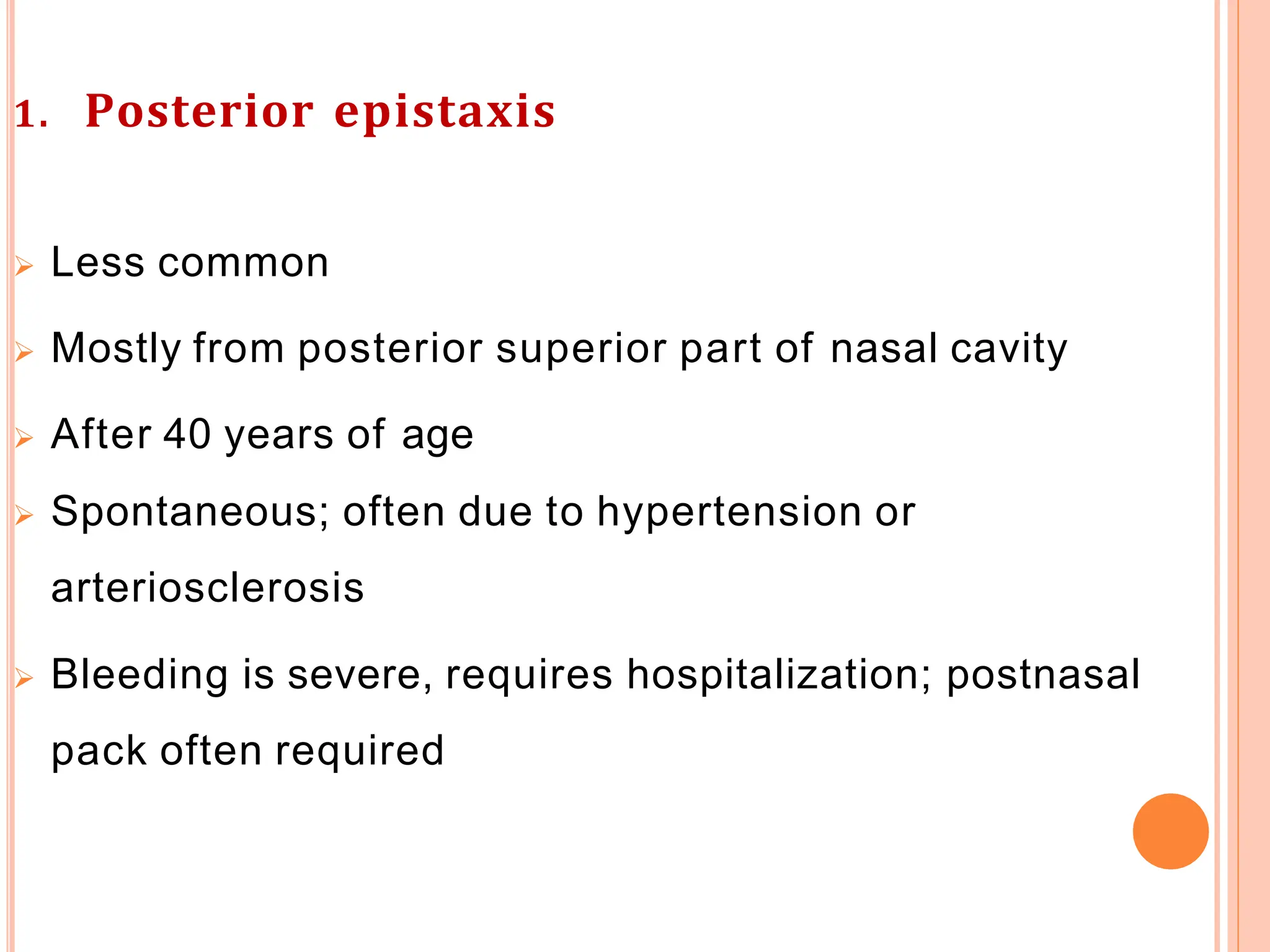
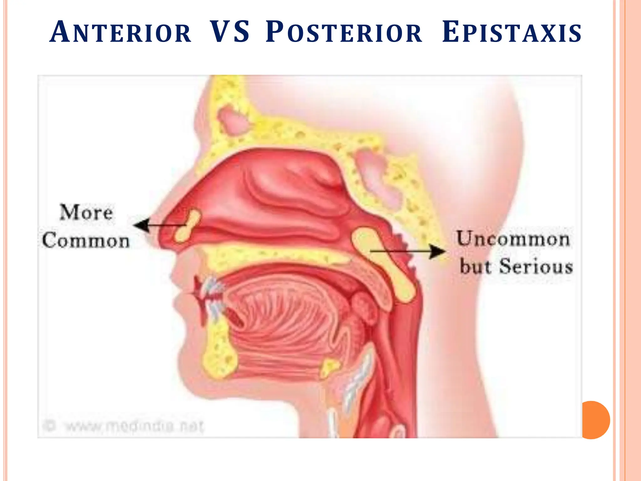
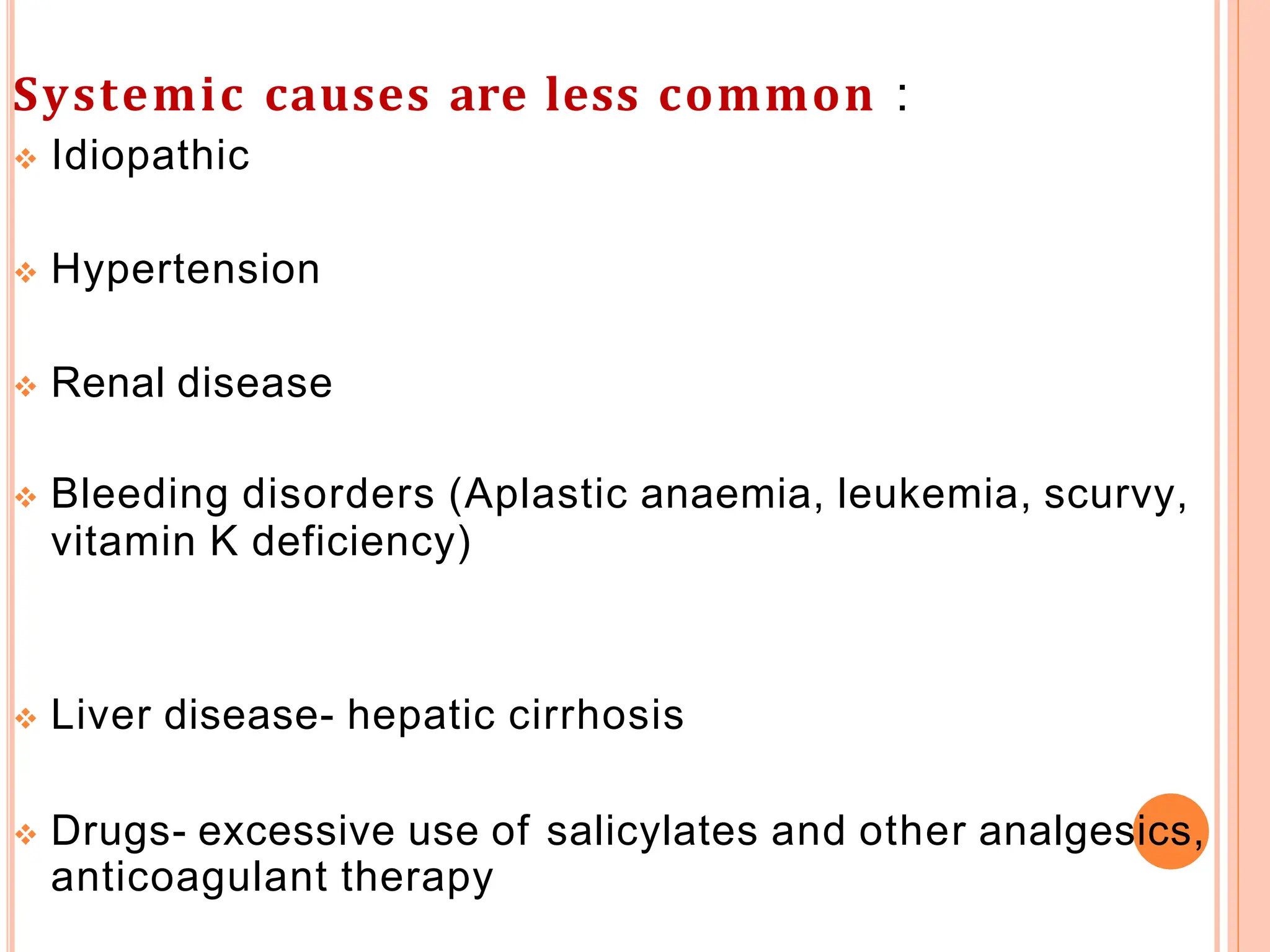
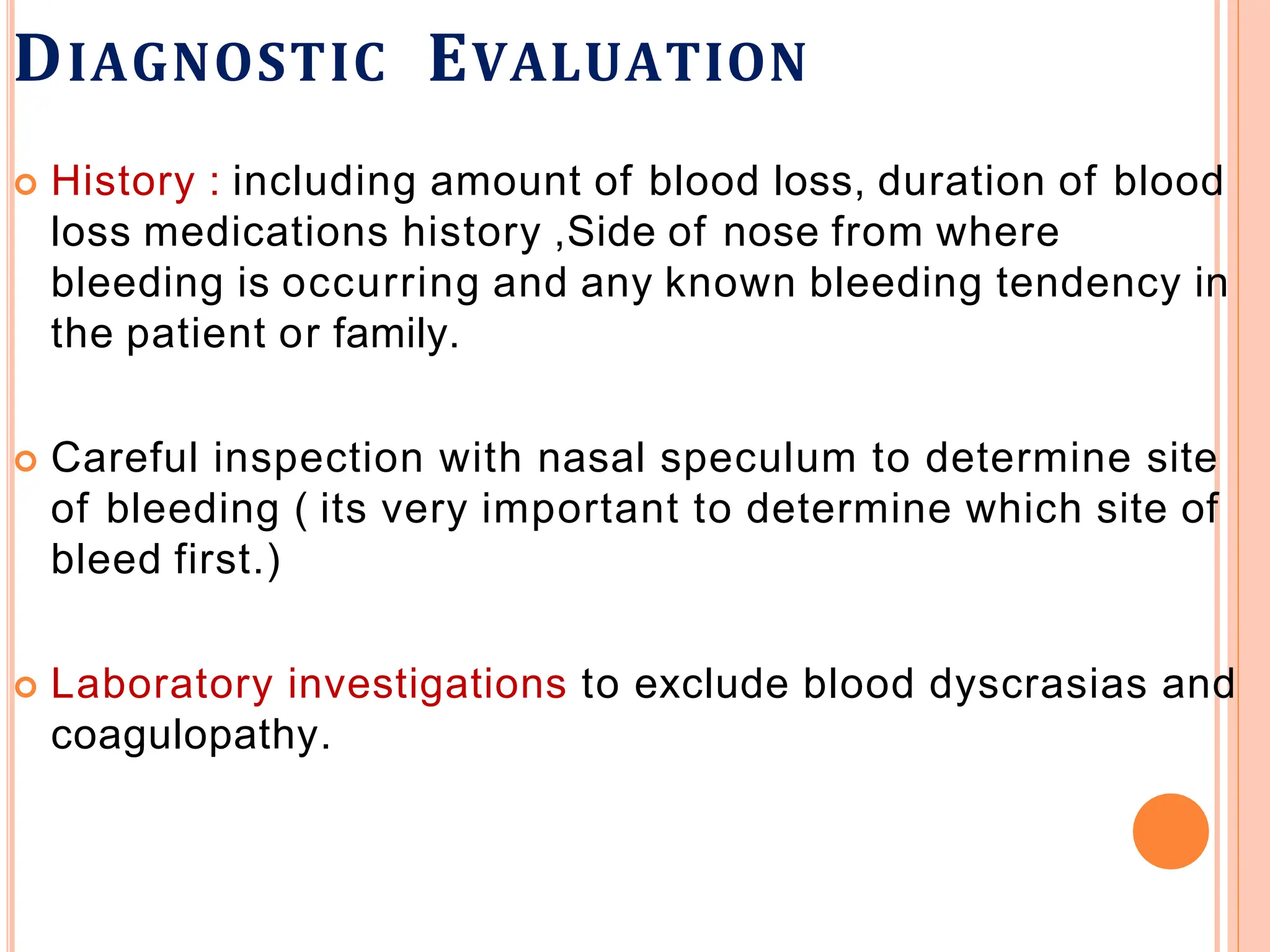
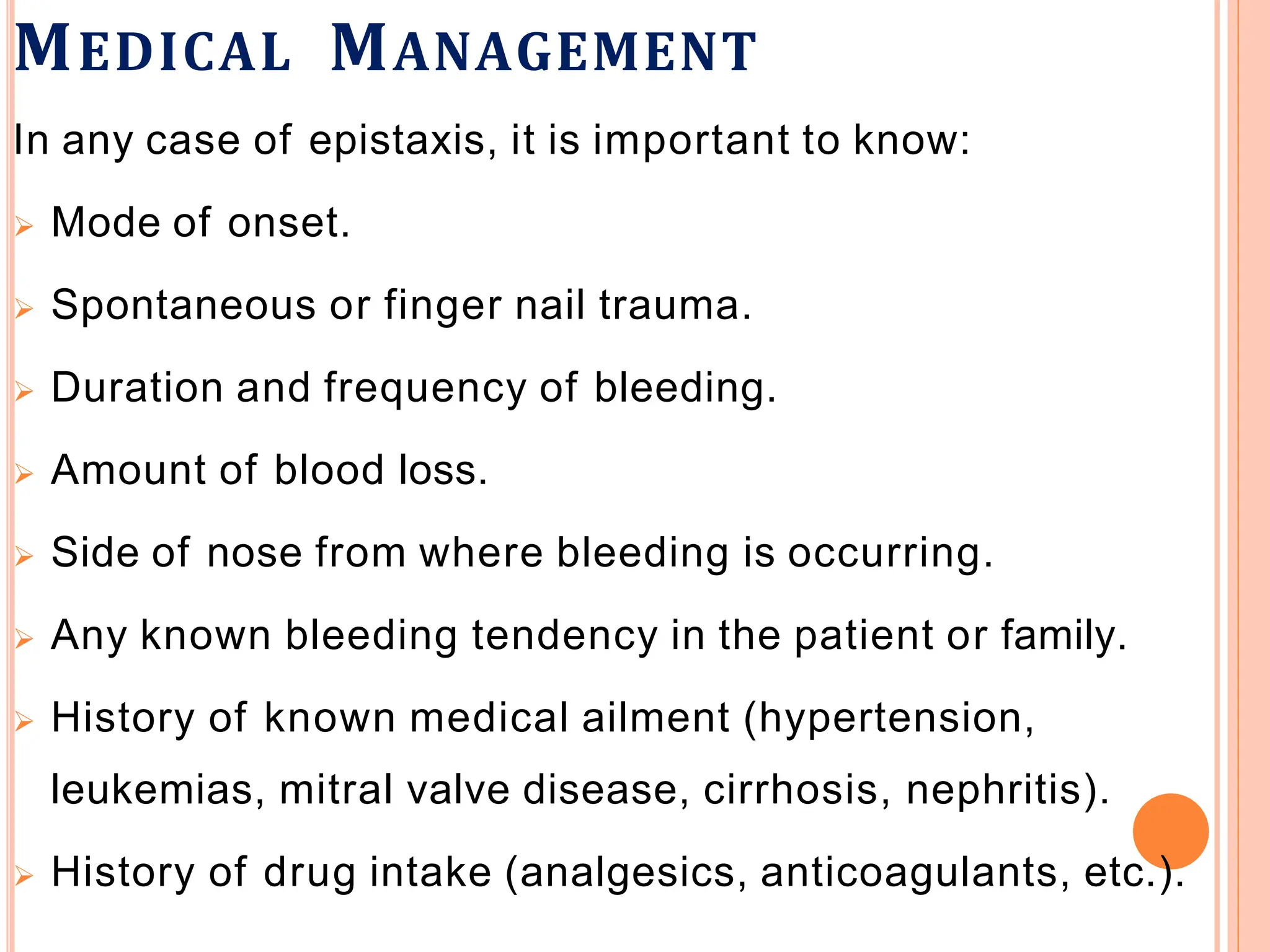
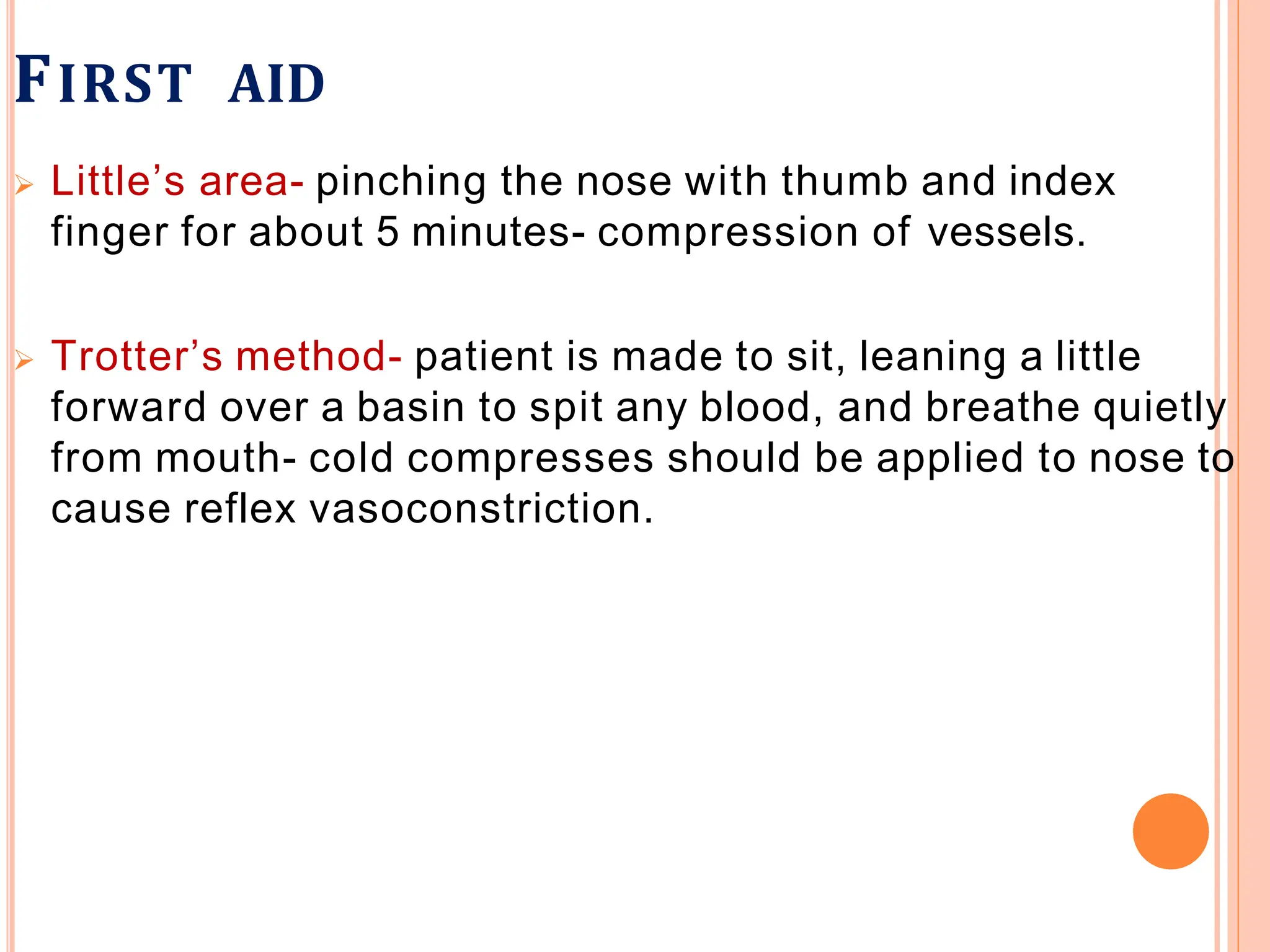
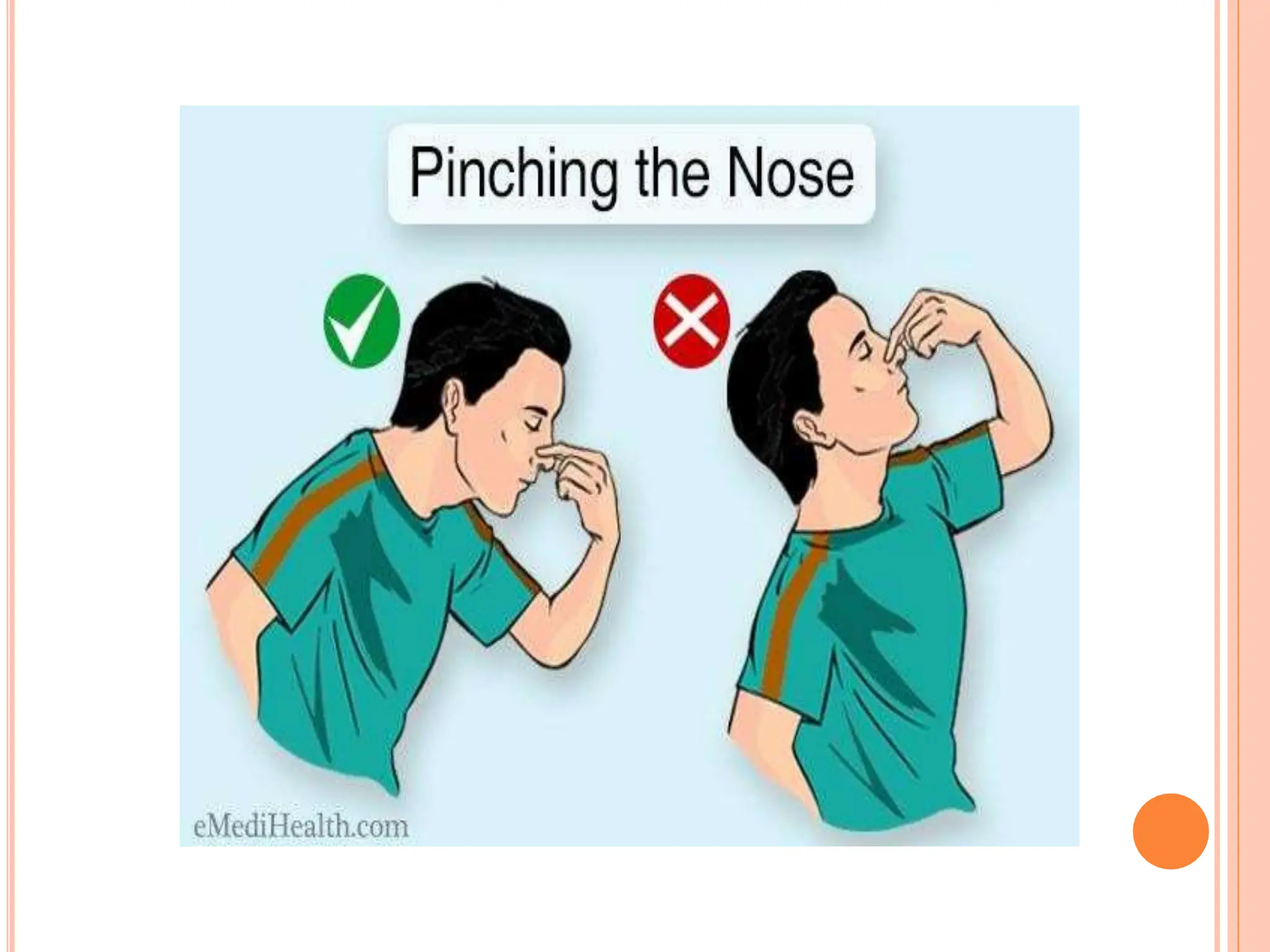
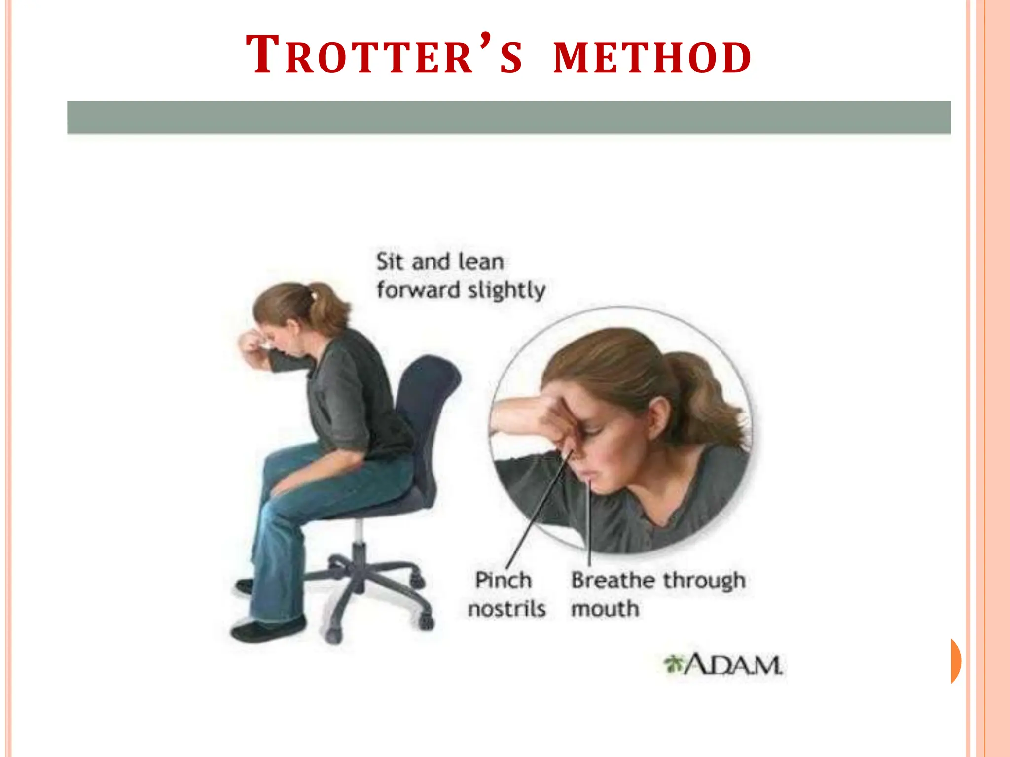
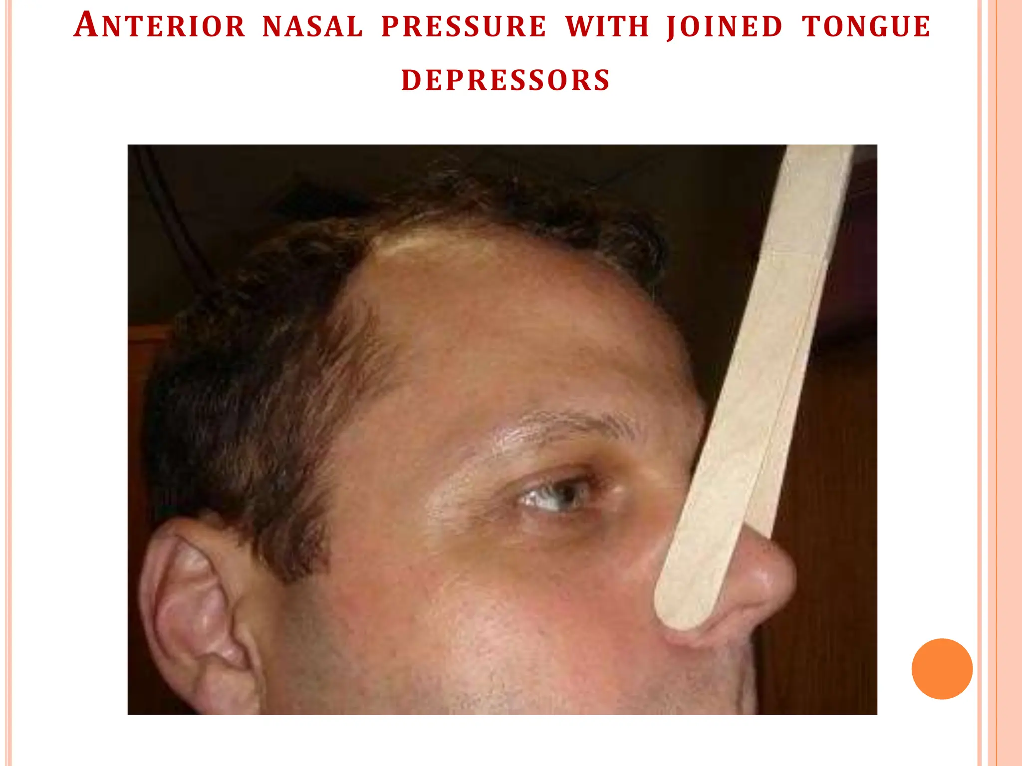
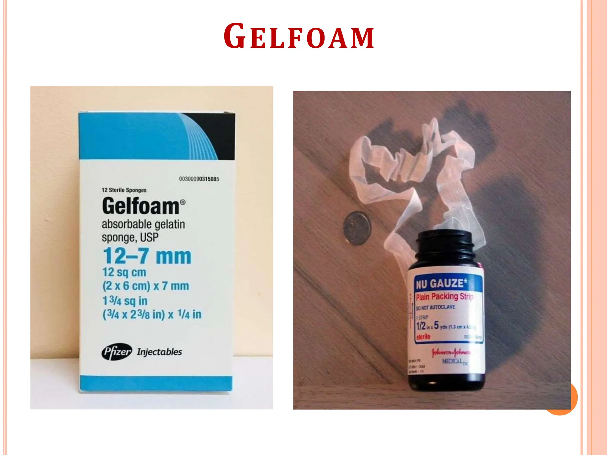
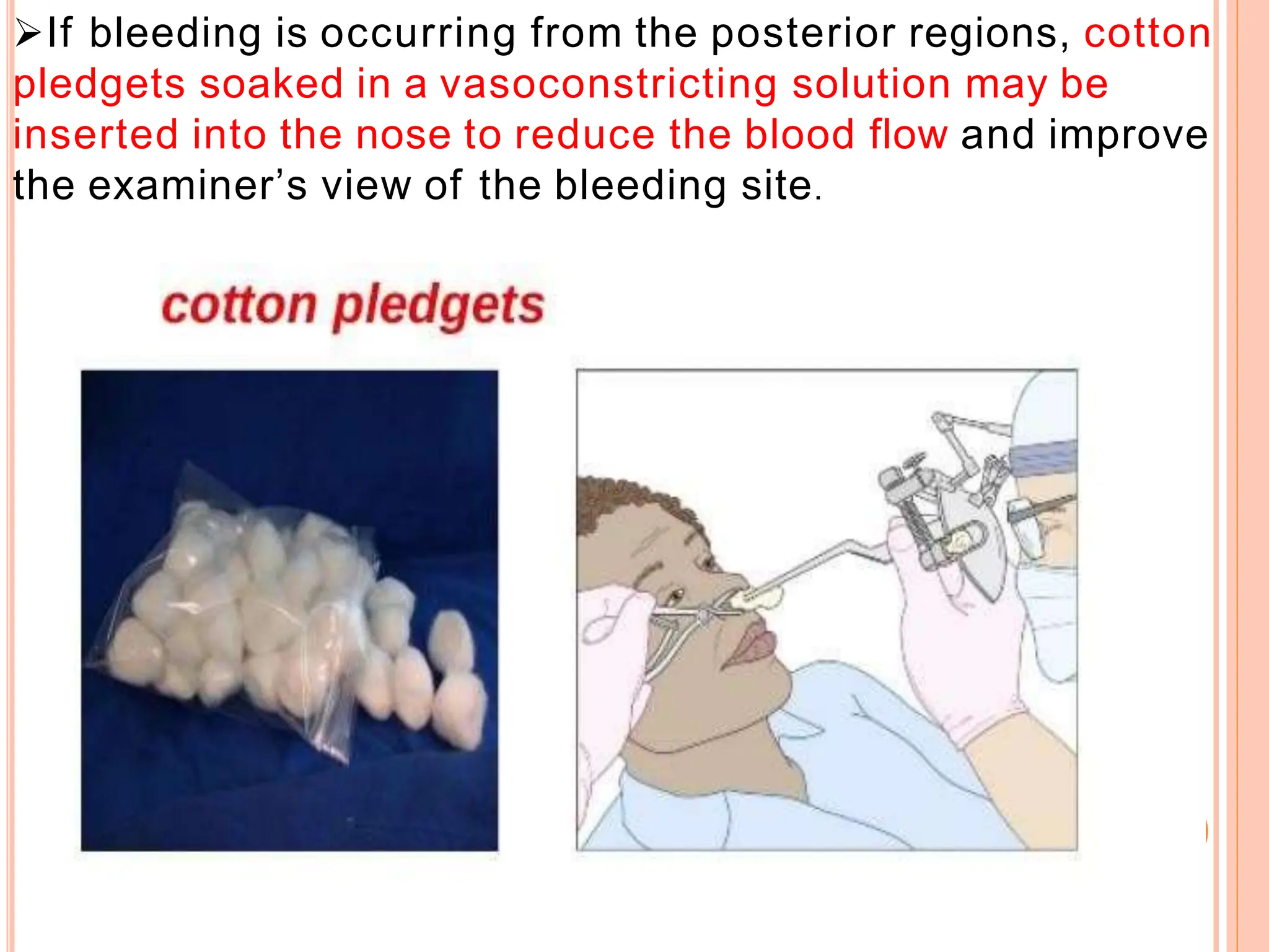
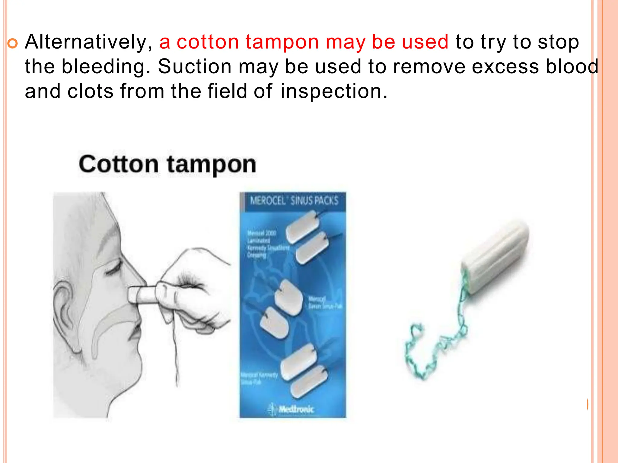
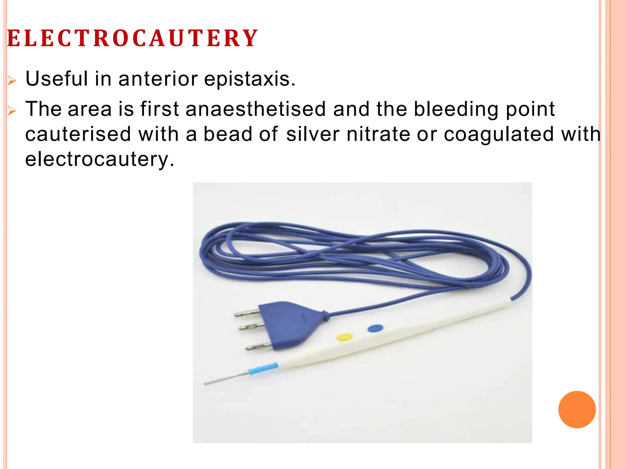
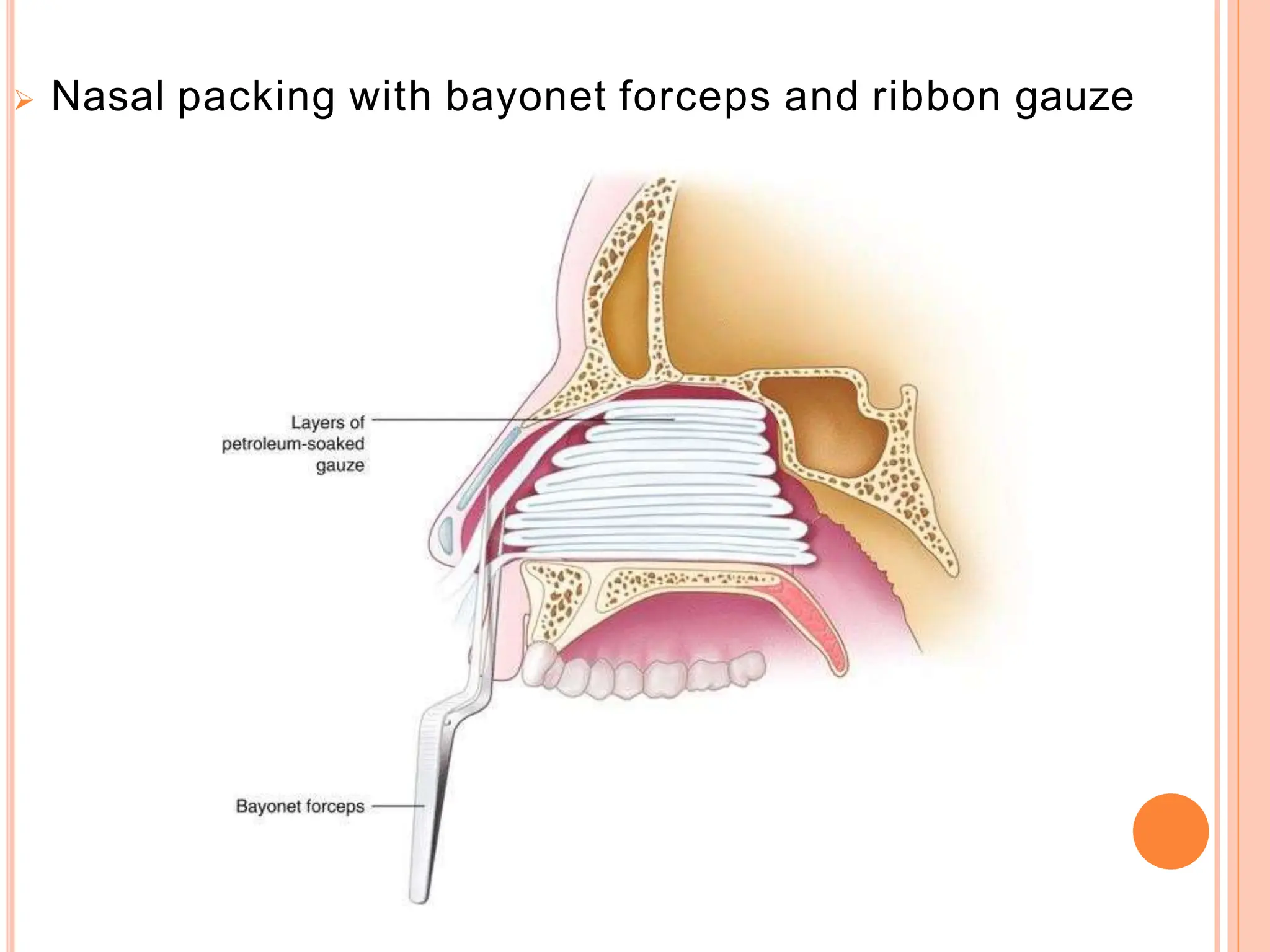
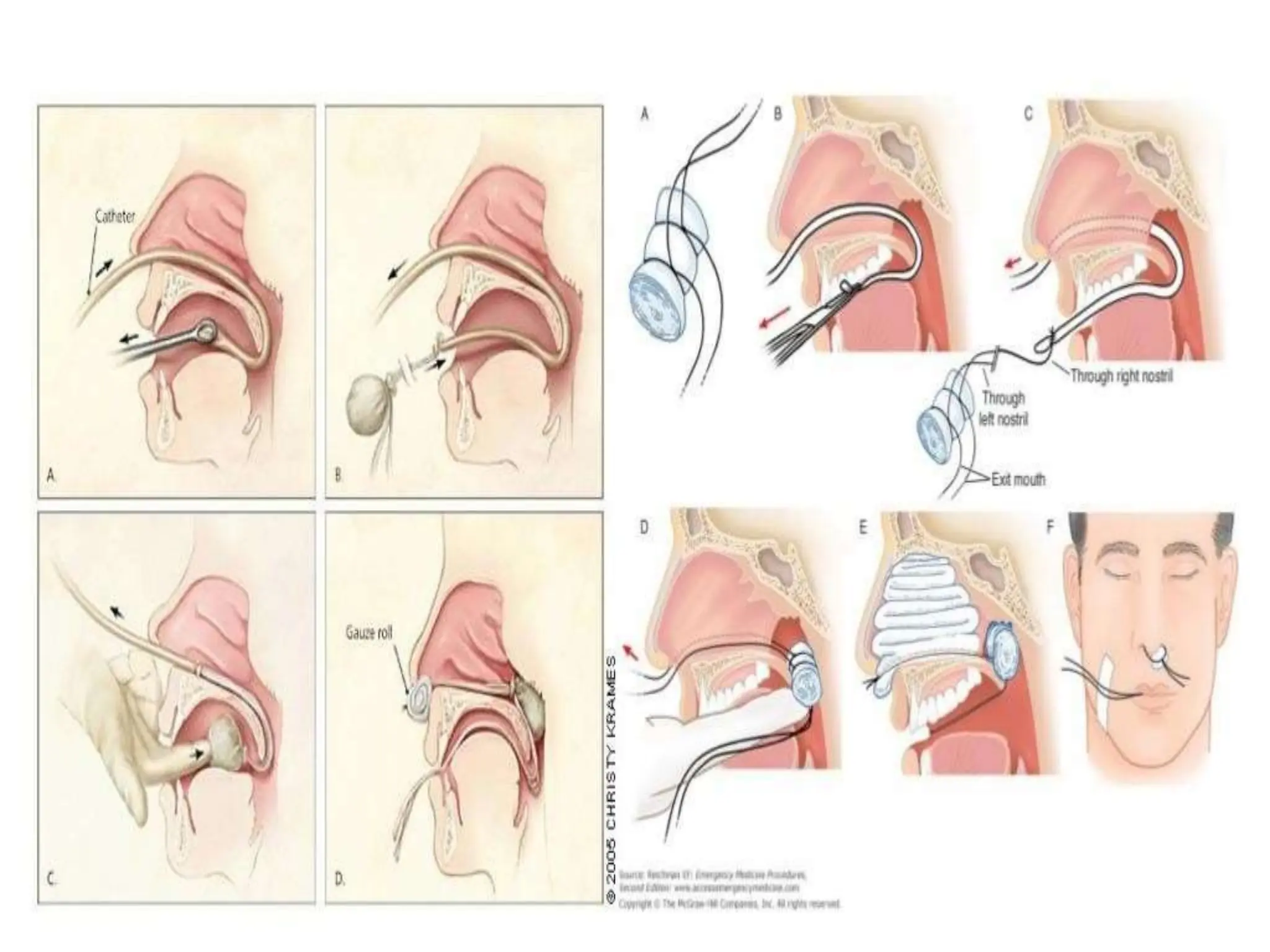
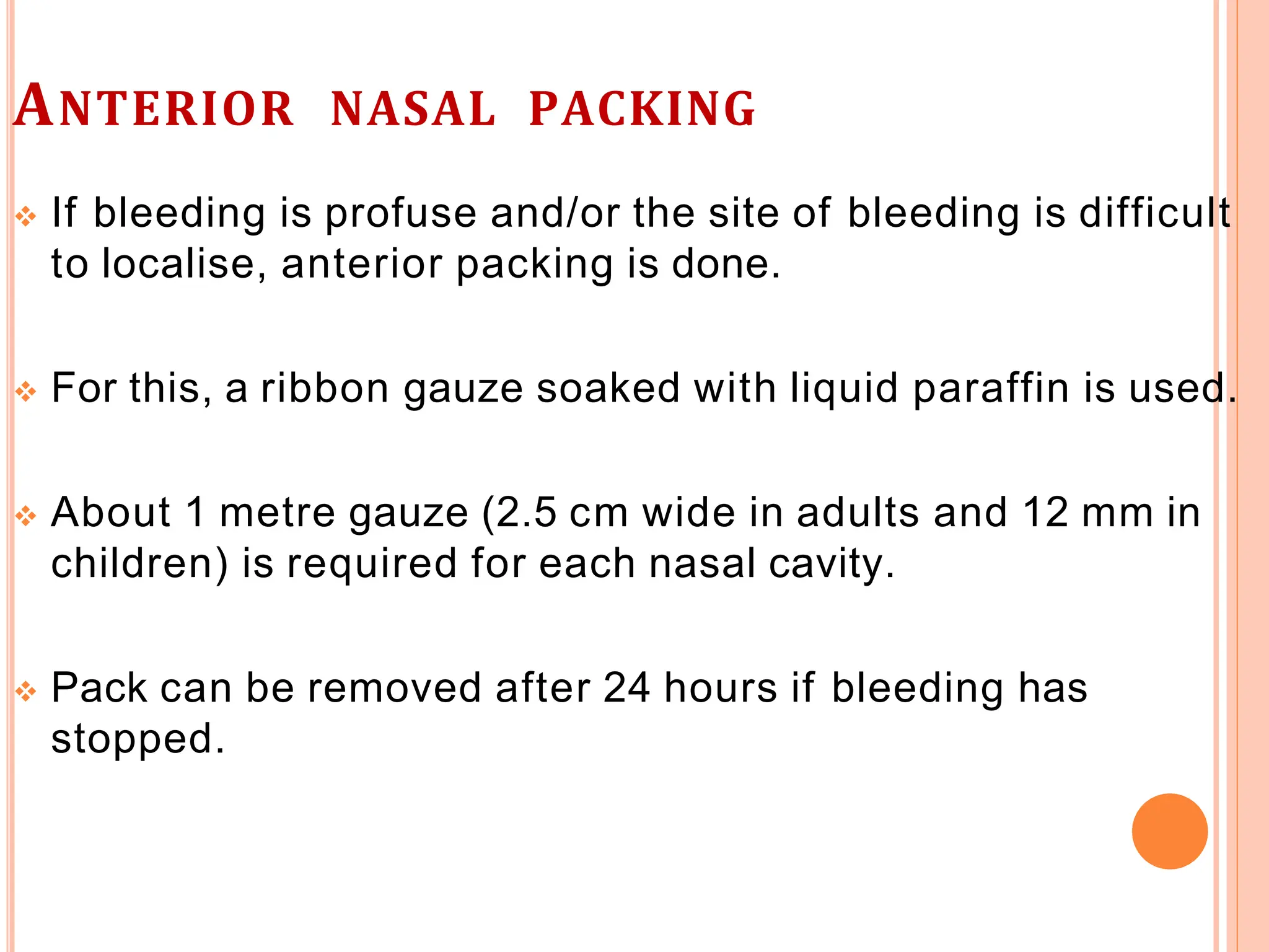
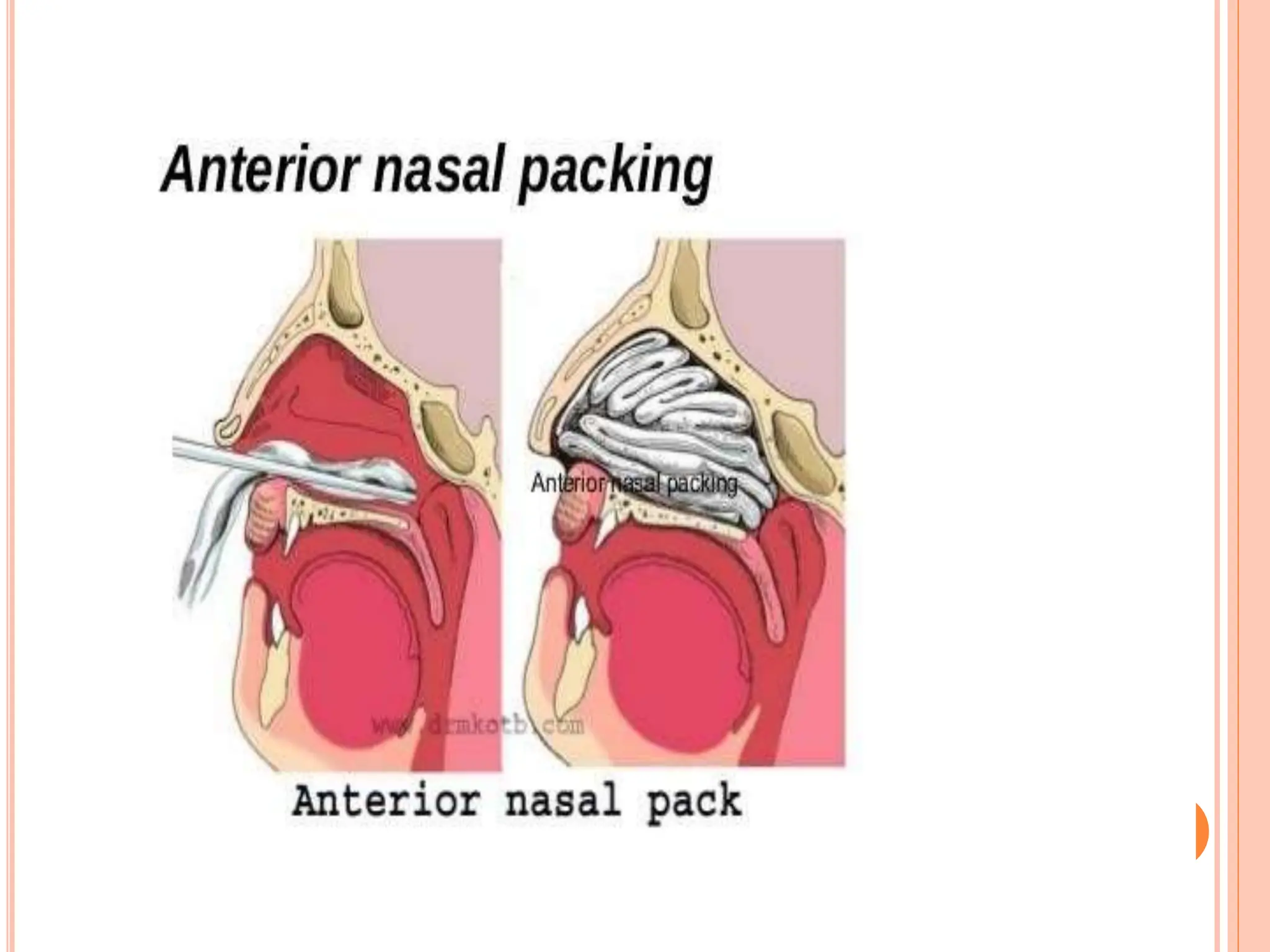
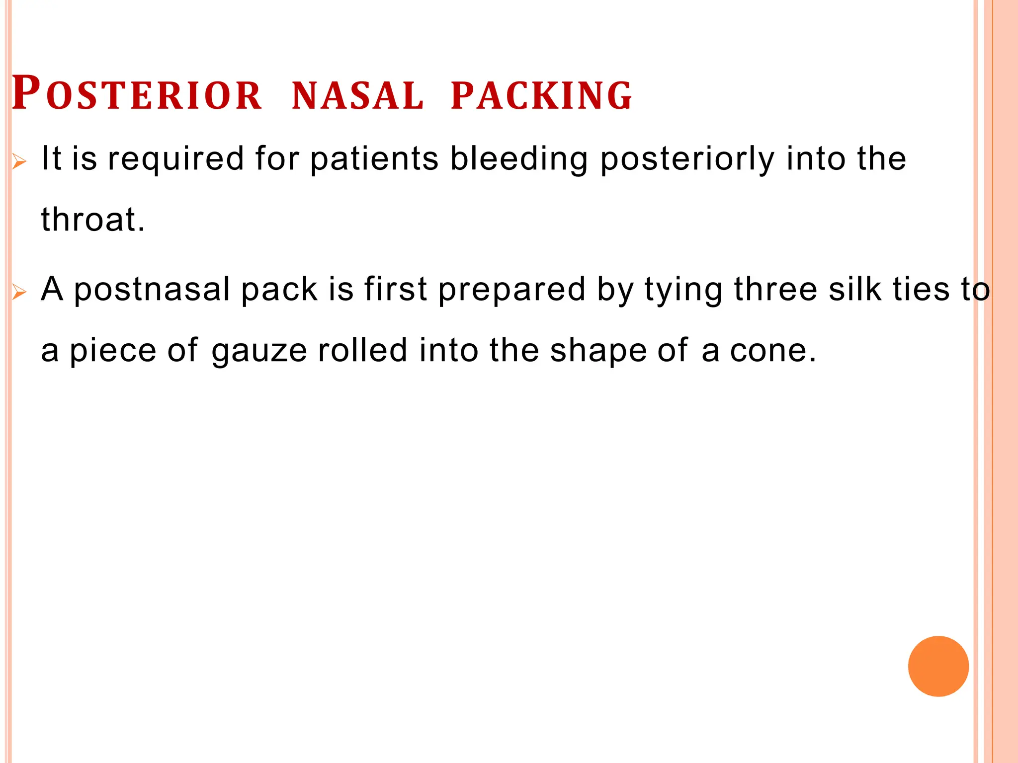
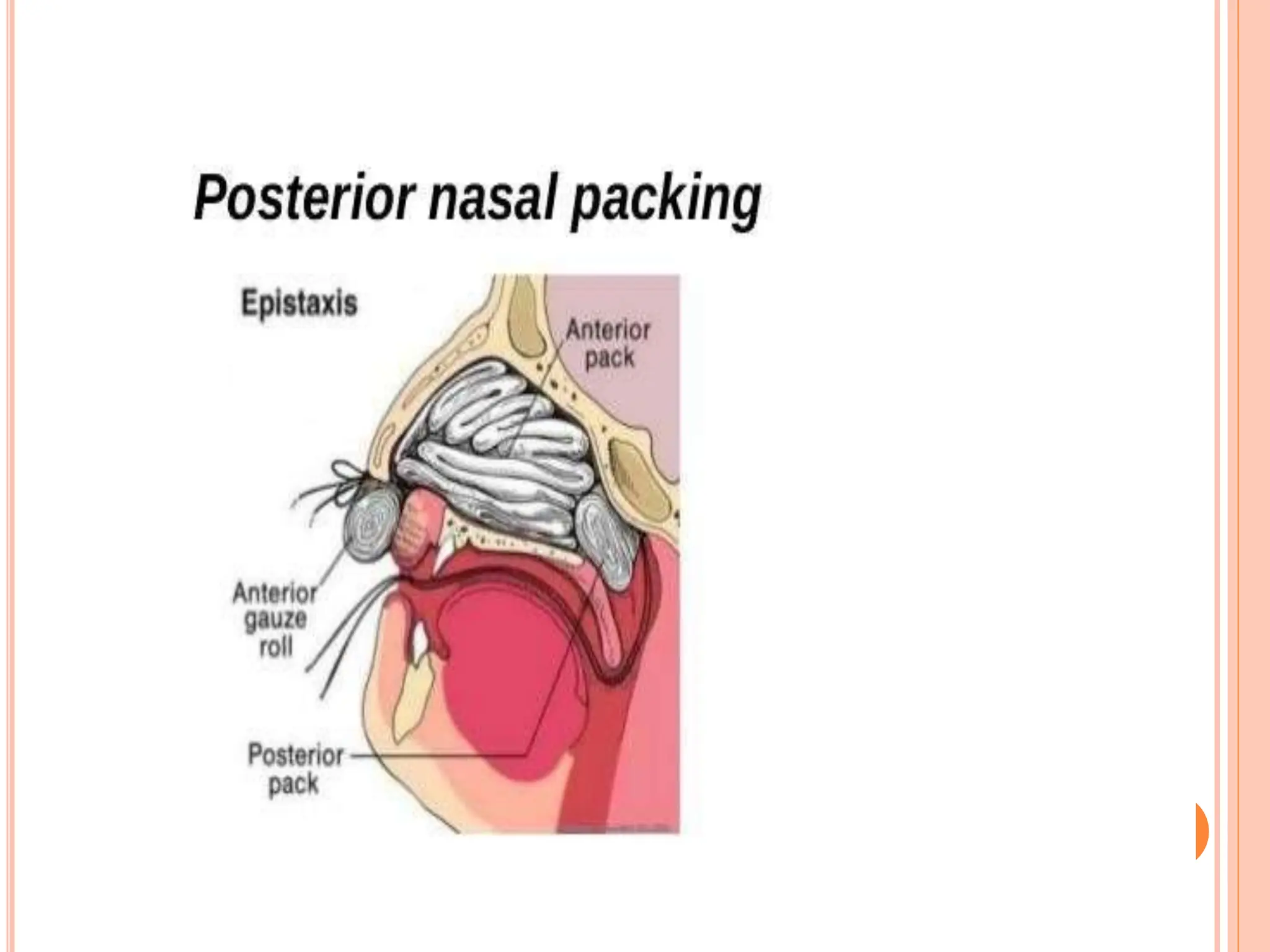
EPISTAXIS
GROUP 2
LAILA PATRICK OKUSA
SEKAGYA
 “Epistaxis refers to nose bleed or hemorrhage
from the nose”.
 Fairly common and is seen in all age groups
 It‘s mostly commonly originates in the anterior
portion of the nasal cavity.
DEFINITION
 A hemorrhage from the nose, referred to as
epistaxis, is caused by the rupture of tiny,
distended vessels in the mucous membrane of
any area of the nose.
LITTLE’S AREA
 It is situated in the anterior part of nasal septum, just
above the vestibule.
 Four arteries- anterior ethmoidal, septal branch of
superior labial, septal branch of sphenopalatine,
posterior ethmoidal and the greater palatine,
anastomose here to form a vascular plexus called
“Kiesselbach’s plexus”.
 Usual site for epistaxis in children and young adults.
LITTLE’S AREA
CLASSIFICATION
1. Anterior epistaxis
 More common
 Mostly from Little’s area or anterior part of lateral wall
 Mostly occurs in children or young adults
 Mostly trauma is the reason
 Usually mild, can be easily controlled by local pressure or
anterior pack
1. Posterior epistaxis
 Less common
 Mostly from posterior superior part of nasal cavity
 After 40 years of age
 Spontaneous; often due to hypertension or
arteriosclerosis
 Bleeding is severe, requires hospitalization; postnasal
pack often required
ANTERIOR VS POSTERIOR EPISTAXIS
CAUSES OF EPISTAXIS
Local causes:
 Dryness leading to crust formation-bleeding occurs with
removal of crusts by nose picking, rubbing, or blowing.
 Trauma – direct blows
 Infections (Acute: viral rhinitis, nasal diphtheria, acute
sinusitis.)
 Foreign bodies (Non-living: any neglected foreign body)
 Atmospheric changes. High altitudes, sudden
decompression (Caisson’s disease).
 Deviated nasal septum (DNS).
Systemic causes are less common :
 Idiopathic
 Hypertension
 Renal disease
 Bleeding disorders (Aplastic anaemia, leukemia, scurvy,
vitamin K deficiency)
 Liver disease- hepatic cirrhosis
 Drugs- excessive use of salicylates and other analgesics,
anticoagulant therapy
DIAGNOSTIC EVALUATION
 History : including amount of blood loss, duration of blood
loss medications history ,Side of nose from where
bleeding is occurring and any known bleeding tendency in
the patient or family.
 Careful inspection with nasal speculum to determine site
of bleeding ( its very important to determine which site of
bleed first.)
 Laboratory investigations to exclude blood dyscrasias and
coagulopathy.
MEDICAL MANAGEMENT
In any case of epistaxis, it is important to know:
 Mode of onset.
 Spontaneous or finger nail trauma.
 Duration and frequency of bleeding.
 Amount of blood loss.
 Side of nose from where bleeding is occurring.
 Any known bleeding tendency in the patient or family.
 History of known medical ailment (hypertension,
leukemias, mitral valve disease, cirrhosis, nephritis).
 History of drug intake (analgesics, anticoagulants, etc.).
FIRST AID
 Little’s area- pinching the nose with thumb and index
finger for about 5 minutes- compression of vessels.
 Trotter’s method- patient is made to sit, leaning a little
forward over a basin to spit any blood, and breathe quietly
from mouth- cold compresses should be applied to nose to
cause reflex vasoconstriction.
TROTTER’S METHOD
ANTERIOR NASAL PRESSURE WITH JOINED TONGUE
DEPRESSORS
If first aid measures are unsuccessful, additional treatment
is indicated:-
 In anterior nosebleeds, the area may be treated with a
silver nitrate applicator and Gelfoam, or by electrocautery.
 Topical vasoconstrictors, such as adrenaline or cocaine
(0.5%), and phenylephrine may be prescribed.
SILVER NITRATE APPLICATOR
GELFOAM
If bleeding is occurring from the posterior regions, cotton
pledgets soaked in a vasoconstricting solution may be
inserted into the nose to reduce the blood flow and improve
the examiner’s view of the bleeding site.
 Alternatively, a cotton tampon may be used to try to stop
the bleeding. Suction may be used to remove excess blood
and clots from the field of inspection.
ELECTROCAUTERY
 Useful in anterior epistaxis.
 The area is first anaesthetised and the bleeding point
cauterised with a bead of silver nitrate or coagulated with
electrocautery.
 Nasal packing with bayonet forceps and ribbon gauze
 When the origin of the bleeding cannot be identified, the
nose may be packed with gauze soaked with petroleum
jelly or antibiotic ointment;
a topical anesthetic spray and decongestant agent may be
used prior to inserting the gauze packing
 The packing may remain in place for 48 hours or up to 5 or
6 days if necessary to control bleeding.
 Antibiotics may be prescribed because of the risk of
iatrogenic sinusitis
ANTERIOR NASAL PACKING
 If bleeding is profuse and/or the site of bleeding is difficult
to localise, anterior packing is done.
 For this, a ribbon gauze soaked with liquid paraffin is used.
 About 1 metre gauze (2.5 cm wide in adults and 12 mm in
children) is required for each nasal cavity.
 Pack can be removed after 24 hours if bleeding has
stopped.
POSTERIOR NASAL PACKING
 It is required for patients bleeding posteriorly into the
throat.
 A postnasal pack is first prepared by tying three silk ties to
a piece of gauze rolled into the shape of a cone.
COMPLICATIONS OF EPISTAXIS
 Hemorrhagic shock
 Septic shock
 Pneumocephalus (is the presence of air or gas
within the cranial cavity. )
 Sinusitis
 Septal pressure necrosis
 Neurogenic syncope during packing (also called:
vasovagal syncope. It is a sudden drop in heart rate and
blood pressure leading to fainting, often in reaction to a
stressful trigger.)
 Epiphora ( is an overflow of tears onto the face).
NURSING MANAGEMENT
 The nurse monitors the vital signs, assists in the control of
bleeding, and provides tissues and an emesis basin to
allow the patient to expectorate any excess blood. It is not
uncommon for patients to be anxious in response to a
nosebleed.
 Monitor vital sings & assist with control of bleeding.
Assess for changes in BP and pulse indicative of
hypovolemia.
 Assuring the patient in a calm, efficient manner that
bleeding can be controlled can help reduce anxiety.
 Instructs to the client to avoid blowing or picking nose
after a nose bleed.
 Discharge teaching includes reviewing ways to prevent
epistaxis: avoiding forceful nose blowing, straining, high
altitudes, and nasal trauma (including nose picking).
 Monitor the client with posterior packing for hypoxia.
 Adequate humidification may prevent drying of the nasal
passages.
 The nurse instructs the patient how to apply direct
pressure to the nose with the thumb and the index finger
for 15 minutes in the case of a recurrent nosebleed. If
recurrent bleeding cannot be stopped, the patient is
instructed to seek additional medical attention.
epistaxis.pptxslidesharethankyousomuchii

epistaxis.pptxslidesharethankyousomuchii

  • 1.
  • 2.
     “Epistaxis refersto nose bleed or hemorrhage from the nose”.  Fairly common and is seen in all age groups  It‘s mostly commonly originates in the anterior portion of the nasal cavity.
  • 3.
    DEFINITION  A hemorrhagefrom the nose, referred to as epistaxis, is caused by the rupture of tiny, distended vessels in the mucous membrane of any area of the nose.
  • 4.
    LITTLE’S AREA  Itis situated in the anterior part of nasal septum, just above the vestibule.  Four arteries- anterior ethmoidal, septal branch of superior labial, septal branch of sphenopalatine, posterior ethmoidal and the greater palatine, anastomose here to form a vascular plexus called “Kiesselbach’s plexus”.  Usual site for epistaxis in children and young adults.
  • 5.
  • 6.
    CLASSIFICATION 1. Anterior epistaxis More common  Mostly from Little’s area or anterior part of lateral wall  Mostly occurs in children or young adults  Mostly trauma is the reason  Usually mild, can be easily controlled by local pressure or anterior pack
  • 7.
    1. Posterior epistaxis Less common  Mostly from posterior superior part of nasal cavity  After 40 years of age  Spontaneous; often due to hypertension or arteriosclerosis  Bleeding is severe, requires hospitalization; postnasal pack often required
  • 8.
  • 9.
    CAUSES OF EPISTAXIS Localcauses:  Dryness leading to crust formation-bleeding occurs with removal of crusts by nose picking, rubbing, or blowing.  Trauma – direct blows  Infections (Acute: viral rhinitis, nasal diphtheria, acute sinusitis.)  Foreign bodies (Non-living: any neglected foreign body)  Atmospheric changes. High altitudes, sudden decompression (Caisson’s disease).  Deviated nasal septum (DNS).
  • 10.
    Systemic causes areless common :  Idiopathic  Hypertension  Renal disease  Bleeding disorders (Aplastic anaemia, leukemia, scurvy, vitamin K deficiency)  Liver disease- hepatic cirrhosis  Drugs- excessive use of salicylates and other analgesics, anticoagulant therapy
  • 11.
    DIAGNOSTIC EVALUATION  History: including amount of blood loss, duration of blood loss medications history ,Side of nose from where bleeding is occurring and any known bleeding tendency in the patient or family.  Careful inspection with nasal speculum to determine site of bleeding ( its very important to determine which site of bleed first.)  Laboratory investigations to exclude blood dyscrasias and coagulopathy.
  • 12.
    MEDICAL MANAGEMENT In anycase of epistaxis, it is important to know:  Mode of onset.  Spontaneous or finger nail trauma.  Duration and frequency of bleeding.  Amount of blood loss.  Side of nose from where bleeding is occurring.  Any known bleeding tendency in the patient or family.  History of known medical ailment (hypertension, leukemias, mitral valve disease, cirrhosis, nephritis).  History of drug intake (analgesics, anticoagulants, etc.).
  • 13.
    FIRST AID  Little’sarea- pinching the nose with thumb and index finger for about 5 minutes- compression of vessels.  Trotter’s method- patient is made to sit, leaning a little forward over a basin to spit any blood, and breathe quietly from mouth- cold compresses should be applied to nose to cause reflex vasoconstriction.
  • 15.
  • 16.
    ANTERIOR NASAL PRESSUREWITH JOINED TONGUE DEPRESSORS
  • 17.
    If first aidmeasures are unsuccessful, additional treatment is indicated:-  In anterior nosebleeds, the area may be treated with a silver nitrate applicator and Gelfoam, or by electrocautery.  Topical vasoconstrictors, such as adrenaline or cocaine (0.5%), and phenylephrine may be prescribed.
  • 18.
  • 19.
  • 20.
    If bleeding isoccurring from the posterior regions, cotton pledgets soaked in a vasoconstricting solution may be inserted into the nose to reduce the blood flow and improve the examiner’s view of the bleeding site.
  • 21.
     Alternatively, acotton tampon may be used to try to stop the bleeding. Suction may be used to remove excess blood and clots from the field of inspection.
  • 22.
    ELECTROCAUTERY  Useful inanterior epistaxis.  The area is first anaesthetised and the bleeding point cauterised with a bead of silver nitrate or coagulated with electrocautery.
  • 23.
     Nasal packingwith bayonet forceps and ribbon gauze
  • 24.
     When theorigin of the bleeding cannot be identified, the nose may be packed with gauze soaked with petroleum jelly or antibiotic ointment; a topical anesthetic spray and decongestant agent may be used prior to inserting the gauze packing  The packing may remain in place for 48 hours or up to 5 or 6 days if necessary to control bleeding.  Antibiotics may be prescribed because of the risk of iatrogenic sinusitis
  • 26.
    ANTERIOR NASAL PACKING If bleeding is profuse and/or the site of bleeding is difficult to localise, anterior packing is done.  For this, a ribbon gauze soaked with liquid paraffin is used.  About 1 metre gauze (2.5 cm wide in adults and 12 mm in children) is required for each nasal cavity.  Pack can be removed after 24 hours if bleeding has stopped.
  • 28.
    POSTERIOR NASAL PACKING It is required for patients bleeding posteriorly into the throat.  A postnasal pack is first prepared by tying three silk ties to a piece of gauze rolled into the shape of a cone.
  • 30.
    COMPLICATIONS OF EPISTAXIS Hemorrhagic shock  Septic shock  Pneumocephalus (is the presence of air or gas within the cranial cavity. )  Sinusitis  Septal pressure necrosis
  • 31.
     Neurogenic syncopeduring packing (also called: vasovagal syncope. It is a sudden drop in heart rate and blood pressure leading to fainting, often in reaction to a stressful trigger.)  Epiphora ( is an overflow of tears onto the face).
  • 32.
    NURSING MANAGEMENT  Thenurse monitors the vital signs, assists in the control of bleeding, and provides tissues and an emesis basin to allow the patient to expectorate any excess blood. It is not uncommon for patients to be anxious in response to a nosebleed.  Monitor vital sings & assist with control of bleeding. Assess for changes in BP and pulse indicative of hypovolemia.  Assuring the patient in a calm, efficient manner that bleeding can be controlled can help reduce anxiety.
  • 33.
     Instructs tothe client to avoid blowing or picking nose after a nose bleed.  Discharge teaching includes reviewing ways to prevent epistaxis: avoiding forceful nose blowing, straining, high altitudes, and nasal trauma (including nose picking).  Monitor the client with posterior packing for hypoxia.  Adequate humidification may prevent drying of the nasal passages.  The nurse instructs the patient how to apply direct pressure to the nose with the thumb and the index finger for 15 minutes in the case of a recurrent nosebleed. If recurrent bleeding cannot be stopped, the patient is instructed to seek additional medical attention.