Download as PDF, PPTX

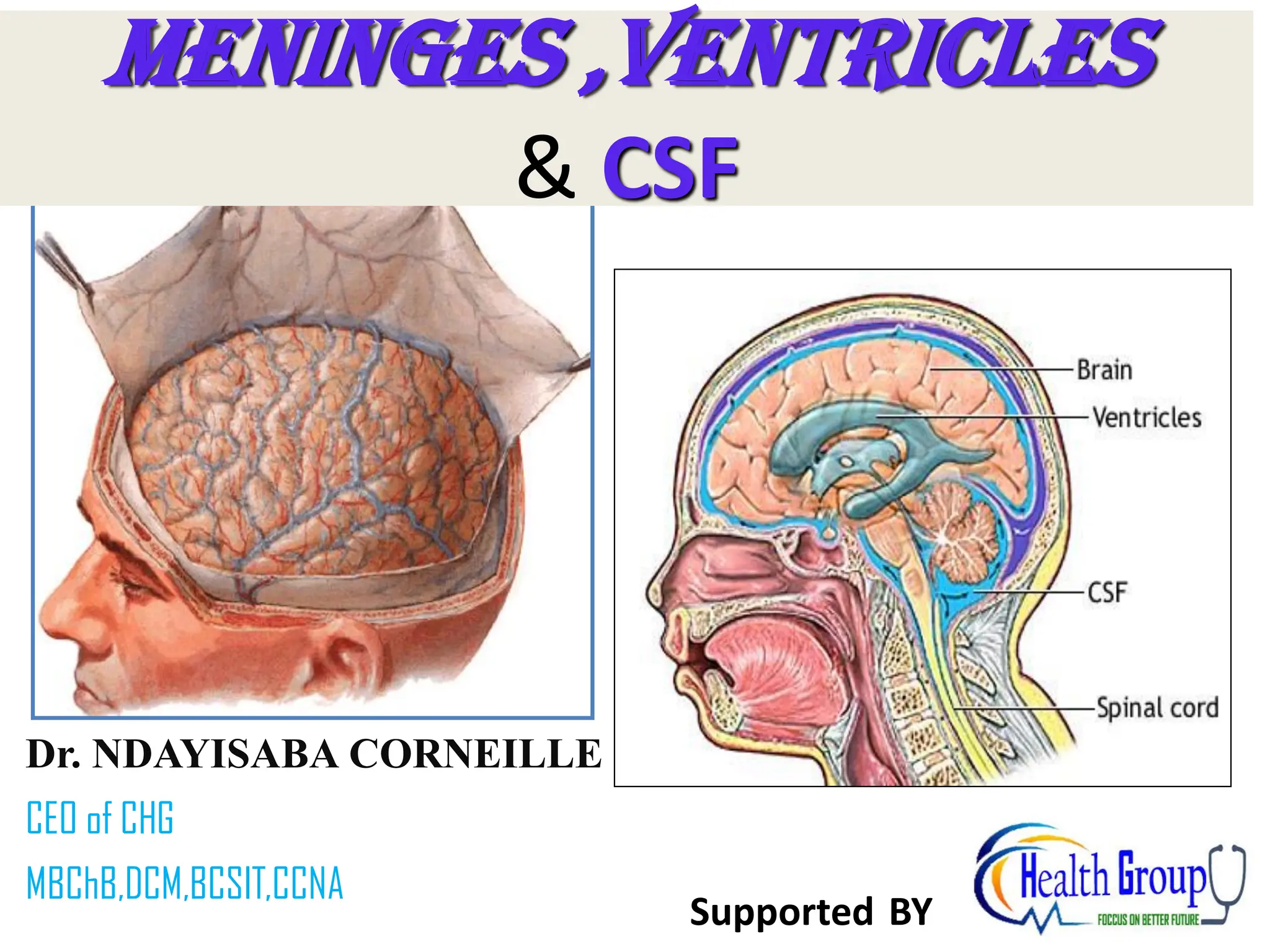
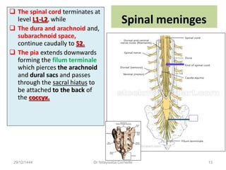
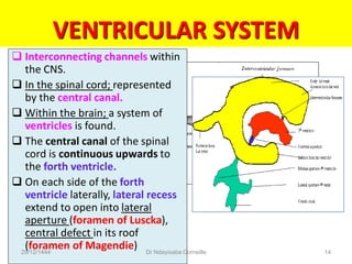
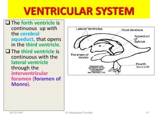

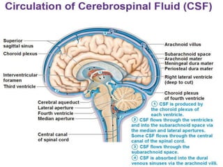
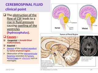
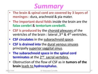


The presentation by Dr. Ndayisaba Corneille covers the anatomy and functions of the brain and spinal cord meninges, detailing the dura, arachnoid, and pia mater, as well as the cerebrospinal fluid (CSF) system. Key points include the structure and function of the dural folds, subarachnoid space, and the ventricular system, along with the formation and circulation of CSF. Clinical considerations related to CSF obstruction, including hydrocephalus, are also addressed.


