Anatomy of airway
•Download as PPT, PDF•
6 likes•1,632 views

upper/lower airway anatomy

Recommended
More Related Content
What's hot
What's hot (20)
Similar to Anatomy of airway
Similar to Anatomy of airway (20)
More from ZIKRULLAH MALLICK
More from ZIKRULLAH MALLICK (20)
Recently uploaded
Recently uploaded (20)
Anatomy of airway
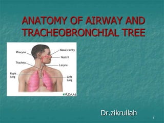
- 1. ANATOMY OF AIRWAY AND TRACHEOBRONCHIAL TREE Dr.zikrullah 1
- 2. 2 THE UPPER AIRWAY The upper airway starts : At the nostrils, extends through the nasal conchae to the nasopharynx, over the uvula to the hypo pharynx and larynx At the lips, extends through the oral cavity, over the tongue and below the hard and soft palates, to the hypo pharynx and larynx.
- 3. UPPER AIRWAY Nose Pharynx Larynx 3
- 4. NOSE Airway functionally begins at the nares , where air first enters the body. Septal cartilage divides nasal cavity into two nasal fossae ROOF-cribriform plate of the ethmoid FLOOR-perpendicular to the face LATERAL-3 turbinates Little’s area on anterior & inferior part of septum; may bleed during nasal intubation or introducing nasal airway. 4
- 5. Nasal septum is often deviated from the midline causing one cavity to be larger than the other . It is therefore essential for anaesthetist to visualize the nasal cavity before attempting nasal intubation 5
- 6. ORAL CAVITY Alternate respiratory passage Extends from mouth opening to anterior tonsillar pillars. Contracture of mouth & lips-difficult laryngoscopy. Teeth loose or buck-difficult intubation 6
- 7. PHARYNX Extends from base of skull to lower border of cricoid cartilage. Subdivided into: nasopharynx, oropharynx, laryngopharynx 7
- 8. NASOPHARYNX Extends from posterior end of turbinates to posterior pharyngeal wall above soft palate. Filters bacteria and foreign particles from inspired air Eustachian tube open into lateral surfaces, and connect nasopharynx to middle ear, each equalizes pressure of middle ear 8
- 9. NASOPHARYNGEAL AIRWAYS Measuring an Airway Measured against the distance from the patient's nose to the patient's earlobe.
- 10. OROPHARYNX Extends from soft palate above to epiglottis below& anteriorly from anterior tonsillar pillar to posterior pharyngeal wall. Mainly has a digestive function Ring of waldeyer 10
- 11. OROPHARYNGEAL AIRWAYS Measuring an Airway Corner of the mouth to tip of the ear
- 12. LARYNGOPHARYNX Lies between the fourth and sixth cervical vertebrae. Starts at the superior border of the epiglottis, and extends to the inferior border of the cricoid cartilage, where it narrows and becomes continuous with the oesophagus . 12
- 13. LARYNX Lies between base of tongue and trachea Primary function is to serve as the “watchdog” of the respiratory tract, allowing passage only to air Houses the vocal cords, and helps in vocalization Connection point-upper and lower airways Extends from C3 to C6 13
- 14. 14 Composed of 3 single cartilaginous structures: Epiglottis-flap, swings down to meet larynx during swallowing Thyroid-bulk of this forms larynx Cricoid-circular
- 15. EPIGLOTTIS Covers the rima glottidis during swallowing (glottis=cords & space) 15
- 16. THYROID CARTILAGE Largest of the laryngeal cartilages Inner side are attached the vocal cords Its two alae meet ant. at 900angle in males 1200angle in females 16
- 17. CRICOID A complete cartilaginous ring Narrowest portion of the lower airway in neonate and infant Actual start of the lower airway 17
- 19. 19 LARYNX The thyrohyoid membrane forms a C- shaped barrier around the anterior and lateral walls of the supraglottis Cricothyroid membrane- easily palpable,avascular,site for surgical cricothyrotomy.
- 20. The Larynx: Anatomy The six smaller cartilages of the larynx (3 pairs) are functionally involved with the movements of the vocal cords.These are: The Arytenoids The Corniculates The Cuneiforms 20
- 21. 21 The arytenoid cartilages are pyramid- shaped and articulate with the superior margin of the cricoid lamina. On their summit, are the corniculate cartilages; on their anterior aspect, are the cuneiform cartilages
- 22. 22 The Larynx: Anatomy The vocal ligaments, are attached posteriorly to the apex of the arytenoids and corniculates. The cuneiforms extend laterally, between the layers of the vocal cords, from the anterior aspect of the arytenocorniculate complex.
- 23. 23 View of the larynx at laryngoscopy.
- 24. INTRINSIC MUSCULATURE Posterior cricoarytenoid Abductor of vocal cords Recurrent laryngeal Lateral cricoarytenoid Adducts arytenoids closing glottis Recurrent laryngeal Transverse arytenoid/ posterior cricoarytenoid Adducts arytenoids Recurrent laryngeal Oblique arytenoid Closes glottis Recurrent laryngeal Thyroarytenoid Relaxes cords Recurrent laryngeal Cricothyroid Tensor of the cords External laryngeal 24
- 25. LARYNX NERVE SUPPLY SENSORY: Above vocal cords- internal laryngeal nerve (b. of superior laryngeal n.) Below vocal cords-recurrent laryngeal nerve MOTOR: All muscles which move the larynx are supplied by recurrent laryngeal n.except the cricothyroid. Cricothyroid:supplied by external laryngeal n. (b. of superior laryngeal n.) 25
- 26. Superior Laryngeal nerve Paralysis Unilateral: Voice weak,Pitch can not raised, Anaesthesia of larynx on side may cause aspiration Bilateral: Voice weak ,husky Inhalation of food and Pharyngeal secretions lead to cough & choking . 26
- 27. Superior and Recurrent laryngeal nerve paralysis Unilateral: Hoarsness of voice and aspiration of liquid, cough is ineffective Bilateral: Aphonia, Aspiration,Inability to cough 27
- 28. 28 Laryngeal function: Airway Protection Open Closed The glottis: open for inspiration and closed for swallowing
- 29. 29 Laryngeal function: Phonation Adducted: Talking Abducted: Breathing The vocal cords: Adducted for phonation; abducted for inspiration
- 30. LOWER AIRWAY Trachea Main stem bronchi Segmental bronchi Subsegmental bronchi Bronchioles Terminal bronchioles Respiratory bronchioles Alveolar ducts Alveolar sacs alveoli 30
- 31. Trachea 10-13 cm long Mean distance lips to carina male-28.5cm; female- 25.2cm-----ETT fixation Mean distance base of nose to carina male 31cm; female 28.4cm-----ETT fixation 1.5-2.5 cm wide 31
- 32. Extends from lower border of cricoid cartilage(C6) to carina(T5) where it bifurcates into right & left main bronchus 15-20 C shaped rings Tracheostomy done at 2-3 tracheal ring. ETT is placed above the carina. In Pregnancy due to airway oedema 1cm smaller diameter ETT used. 32
- 33. Tracheobronchial Tree Series of branching airways commonly referred to a “generations” or “orders” The first generation or order is zero (0), the trachea itself. Bifucrates at the carina Touching of carina during intubation may lead to vagal stimulation. 33
- 34. 34
- 35. Main Stem Bronchi Right bronchus Wider More vertical 5 cm long Supported by C shaped cartilages 20-30 degree angle First generation Usually bronchial intubation, secretion & foreign bodies lodged Left bronchus Narrower More angular 5.5 cm Long Supported by C shaped cartilages 40-60 degree angle First generation 35
- 36. 36
- 37. Lobar Bronchi R main stem divides into: Upper lobar bronchus Middle lobar bronchus Lower lobar bronchus L main stem divides into: Upper lobar bronchus Lower lobar bronchus 37
- 38. Segmental Bronchi 3rd generation R lobar divides into Segmental bronchi 10 segments on right L lobar divides into Segmental bronchi 10 segments on left 38
- 39. Subsegmental Bronchi 4th to 9th generations Progressively smaller airways 1-4 mm diameter 39
- 40. Noncartilagenous Airways Bronchioles 10-th to 15th generation Cartilage is absent Surrounded by spiral muscle fiber With no cartilage, airway remains open due to pressure gradients 40
- 41. Terminal Bronchioles 16th to 19th generation Average diameter is 0.5 mm Cilia and mucous glands begin to disappear totally End of the conducting airway 41
- 42. Gas exchange zone Respiratory bronchioles Acinus -respiratory bronchioles to the alveoli Ducts, sacs, alveoli 42
- 43. Alveoli 300 million alveoli Between 75 µ to 300 µ in diameter Most gas exchange takes place at alveolar-capillary membrane 85-95% of alveoli covered by small pulmonary capillaires The cross-sectional area or surface area is approximately 70m2 43
- 44. Intersitium/interstial space Surround, supports, and shapes the alveoli and capillaries Composed of a gel like substance and collagen fibers Contains tight space and loose space areas 44
- 46. 46 THANK YOU…