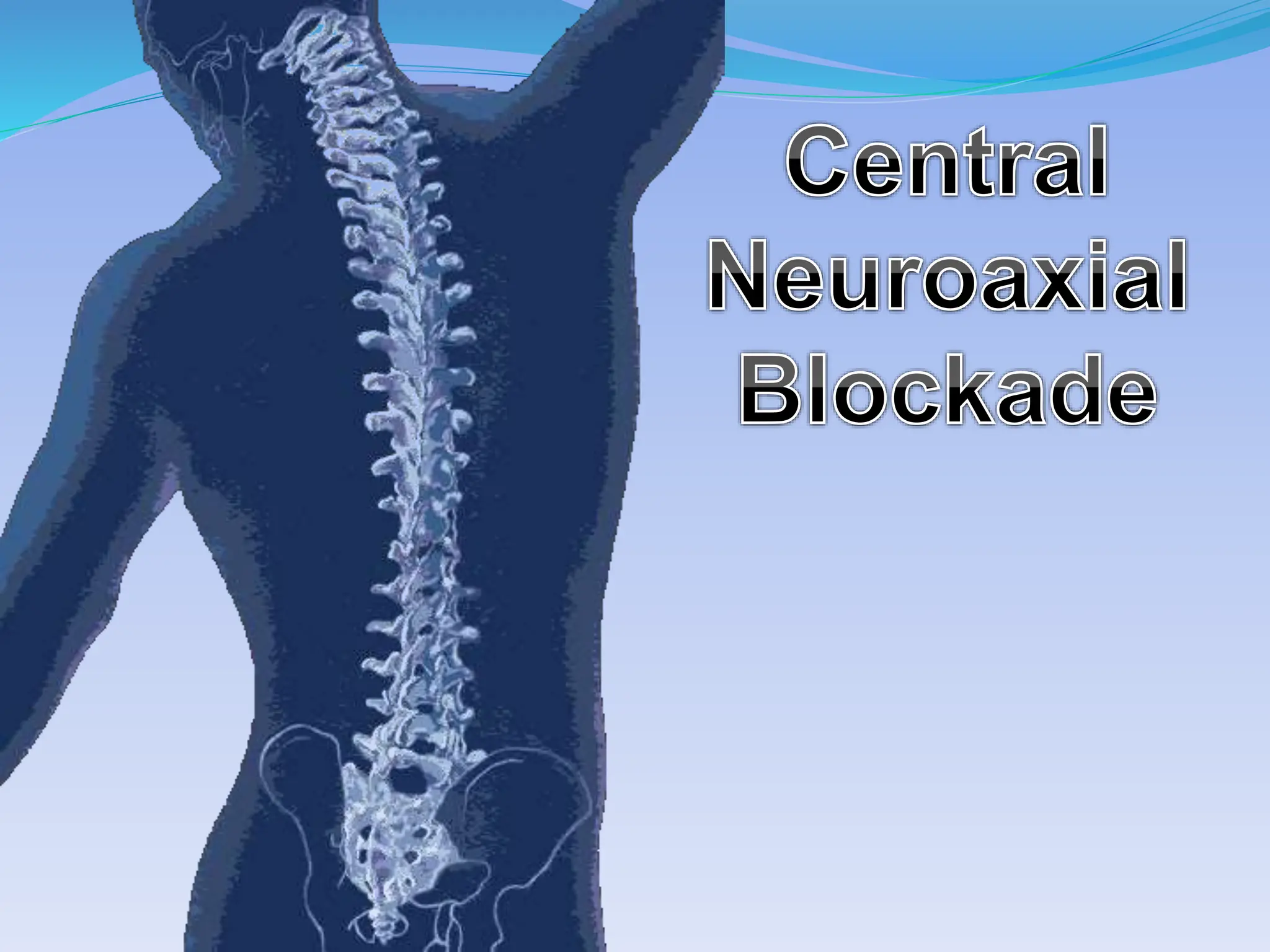
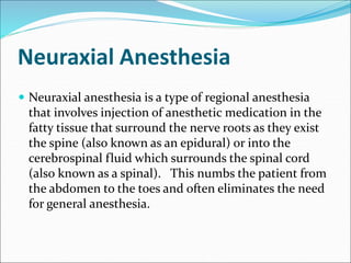
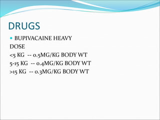
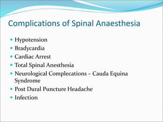
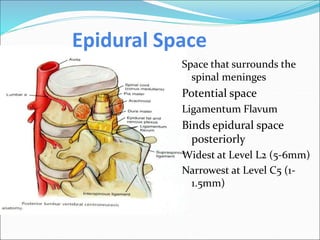
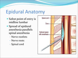
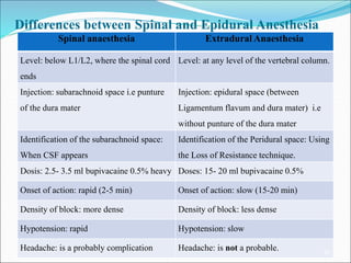
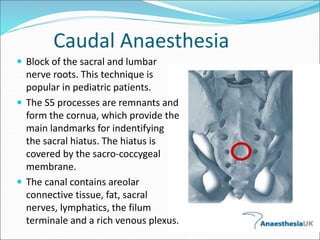
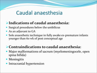
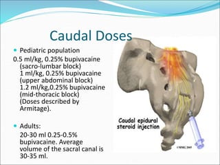
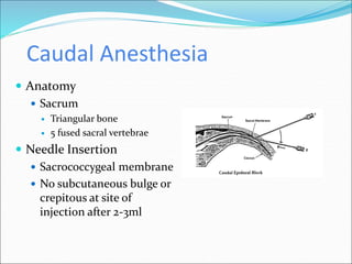
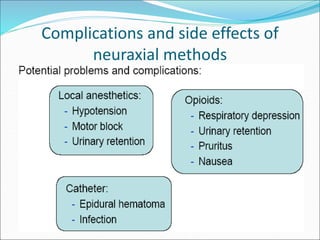
2. Neuraxial Anesthesia
Neuraxial anesthesia is a type of regional anesthesia
that involves injection of anesthetic medication in the
fatty tissue that surround the nerve roots as they exist
the spine (also known as an epidural) or into the
cerebrospinal fluid which surrounds the spinal cord
(also known as a spinal). This numbs the patient from
the abdomen to the toes and often eliminates the need
for general anesthesia.
3. HISTORY
1885 - J. Leonard Corning –
first spinal anesthetic was administered accidentally
The needle was made of gold
1898 - August Bier - first planned spinal anesthesia for
surgery
In 1921, Spanish military surgeon Fidel Pagés (1886–
1923) developed the modern technique of lumbar
epidural anesthesia
Robert Andrew Hingson (1913–1996), working at
the United States Marine Hospital in New York,
developed the technique of continuous caudal
anesthesia.
4. Advantages over Regional
Anaesthesia over GA
Safe, reliable technique in patients at risk of apnoea, bradycardia,
desaturation, cardiac or respiratory complications after GA
Good alternative for day care surgeries
Minimal risk of postoperative respiratory depression
Limited stress response to surgery
Cost effective
7. Spinal Cord
Spinal Cord
Adult
Begins: Foramen Magnum
Ends: L1
Newborn
Begins: Foramen Magnum
Ends: L3
Terminal End: Conus Medullaris
Filum Terminale: Anchors in sacral region
Cauda Equina: Nerve group of lower dural sac
8. Sagittal Section Through Lumber Vertebrae
Supraspinous
Ligament
(Outer most layer)
Intraspinous
Ligament
(Middle layer)
Ligamentum Flavum
(Inner most layer)
9. CONTRAINDICATIONS
Absolute
Patient Refusal
Infection At The Site Of Injection
Coagulopathy And Other Bleeding Disorders
Severe Hypovolemia
Increased Intracranial Pressure
Severe Aortic Stenosis
Severe Mitral Stenosis
10. CONTRAINDICATIONS
Relative
Sepsis
Uncoperative Patient
Preexisting Neurological Deficits
Severe Spinal Deformity
Controversial
Prior Surgery At The Site Of Injection
Complicated Surgery
Prolonged Operation
Major Blood Loss
16. Selection of equipments
Selection of block needles and catheters:
Block procedure Recommended device
Spinal
anaesthesia
Spinal needle (24-25 gauge; 30, 50 or 100 mm long,
Quincke bevel, stylet)
Caudal
anaesthesia
Short (25-30 mm) and short beveled (45-degree)
needle with stylet
Epidural
anaesthesia
Tuohy needle (22, 20, and 19/18 gauge); LOR
syringe and medium epidural catheter
PNB Insulated 21-23 gauge short beveled needles
17. FACTORS AFFECTING LEVEL
BARICITY OF DRUG
POSITION OF PATIENT
DOSE
SITE
AGE
CURVATURE OF SPINE
PATIENT HEIGHT
PREGNANCY
20. Complications of Spinal Anaesthesia
Hypotension
Bradycardia
Cardiac Arrest
Total Spinal Anesthesia
Neurological Complecations – Cauda Equina
Syndrome
Post Dural Puncture Headache
Infection
21. Backache
Inflammatory reaction due to tissue trauma
May result in back spasms
Short lived, analgesics, ice
May last a few weeks
Back ache may be a sign of serious complications
such as epidural/spinal hematoma, abscess
Careful evaluation to determine if a
common/benign complication or something more
serious
22. Postdural Puncture Headache
Caused by disrupting the integrity of the dura
Can occur due to: spinal anesthesia, “wet” tap with
epidural, epidural catheter migration, tip of the
epidural needle “indenting” the dura enough to cause a
leak.
Headache occurs due to leakage of CSF through the
dura
Decrease in intracranial pressure occurs due to the
leak
Upright position in the patient leads to traction on the
dura, tentorium, and blood vessels resulting in pain.
23. Postdural Puncture Headache- Symptoms
Onset is generally within 12-72 hours
Headache associated with upright position (i.e. sitting
or standing). Relief found with a supine position
Headache may be bilateral, frontal, retroorbital and/or
occipital with or without radiation to the neck
Described as “throbbing” or constant
May be associated with nausea and/or photophobia
Traction on the 6th cranial nerve can result in diplopia
and tinnitus
24. Postdural Puncture Headache- Conservative
Treatment
Hydration- theoretically helps to encourage the
production of CSF.
Analgesics- will decrease the severity of symptoms
and include acetaminophen and NSAIDS
Caffeine- Helps to decrease symptoms by
vasoconstriction of the cerebral vessels.
A dose of 300 mg of oral caffeine has been shown
to decrease the intensity of PDPH
Epidural blood patch.
25. Epidural Space
Space that surrounds the
spinal meninges
Potential space
Ligamentum Flavum
Binds epidural space
posteriorly
Widest at Level L2 (5-6mm)
Narrowest at Level C5 (1-
1.5mm)
26. Epidural Anatomy
Safest point of entry is
midline lumbar
Spread of epidural
anesthesia parallels
spinal anesthesia
◦ Nerve rootlets
◦ Nerve roots
◦ Spinal cord
27. Epidural Anesthesia
Order of Blockade
B fibers
C & A delta fibers
Pain
Temperature
Proprioception
A gamma fibers
A beta fibers
A alpha fibers
28. Epidural Anesthesia
Test Dose: 1.5% Lido with Epi 1:200,000
◦ Tachycardia (increase >30bpm over resting HR)
◦ High blood pressure
◦ Light headedness
◦ Metallic taste in mouth
◦ Facial numbness
◦ Note: if beta blocked will only see increase in BP not
HR
Bolus Dose: Preferred Local of Choice
◦ 10 milliliters for labor pain
◦ 20-30 milliliters for C-section
29. Epidural Anesthesia
Distances from Skin to Epidural Space
Average adult: 4-6cm
Obese adult: up to 8cm
Thin adult: 3cm
Assessment of Sensory Blockade
Alcohol swab
Most sensitive initial indicator to assess loss of temperature
Pin prick
Most accurate assessment of overall sensory block
30. Epidural Anesthesia
Complications
Penetration of a blood vessel
Hypotension (nausea & vomiting)
Intravascular catheterization
Back pain
Wet tap
Infection
31. Differences between Spinal and Epidural Anesthesia
Spinal anaesthesia Extradural Anaesthesia
Level: below L1/L2, where the spinal cord
ends
Level: at any level of the vertebral column.
Injection: subarachnoid space i.e punture
of the dura mater
Injection: epidural space (between
Ligamentum flavum and dura mater) i.e
without punture of the dura mater
Identification of the subarachnoid space:
When CSF appears
Identification of the Peridural space: Using
the Loss of Resistance technique.
Dosis: 2.5- 3.5 ml bupivacaine 0.5% heavy Doses: 15- 20 ml bupivacaine 0.5%
Onset of action: rapid (2-5 min) Onset of action: slow (15-20 min)
Density of block: more dense Density of block: less dense
Hypotension: rapid Hypotension: slow
Headache: is a probably complication Headache: is not a probable. 32
32. Caudal Anaesthesia
Block of the sacral and lumbar
nerve roots. This technique is
popular in pediatric patients.
The S5 processes are remnants and
form the cornua, which provide the
main landmarks for indentifying
the sacral hiatus. The hiatus is
covered by the sacro-coccygeal
membrane.
The canal contains areolar
connective tissue, fat, sacral
nerves, lymphatics, the filum
terminale and a rich venous plexus.
33. Caudal anaesthesia
Indications of caudal anaesthesia:
Surgical procedures below the umbilicus
As an adjuvant to GA
Sole anaesthetic technique in fully awake ex-premature infants
younger than 60 wk of post conceptual age
Contraindications to caudal anaesthesia:
Major malformations of sacrum (myelomeningocele, open
spina bifida)
Meningitis
Intracranial hypertension
34. Caudal Doses
Pediatric population
0.5 ml/kg, 0.25% bupivacaine
(sacro-lumbar block)
1 ml/kg, 0.25% bupivacaine
(upper abdominal block)
1.2 ml/kg,0.25% bupivacaine
(mid-thoracic block)
(Doses described by
Armitage).
Adults:
20-30 ml 0.25-0.5%
bupivacaine. Average
volume of the sacral canal is
30-35 ml.
35. Caudal Anesthesia
Anatomy
Sacrum
Triangular bone
5 fused sacral vertebrae
Needle Insertion
Sacrococcygeal membrane
No subcutaneous bulge or
crepitous at site of
injection after 2-3ml
36. Caudal Anesthesia
Post Operative Problems
Pain at injection site is most common
Slight risk of neurological complications
Risk of infection