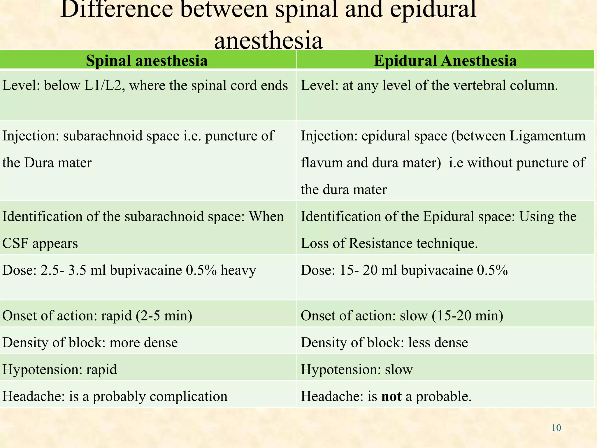
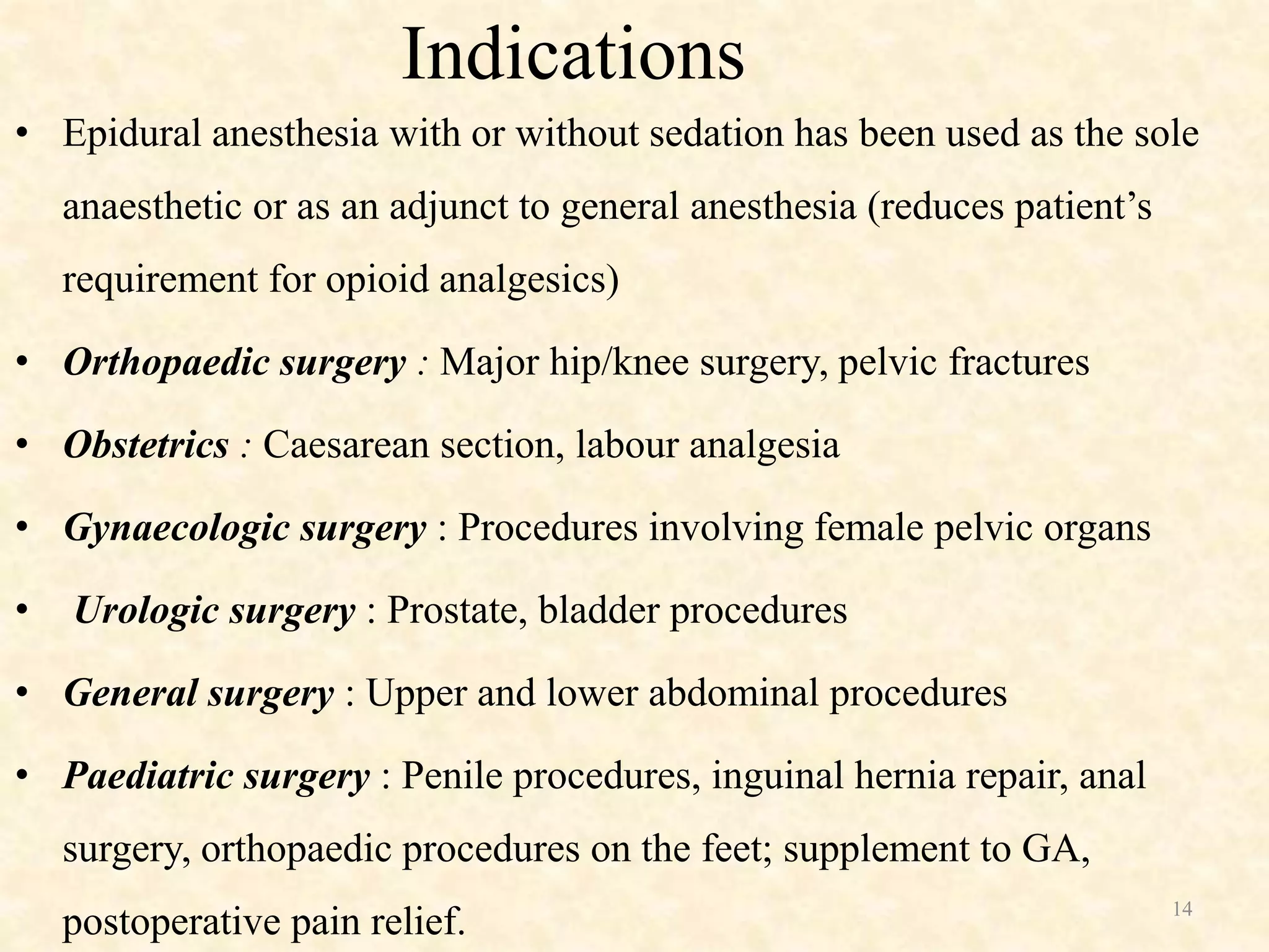
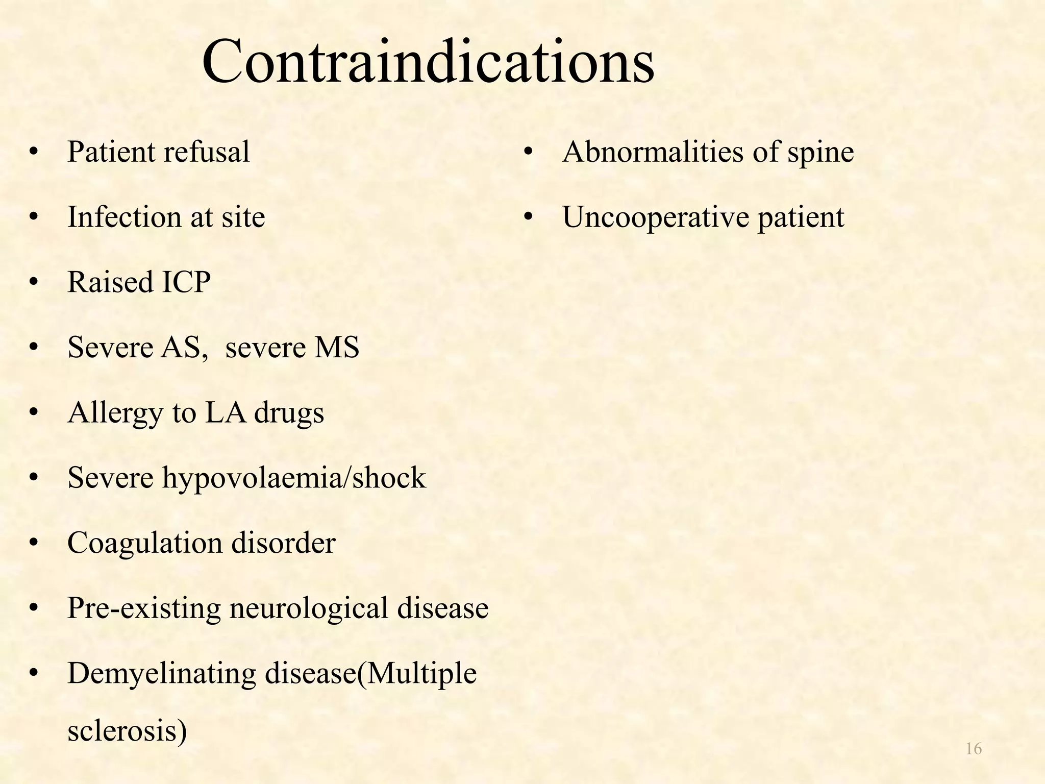
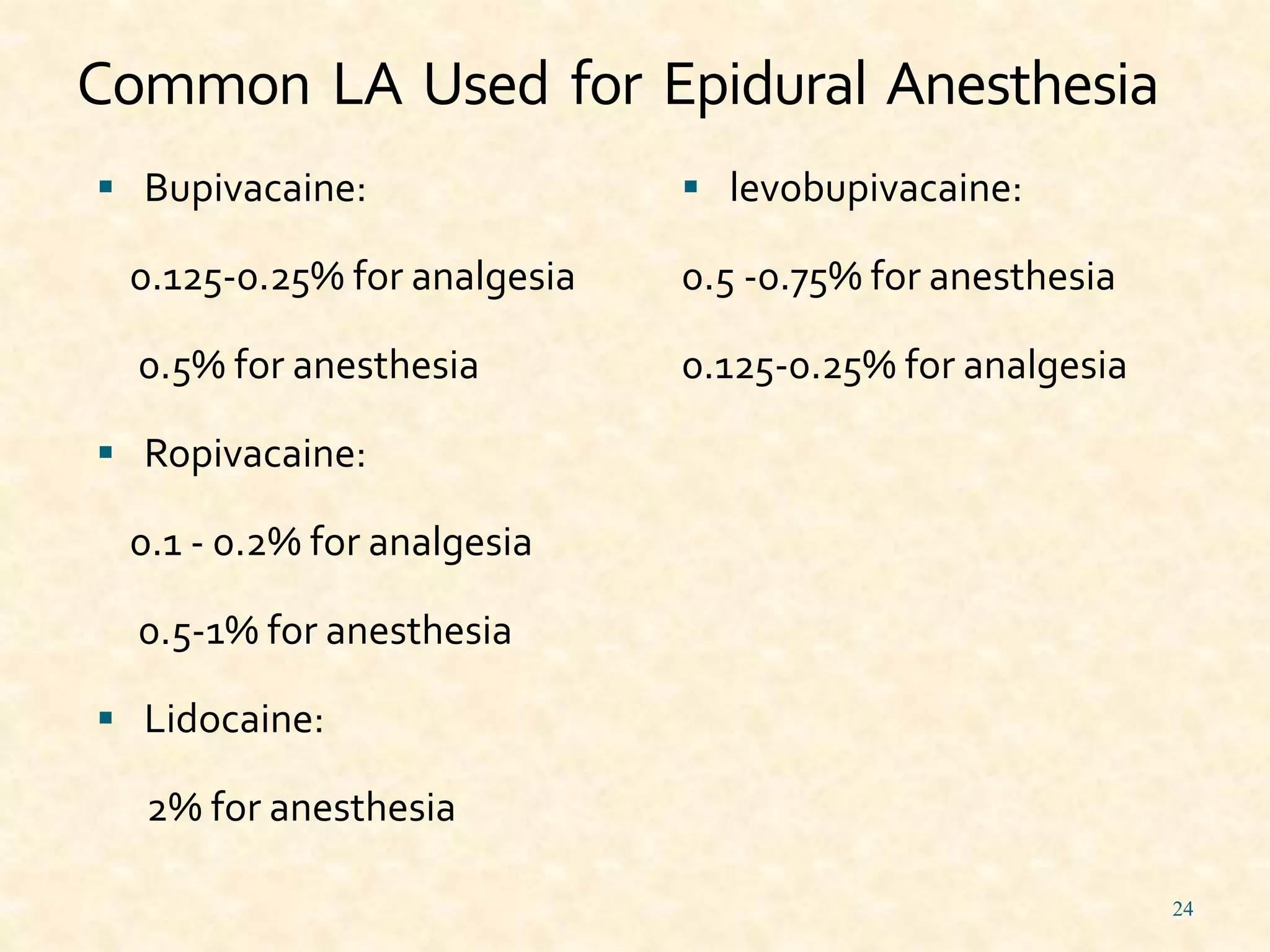
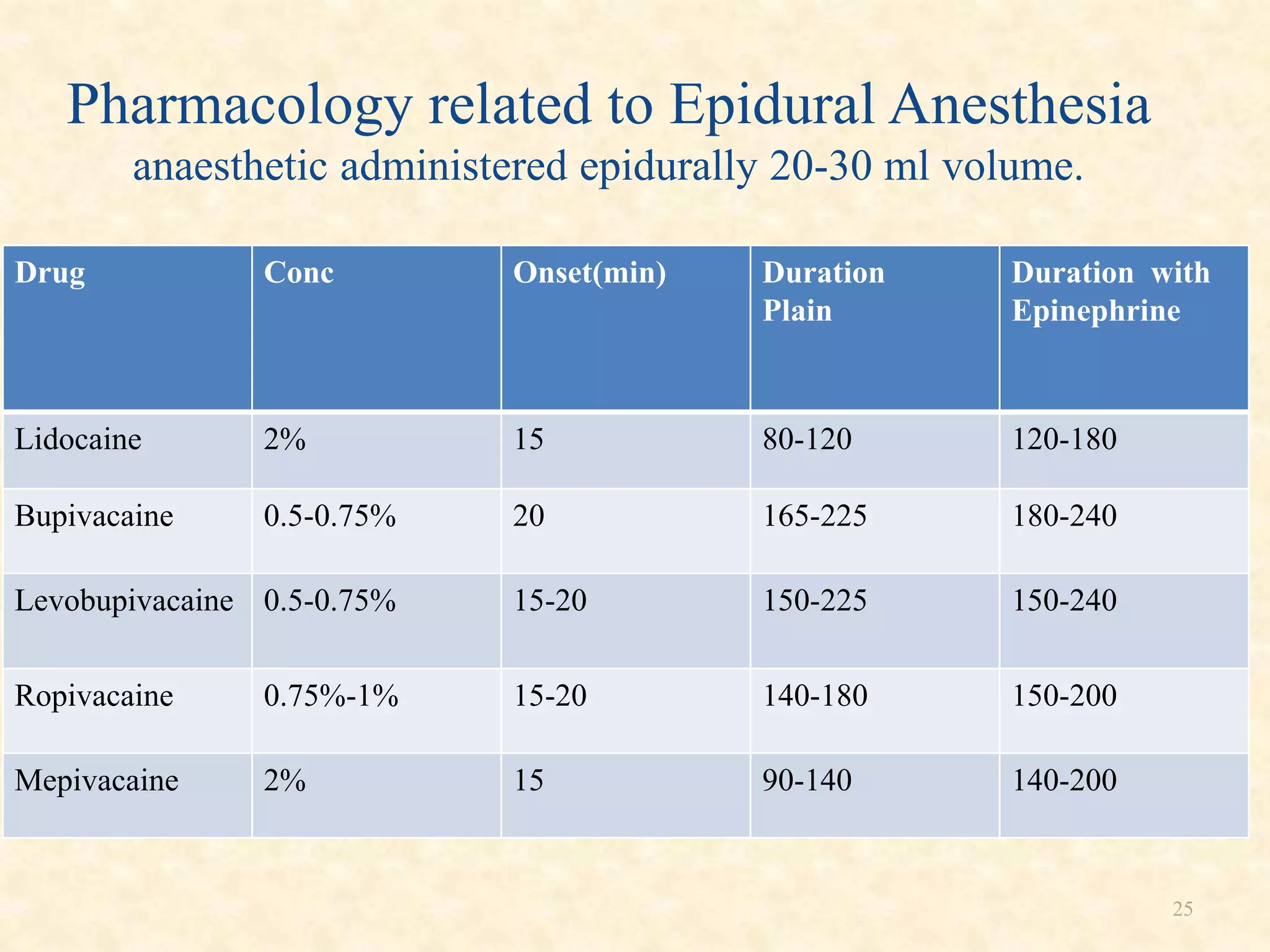
2. Epidural Anesthesia
• A Neuraxial technique that offers a wide range of applications.
• An Epidural block can be performed at the Lumbar, Thoracic,
Cervical and Caudal level
• Wide use of applications; Operative anesthesia, Obstetric
Anesthesia & Analgesia, Postop pain control and Chronic Pain
Management
• It can be used as a “Single Shot” or with a catheter that allows
intermittent boluses or a Continuous Infusion
3. 3
HISTORY
Fidel Pages first used Epidural anesthesia in human in
1921.
Tuohy introduced the needle used in Epidural Anesthesia
in 1945 .
4. Anatomy
• The Epidural space surrounds the Dura Mater posteriorly,
laterally and anteriorly
• Nerve roots travel in this space as they exit the spinal cord
laterally
• They then exit the foramen and travel peripherally to become
peripheral nerves carrying both afferent and efferent pathways
7. Physiology
Local anesthetics or other solutions injected into the epidural
space (steroids, narcotics) spread anatomically
Horizontal spread to the region of the Dural cuffs with
diffusion into the CSF and leakage through the intervertebral
foramen into paravertebral spaces
Longitudinal spread is preferentially cephalad in direction.
8. Physiology
Possible sites of anesthetic action include:
1 Intradural spinal roots
2 Dorsal and Ventral spinal roots
3 Dorsal root ganglia
4 The Spinal Cord
5 The Brain itself (by diffusion)
6 Paravertebral nerve roots
9. Physiology
Initial blockade is PROBABLY a result of anesthetic blockade
at the spinal roots within the Dural sleeves
The Dural Cuffs or Sleeves have a proliferation of arachnoid
villi and granulation tissue that effectively reduce the
THICKNESS of the dura mater facilitating rapid diffusion of
the LA from the Epidural space, through the Dura and into the
CSF surrounding the nerve roots
Then the local anesthetic diffuses into the nerve root itself,
producing anesthesia to that particular dermatome
10. 10
Difference between spinal and epidural
anesthesia
Spinal anesthesia Epidural Anesthesia
Level: below L1/L2, where the spinal cord ends Level: at any level of the vertebral column.
Injection: subarachnoid space i.e. puncture of
the Dura mater
Injection: epidural space (between Ligamentum
flavum and dura mater) i.e without puncture of
the dura mater
Identification of the subarachnoid space: When
CSF appears
Identification of the Epidural space: Using the
Loss of Resistance technique.
Dose: 2.5- 3.5 ml bupivacaine 0.5% heavy Dose: 15- 20 ml bupivacaine 0.5%
Onset of action: rapid (2-5 min) Onset of action: slow (15-20 min)
Density of block: more dense Density of block: less dense
Hypotension: rapid Hypotension: slow
Headache: is a probably complication Headache: is not a probable.
11. 11
Advantage
Minimise effect of surgery on Cardiopulmonary reserve.
patient with compromised Respiratory system
Morbid obesity
COPD
Elderly
Earlier mobilization
12. 12
Effective analgesia without taking systemic opioids.( analgesics
are given through catheter)
Reduce the incidence of Myocardial infarction
Surgery induced Stress response is decreased
Blood loss is less and
Everything can be regulated and changed by:
1) Choice of drug
2) Concentration of LA
3) Dosage
4) Level of Injection
13. 13
Disadvantage
• Risk of block failure
• Onset is slower
• Risk of infection
• Epidural hematoma
• Continous epidural catheter should not be used in the ward
if monitoring is not proper.
14. 14
Indications
• Epidural anesthesia with or without sedation has been used as the sole
anaesthetic or as an adjunct to general anesthesia (reduces patient’s
requirement for opioid analgesics)
• Orthopaedic surgery : Major hip/knee surgery, pelvic fractures
• Obstetrics : Caesarean section, labour analgesia
• Gynaecologic surgery : Procedures involving female pelvic organs
• Urologic surgery : Prostate, bladder procedures
• General surgery : Upper and lower abdominal procedures
• Paediatric surgery : Penile procedures, inguinal hernia repair, anal
surgery, orthopaedic procedures on the feet; supplement to GA,
postoperative pain relief.
15. 15
• Vascular surgery : Vascular reconstruction of the lower limb vessels,
amputations involving the lower extremities.
• Thoracic surgery : Postoperative analgesia, combination with GA to
reduce GA requirements.
• Diagnosis and management of chronic pain : Chronic benign pain-
Cervical & lumbar radiculopathy, vertebral compression fracture ,
degenerative disc disease, peripheral neuropathy, low back pain,
pelvic pain syndrome.
• Cancer related pain- pain secondary to face, neck, shoulder, genital,
pelvic, perineal etc .malignancy. & chemotherapy related peripheral
neuropathy.
16. 16
Contraindications
• Patient refusal
• Infection at site
• Raised ICP
• Severe AS, severe MS
• Allergy to LA drugs
• Severe hypovolaemia/shock
• Coagulation disorder
• Pre-existing neurological disease
• Demyelinating disease(Multiple
sclerosis)
• Abnormalities of spine
• Uncooperative patient
17. 17
Physiologic Effects of Epidural Block
• Most physiologic effects of epidural block stem from Autonomic
Blockade due to action of LA on autonomic nerve fibres of the
spinal cord.
• The actions mostly pertain to either Blockage of Sympathetic
outflow or Unopposed dominance of Parasympathetic outflow.
18. 18
Cardiovascular system
Block below T5
• Sympathetic blockade venodilation in blocked segments
venous return CO hypotension
• The compensatory mechanism for the decrease in mean arterial
pressure causes
1)reflex vasoconstriction above the level of the block
2)release in catecholamines from the adrenal medulla.
Block above T4 (cardiac sym fibres T1-T4)
so profound hypotension and bradycardia ,d/t high level of
sympathetic blockade.
19. 19
Respiratory system
Minimal impact on Pulmonary function in normal & healthy patient,
even in case of high thoracic block
There is concern regarding the use of epidural blockade in patients
with severe chronic lung disease dependent on accessory muscle
function to maintain adequate ventilation, because, paralysis of
respiratory muscles from epidural analgesia can occur.
20. 20
Gastrointestinal system
Lumbar segments (T5-L1) for major abdominal surgeries
vagal dominance leads to increased secretions, inc peristalsis and
contracted gut
Stable visceral perfusion prevents decrease in intramucosal pH,
post-op period epidural analgesia has a protective effect on gastric
mucosa
Thoracic segments (T1-T5) as adjunct to GA in thoracic / cardiac /
abdominal surgeries
Segmental sympatholysis creating an increase of sympathetic
activity in segments below the block leading to impaired splanchnic
blood flow has been a concern.
Nausea is a common problem (20%) – treat with atropine
21. 21
Renal system
Block both Sympathetic & Parasympathetic fibres
Loss of control of bladder function
Urinary retention
Endocrine system
• Abolishes stress response to surgery.
• Decreased release of catecholamines, GH, cortisol, TSH,
ADH, vasopressin, renin, angiotensin, norepinephrine
22. Epidural needle
Epidural needles have larger diameter than Spinal needle.
Typically sized of 16-19 gauge.
1 Tuohy & Hustead needle –with gently curve of 15-30°
degree.
2 Crawford needle- with straight tip.
22
23. 23
Epidural needle
Epidural needles have larger diameter than Spinal
needle.
Typically sized of 16-19 gauge.
1 Tuohy & Hustead needle –with curved tip
2 Crawford needle- with straight tip
24. Common LA Used for Epidural Anesthesia
Bupivacaine:
0.125-0.25% for analgesia
0.5% for anesthesia
Ropivacaine:
0.1 - 0.2% for analgesia
0.5-1% for anesthesia
Lidocaine:
2% for anesthesia
levobupivacaine:
0.5 -0.75% for anesthesia
0.125-0.25% for analgesia
24
25. 25
Pharmacology related to Epidural Anesthesia
anaesthetic administered epidurally 20-30 ml volume.
Drug Conc Onset(min) Duration
Plain
Duration with
Epinephrine
Lidocaine 2% 15 80-120 120-180
Bupivacaine 0.5-0.75% 20 165-225 180-240
Levobupivacaine 0.5-0.75% 15-20 150-225 150-240
Ropivacaine 0.75%-1% 15-20 140-180 150-200
Mepivacaine 2% 15 90-140 140-200
26. 26
Pre-op preparation
• PAC
• Informed consent
• Equipped for airway management & resuscitation
• INR & aPTT, Platelets counts should be with in normal range
• Monitor BP & HR
• IV access
27. 27
Performing the procedure
Position of patient- Careful attention to the patient’s position is
essential to successful placement of the epidural needle and
catheter.
Depending on the patient’s medical status, weight, and ability to
cooperate, the sitting or lateral decubitus position can be used.
Easier in sitting position.
Approach - Four common approaches to the epidural space are
possible:
1. Midline,
2. Paramedian,
3. Taylor (modified paramedian),
4. Caudal
28. Technique
• The most commonly performed Epidural is a Lumbar Epidural,
followed by a Caudal, then Thoracic and finally Cervical.
• Today most high thoracic and cervical epidurals are performed under
flouroscopic guidance by pain specialists as it takes a greater level of
skill to successfully perform those procedures.
31. 31
• Needle angulation required to accomplish epidural blockade
in the high thoracic/low thoracic/lumbar regions.
A: High thoracic region. B: Low thoracic region. C: Lumbar
region.
32. 32
Locating the Epidural space
• All aseptic precaution is taken.
• Skin is infiltrated with local anesthetic in to desired space(identified).
• Needle is advanced slowly, feel of increase resistance.
• 3 methods are used to identify Epidural space-
Loss of resistance (to with air or saline):-As needle
reaches Epidural space Loss of Resistance is felt less.
LORS Vs LORA:
• LORA is associated with nerve root compression, pneumocephalus and
greater incidence of incomplete analgesia, paresthesia and venous air
embolism.
• LORS is associate with reduces incidence of dural puncture in adult, while
in pediatric patients, dural puncture incidence are more.
• LORA is safer than LORS in children less than 2 yrs old.
33. Hanging drop method:-As needle reaches Epidural space
Hanging drop is sucked in d/t negative pressure.
In cervical region, negative pressure poorly reliable and
only useful in sitting position.
The negative intra-thoracic pressure may influence the
pressure in epidural spaces in thoracic region and should
be maximal during inspiration.
Ultrasonography / Fluoroscopy
36. Single –end hole catheter Spring wire-reinforced
catheter
Closed tip, multiple-side
hole catheter
Types of epidural catheter
37. 37
Catheter placement
• The catheter is made of a flexible, calibrated, durable,
radiopaque plastic .
• Typically, 19-or 20- gauge catheter is introduced through 17-or
18- gauge epidural needle.
• Catheter is threaded through needle after placing in space.
• Needle is withdrawn over the catheter.
• 4-6 cms catheter remain in epidural space. Threading more
catheter may increase the likelihood of catheter malposition.
• Catheter is firmly secured to skin with surgical tape.
40. CAUTION
NEVER pull the catheter back through the needle
once it has been inserted
It is possible to catch the catheter on the needle tip and shear or
cut the tip off
Then it becomes a permanent new addition to the epidural space
and will be there for the rest of the patient’s life!!!!
41. 41
Epidural Dosing
As a general guideline,
1. 1–2 mL per segment in a lumbar epidural,
2. 0.7 mL per segment in a thoracic epidural, and
3. 3 mL per segment for a sacral/caudal epidural
is used as an initial loading dose.
• Test Dose
• Incremental Dosing
• Aspiration to check for blood or CSF before each dose.
• After the initial loading dose, one quarter to one third of the amount
can be administered 10–15 min later to intensify the sensory block.
The overall level of the block will not be significantly increased
with this method.
42. 42
Test Dose
• The purpose of the “test dose” is to make sure that the catheter is not in
the subarachnoid, intravascular, or subdural space.
• The classic test dose combines 3 mL of 1.5% lidocaine with 15 mcg of
epinephrine.
• The intrathecal injection of 45 mg of lidocaine will produce a
significant motor block consistent with spinal anesthesia.
• A change in heart rate of 20% or greater is an indication of
intravascular injection warranting the removal and replacement of the
catheter.
• If the heart rate does not increase by 20% or greater, or if a significant
motor block does not develop within 5 min of administering the test
dose, it is considered negative.
• False-ve if pt is on β blocker, false +ve in pregnancy if coincides with
labour pain.
43. 43
Incremental Dosing
• Its purpose is to avoid excessively high anaesthetic levels.
• The loading dose should be given in 5-mL aliquots through the
catheter, repeated at 3- to 5-min intervals, giving the clinician time to
assess the patient’s response to dosing.
• If at any time the patient demonstrates an exaggerated response,
further incremental doses should be withheld and the patient
reassessed.
44. 44
Factors affecting Epidural Anesthesia
Site of injection-
Lumbar- spread cranially more than caudally
Thoracic- spread evenly from site of injection
Upper thoracic & lower cervical fibers are comparatively resistant
d/t larger size of nerve roots-requires larger dose of LA.
Thoracic epidural space is smaller, require lower volume of drug.
Dose- 1-2 ml /segment.
Depends on volume & concentration of drug. Higher conc.
produces a profound motor and sensory block, whereas low conc. a
selective sensory block.
45. 45
Age - as patient age increases reduced size of intervertebral
foramina decreased epidural space size and compliance.
Decreased epidural fat necessitates decrease of dose in elderly.
Weight - There is little correlation between the spread of analgesia
and the weight of the patient.
In morbidly obese patients, there may be compression of the
epidural space secondarily to increased intra-abdominal pressure,
creating a higher block for a given dose of local anesthetic.
46. 46
Height - The correlation with height is usually not clinically
significant.
Ht. <5 ft 2 inch, reduce the dose to 1 ml/segment to be blocked.
Bromage dosing regime - Increasing the dose of local anaesthetic by
0.1 mL per segment for each 2 inch over 5 ft of height.
Addition of Vasoconstrictors - Epinephrine 5 mcg/ml (1:200000)
is most commonly added.
Prolongs duration of action by reducing the vascular absorption of
drug.
47. 47
Posture-
Block Ht. - Whether the patient is sitting or in the lateral position,
there is no significant difference in block height. This is explained by
the fact that gravity and soln. baricity are not intimately related to
block spread.
Onset, Duration & Density - slightly faster on the dependent side
when the epidural in placed with the patient in the lateral position
Pregnancy- Increased sensitivity to regional anesthetics leads to
faster onset time.
Engorgement of Epidural veins from caval compression leads to
increased incidence of blood vessel puncture during procedure.
48. Complications
Drug Related Complications-
• CNS toxicity
• CVS toxicity
Procedure Related Complications-
• Minor Back Pain
• Postdural Puncture Headache
• Subarachnoid Injection/High or Total Spinal
• Major Subdural Injection
• Sheering of catheter
Neurologic Complications-
• Spinal nerve neuropathy
• Transient neurological symptoms
• Anterior spinal artery syndrome
• Adhesive arachnoiditis
• Epidural hematoma
• Epidural abscess
49. 49
• Postdural Puncture Headache : Due to inadvertent dural
puncture.
TOC – Epidural blood patch.
Cosyntropin, ( ACTH analogue) .
Postulated mechanisms include increased CSF production via sodium
channels; aldosterone mediated salt and water retention, and possibly
increased β endorphin output.
One of the trials used 1 mg of cosyntropin for the prophylaxis of PDPH.
It showed more than 50% reduction in the incidence of PDPH
Mannitol-
It acts “ acute increases in blood osmolality decreases brain water
content (mainly in healthy brain tissue with intact blood brain
barrier)decrease brain bulk, intracranial pressure, increased intracranial
compliances.” decrease brain bulk, causes brain re-float in contracted
CSF volume.
It believe that re-floatation of brain is an important factor to alleviate
PDPH with other factors.
50. Epidural Analgesia for labour
• Mainstay for many years
• Only therapy providing complete analgesia for both stages
• LA T10-L1 (relieve pain of uterine contractions and cervical dilatation)
S2-S4 (relieve pain of vaginal and perineal distension)
Most commonly: Mid-lumbar midline placement
• Generally initiated when the parturient wants it & the obstetrician
approves it.
• Commonly accepted criteria for placement:
1. No fetal distress
2. Good regular contractions 3-4 min apart & lasting abt. 1 min
3. Adequate cervical dilatation i.e. 3-4cm
4. Engagement of the fetal head.
51. Maternal positioning
Lateral position :
Advantages -
• orthostatic hypotension less likely
• continuous FHR monitoring
Disadvantage –
• concealed aortocaval compression
Sitting position : Preferred in obese
NEVER SUPINE. AVOID AORTOCAVAL COMPRESSION AT ALL
TIMES
52. 52
Combined spinal-epidural anesthesia
• Introduced in 1939 when Soresi et al presented a paper on usage of
CSE in 200 patients.
• Technique combining both spinal and epidural.
Spinal component gives rapid onset and dense predictable block.
Epidural catheter is used to supplement insufficient subarachnoid
block (to increase height or duration of block) and also to provide
long-lasting analgesia.
reduces the incidence of several potential problems associated with
the conventional epidural technique, including incomplete (patchy)
blockade, motor block, and poor sacral spread.
53. 53
The sequential CSE technique may be particularly advantageous in
high-risk patients, such as in those with cardiac disease, when slower
onset of sympathetic blockade is desirable.
• Two approaches are mostly used:
1. Needle Through Needle
2. Needle Below Needle.
T10-L1 sc segments need to be blocked to relieve pain of uterine contractions and cervical dilatation.
S2-S4 sc segments need to be blocked to relieve pain of vaginal and perineal distension