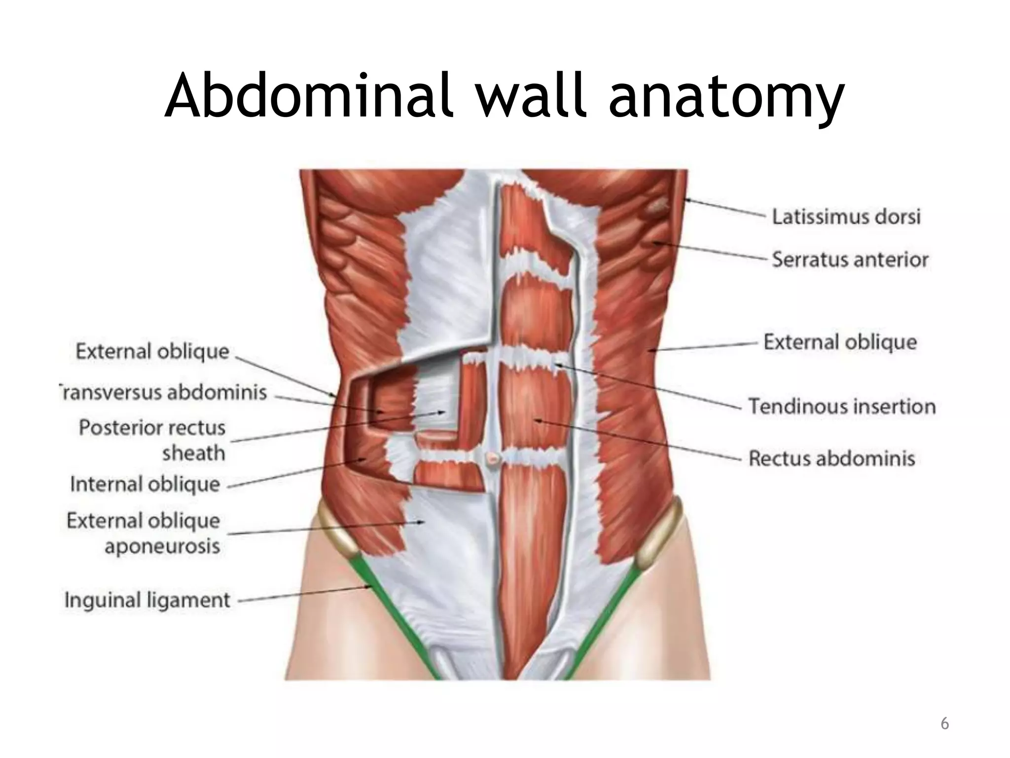
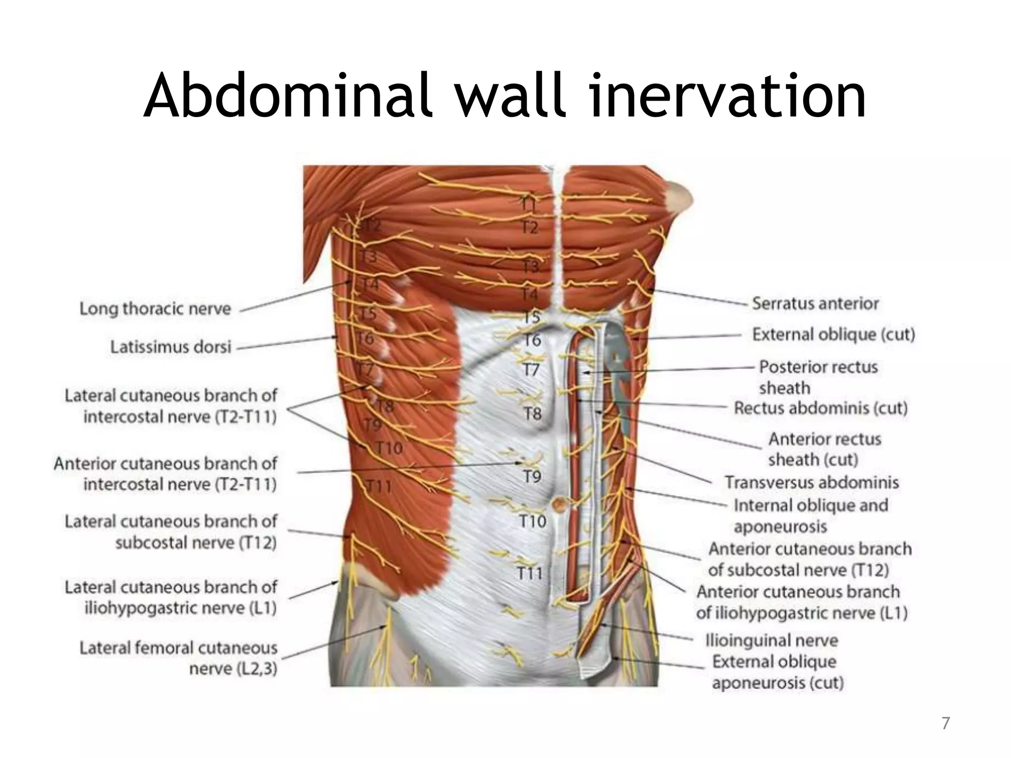
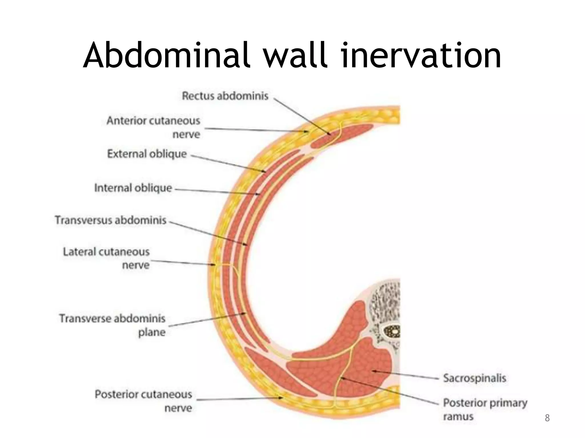
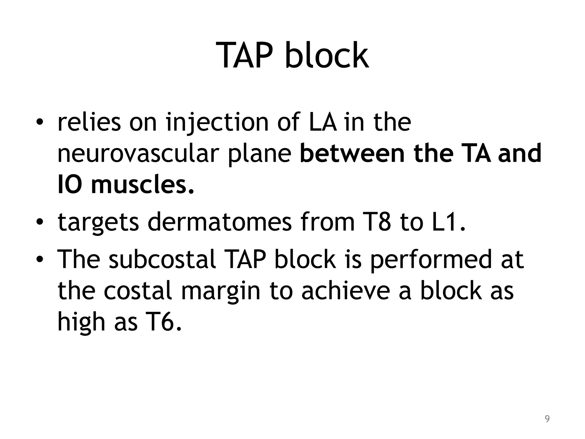
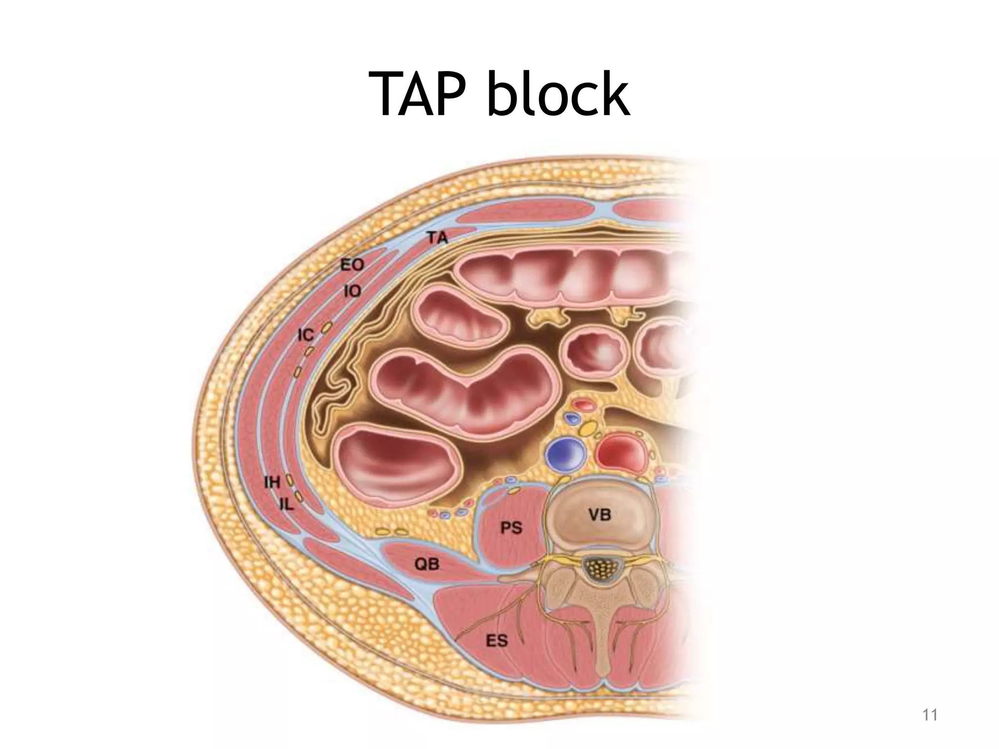
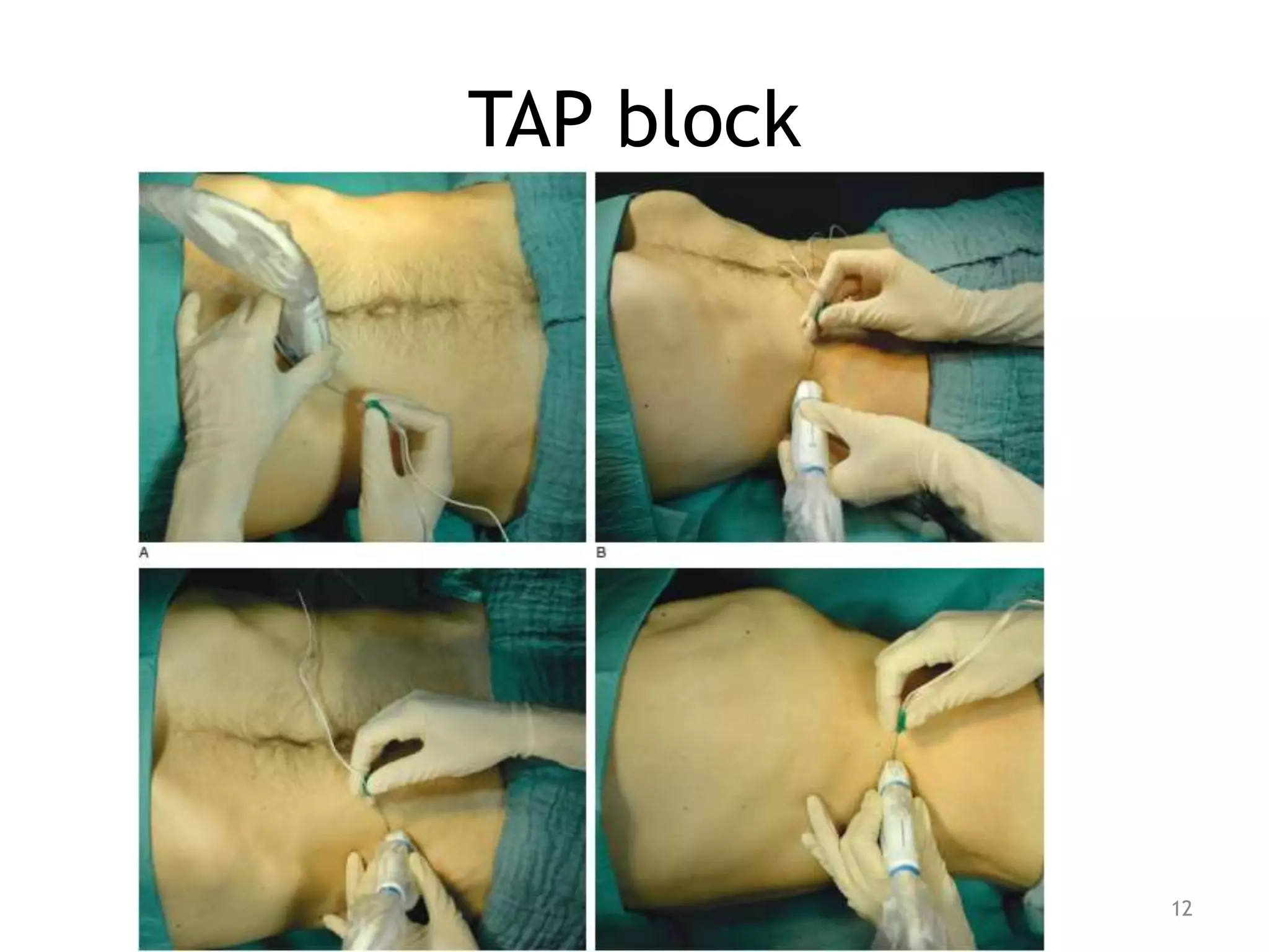
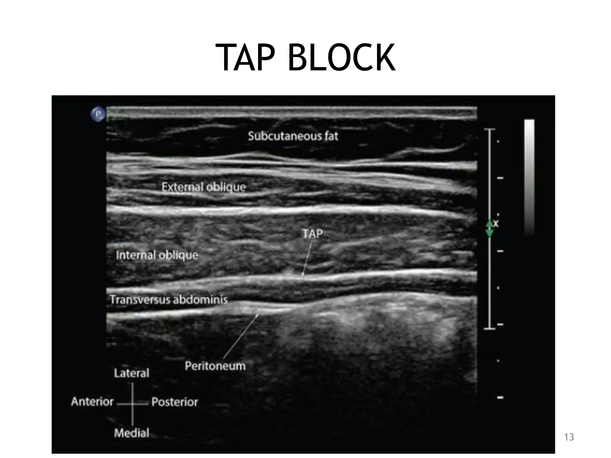
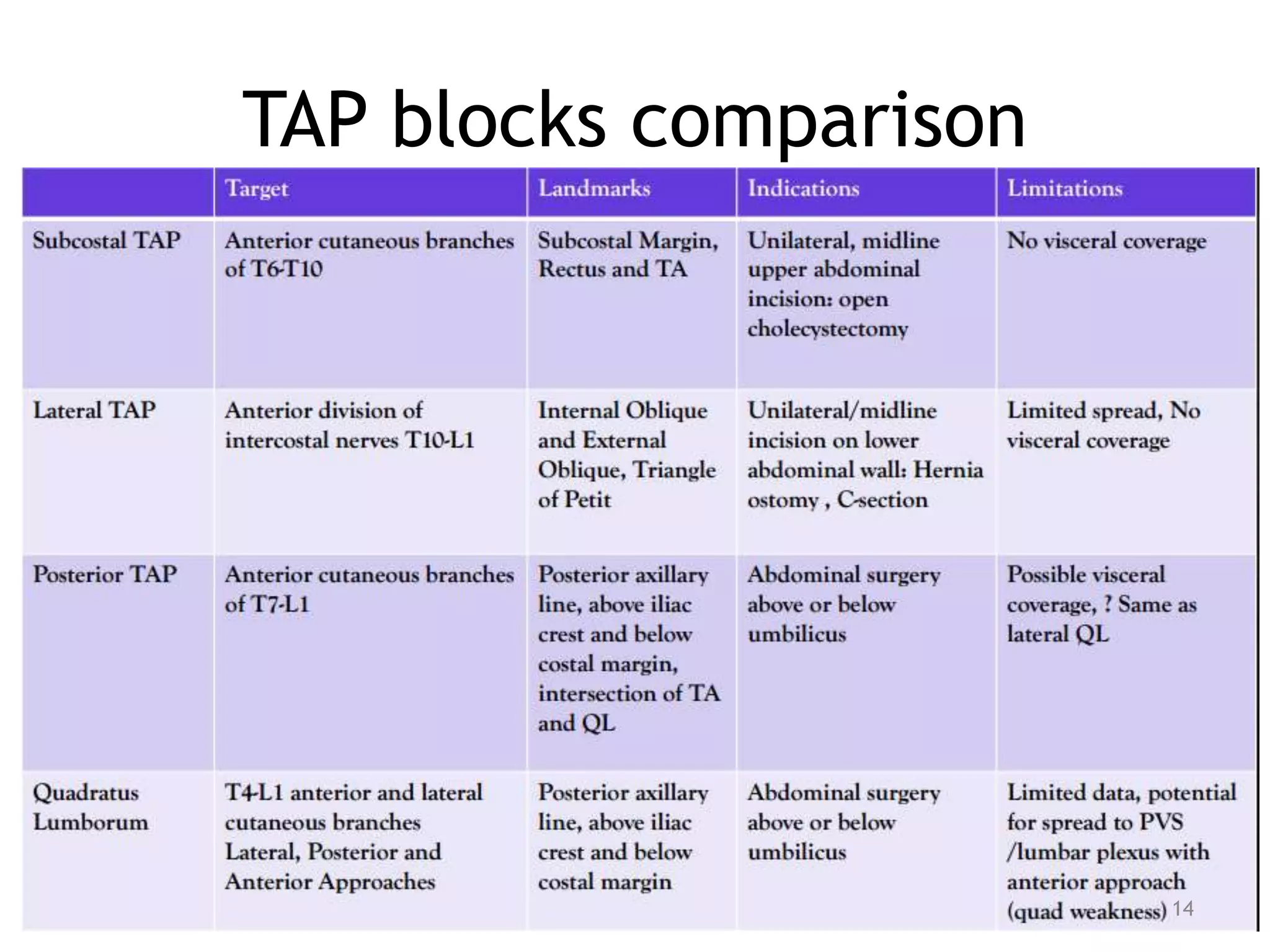
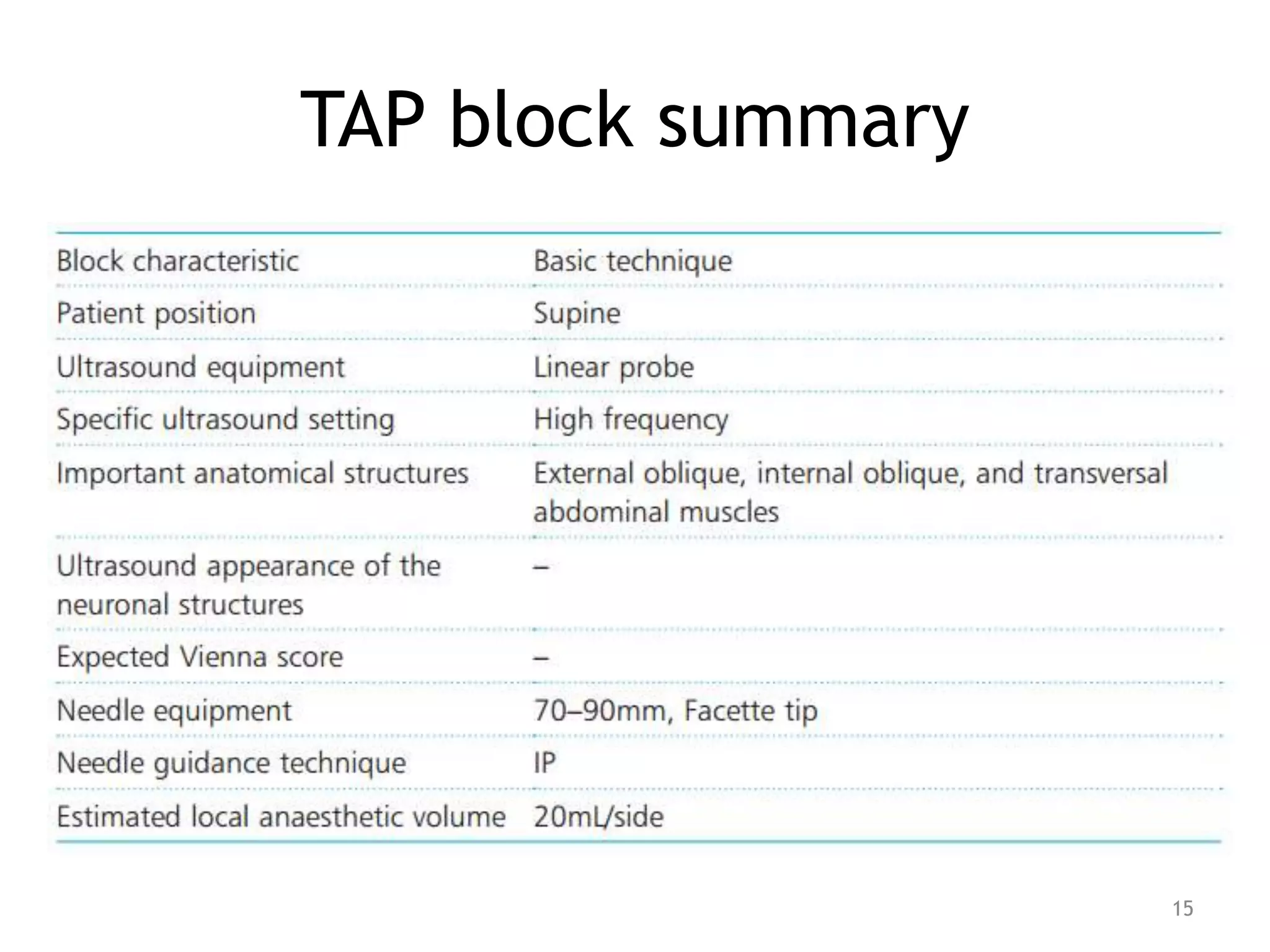
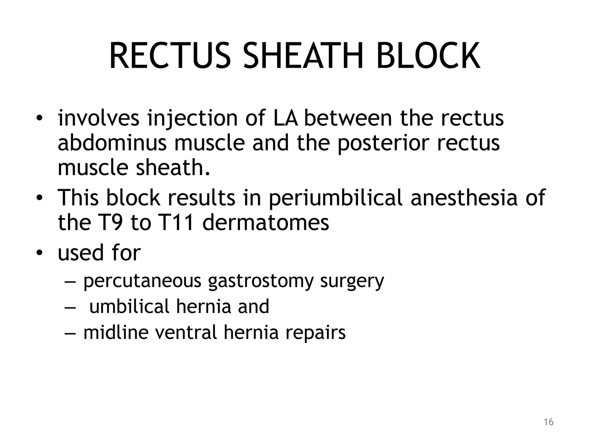
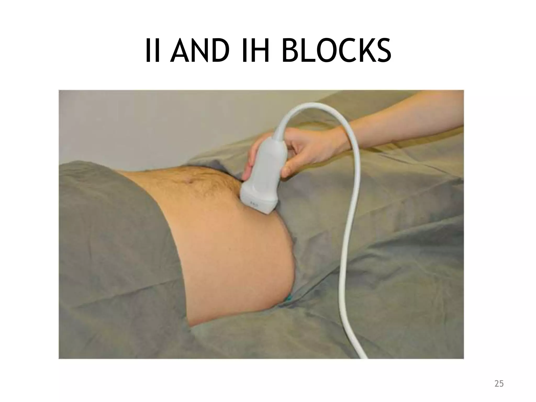
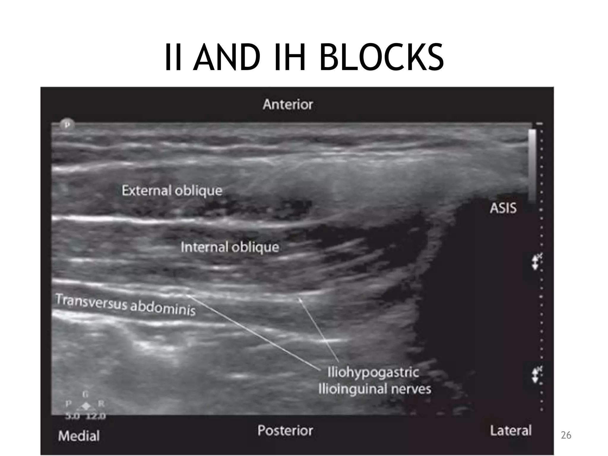
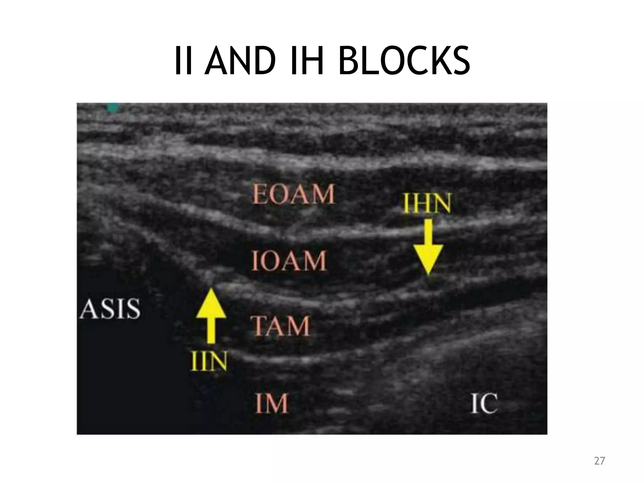
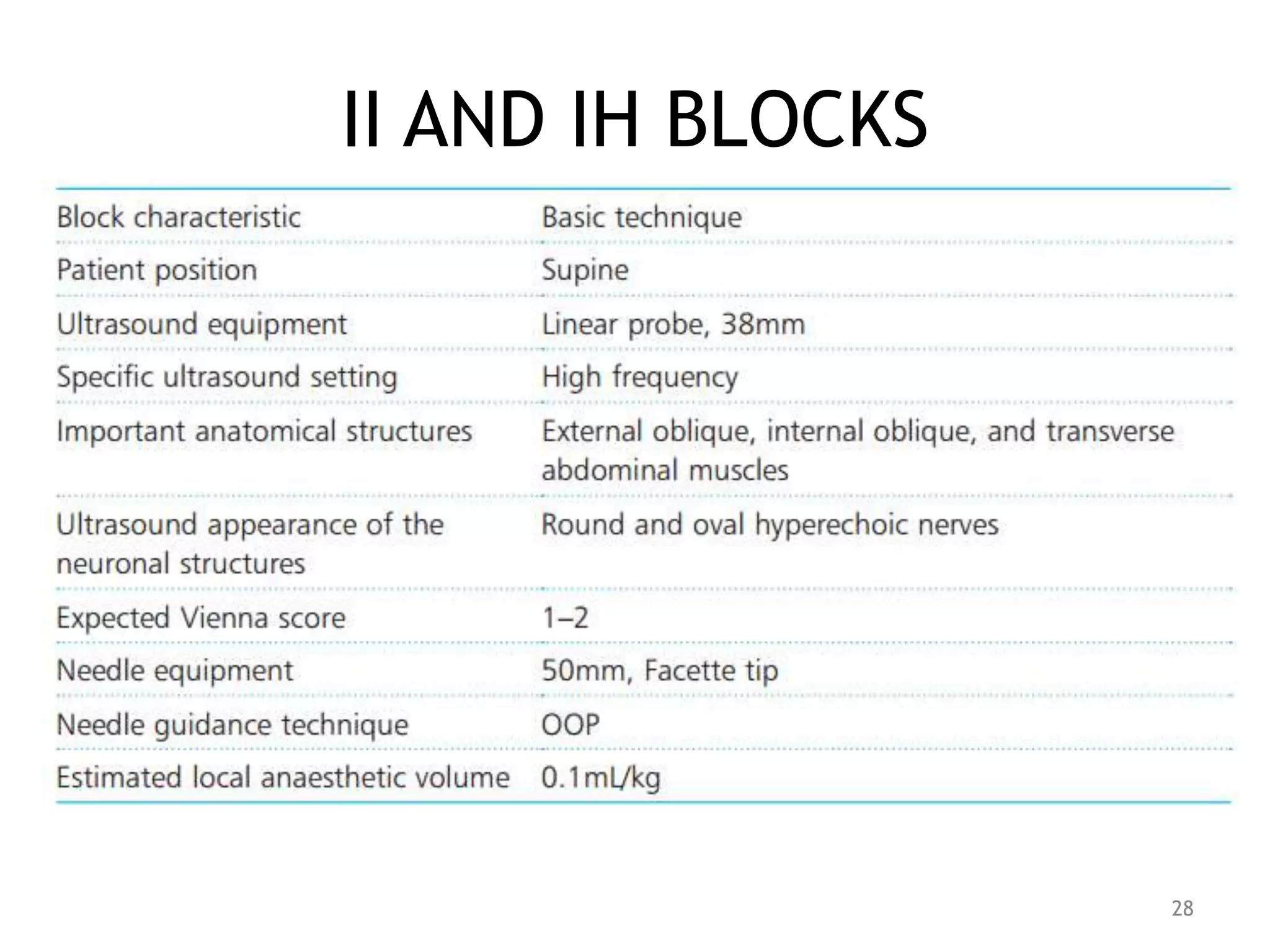
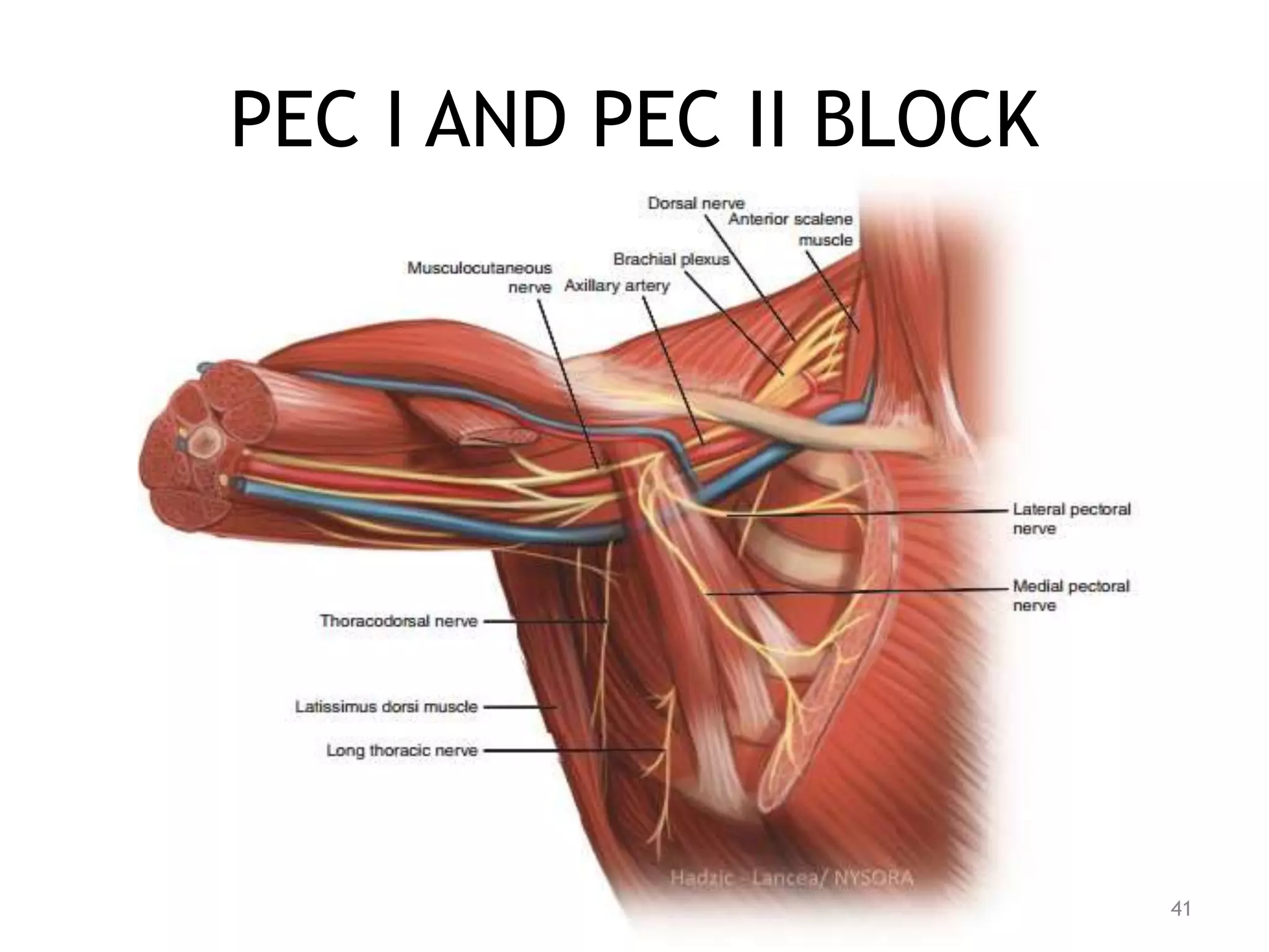
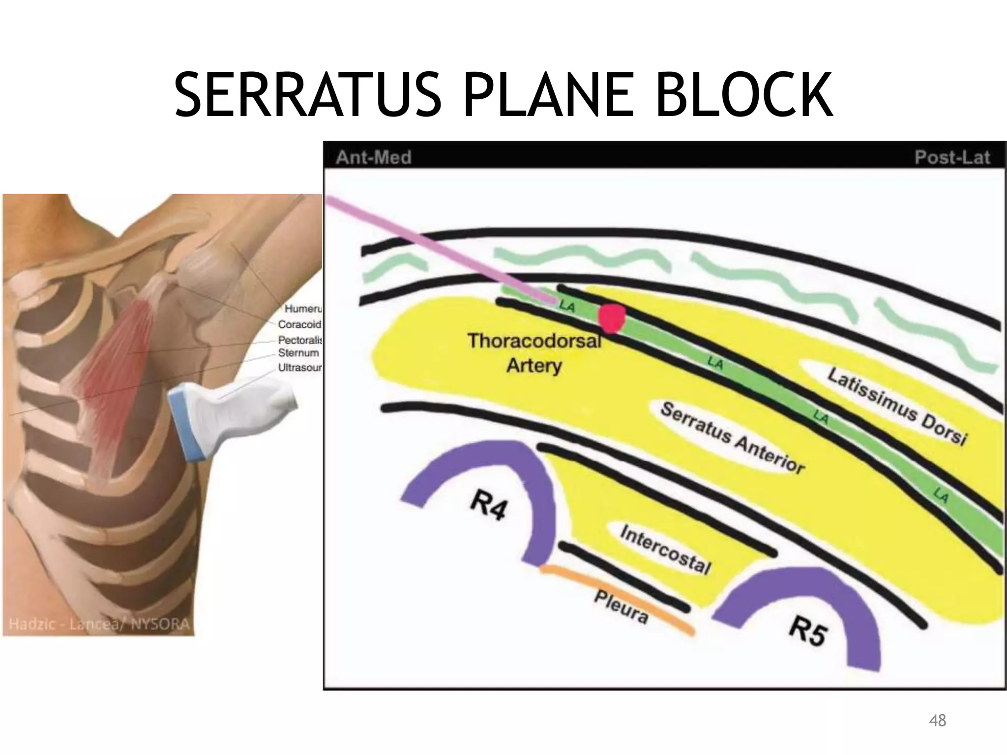
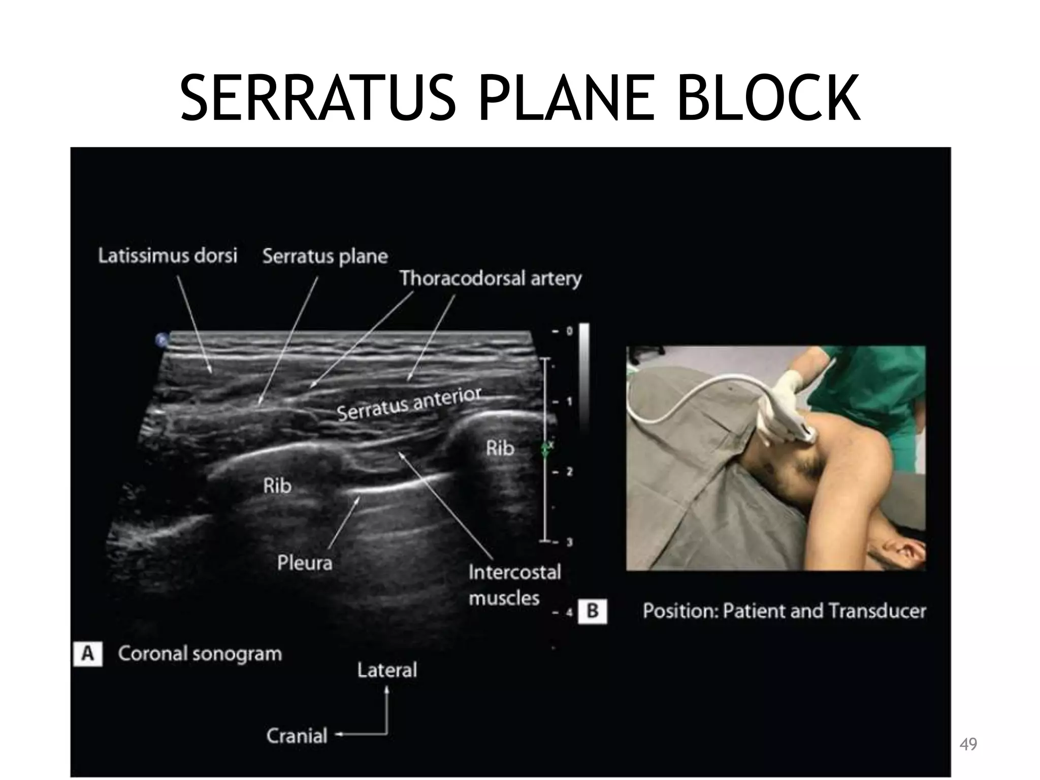
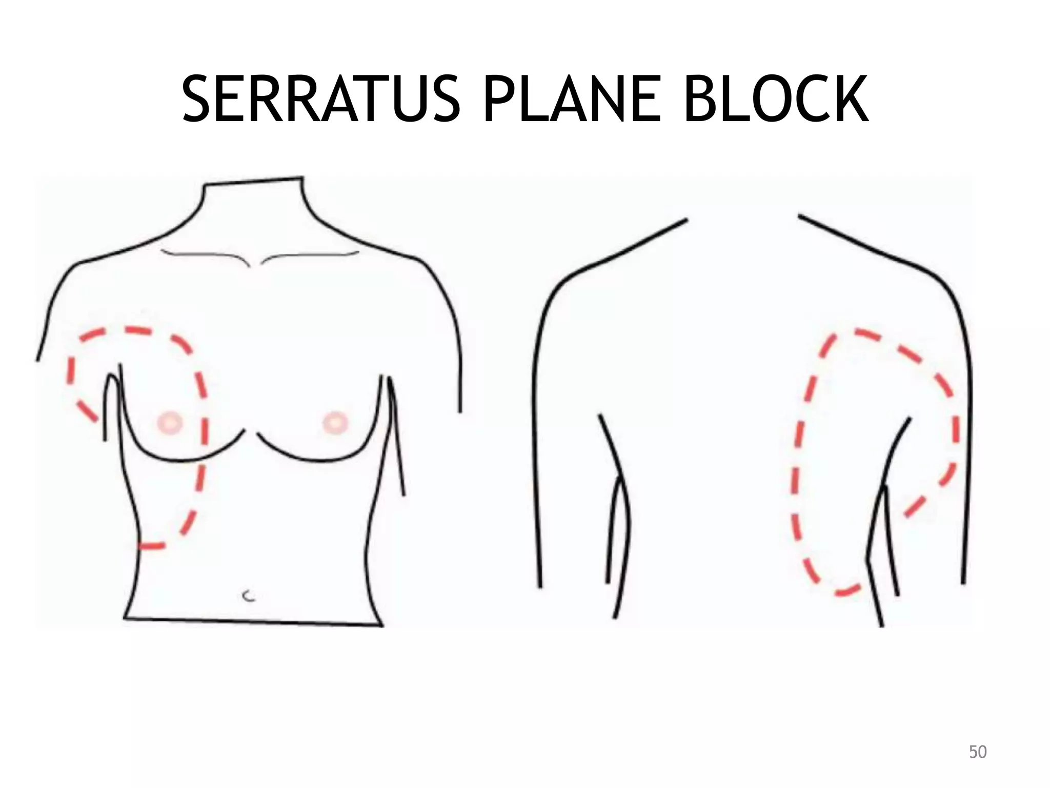
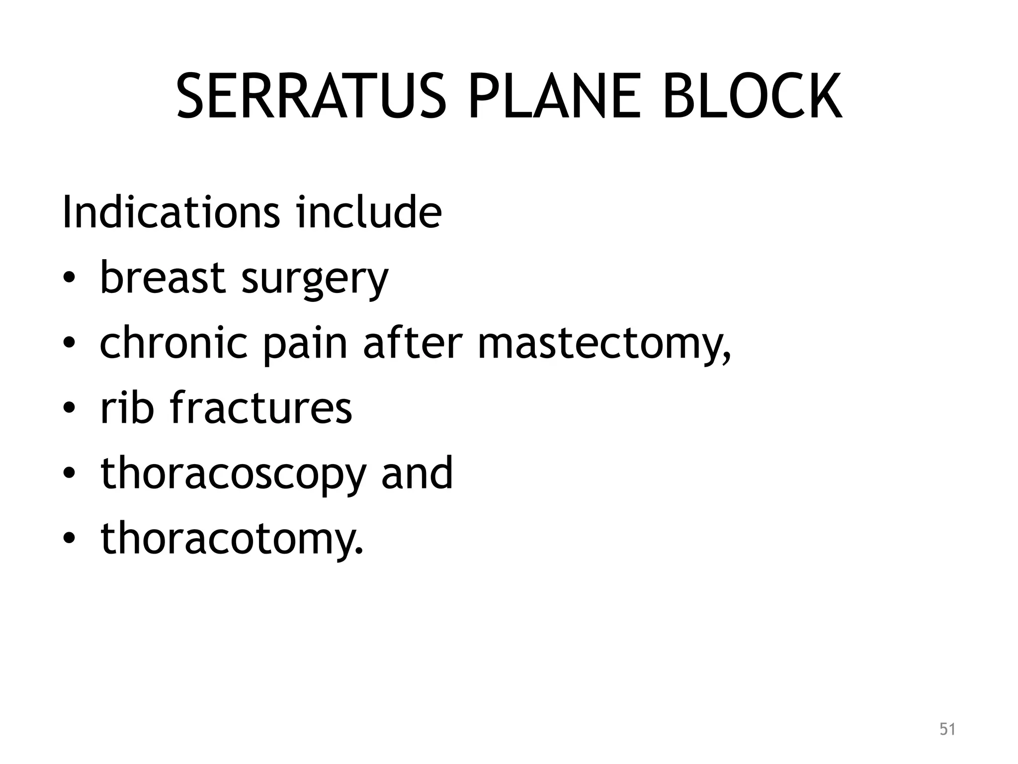
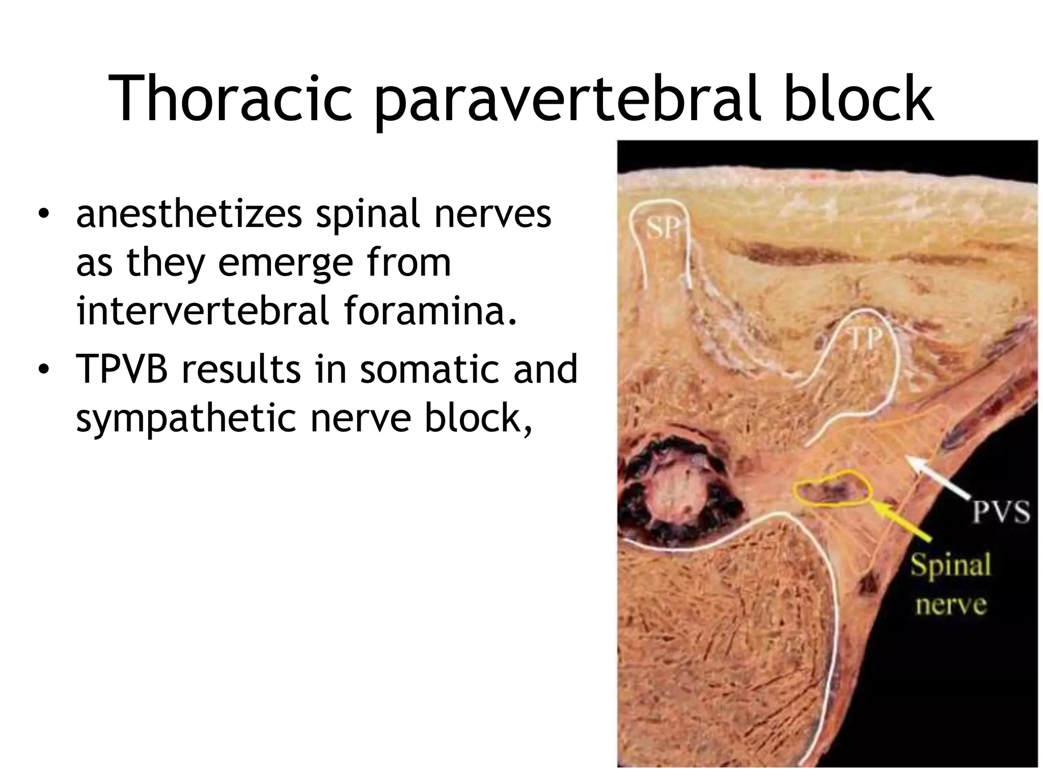
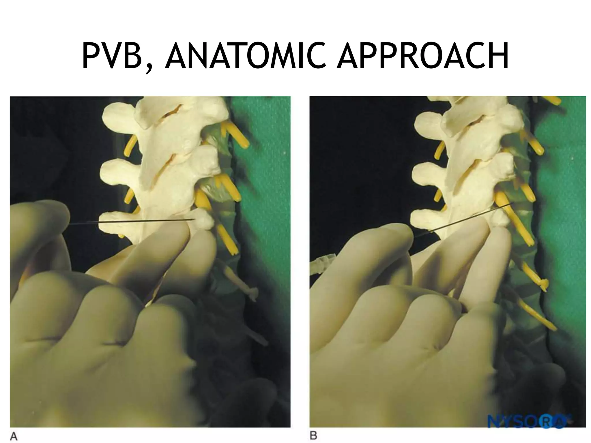
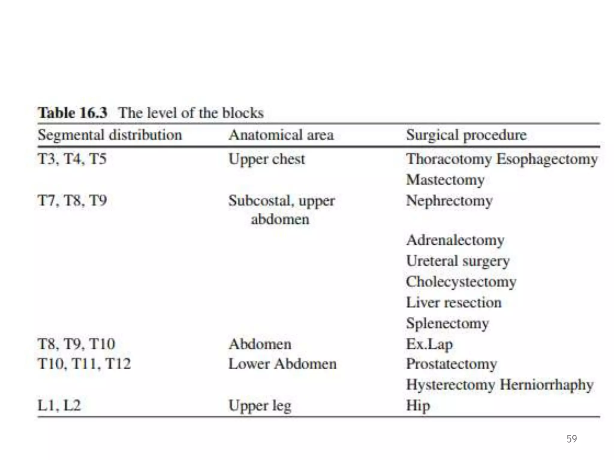
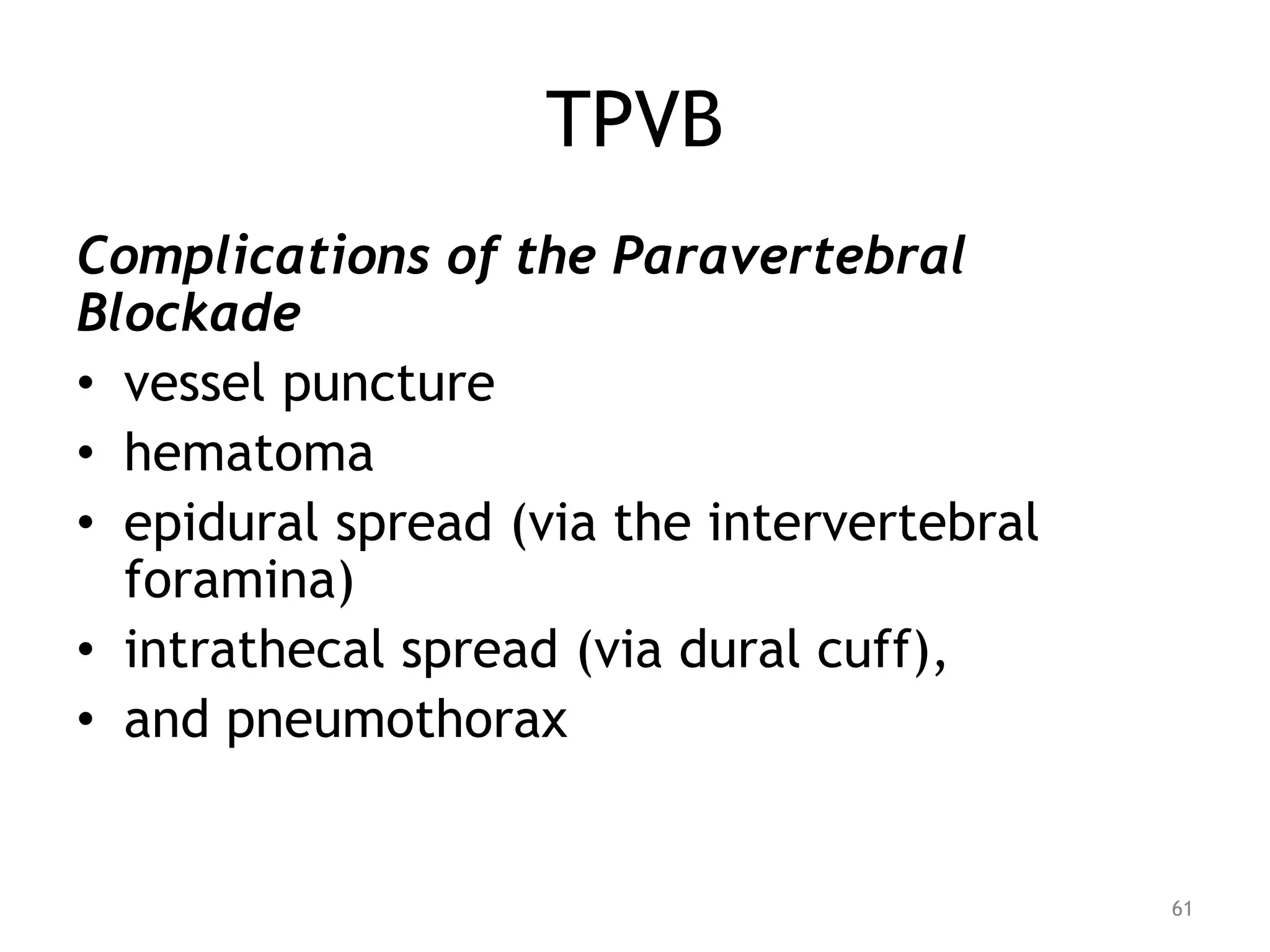
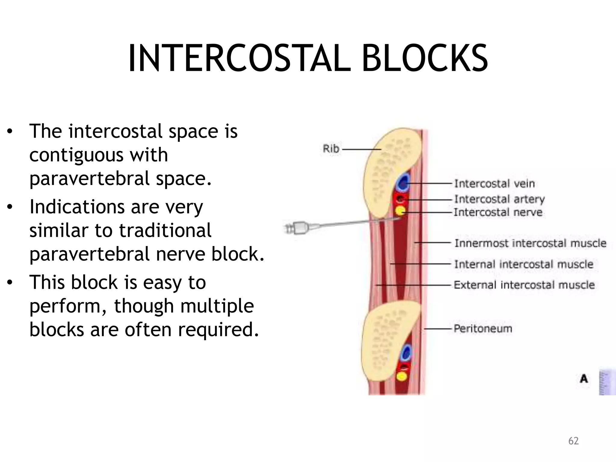
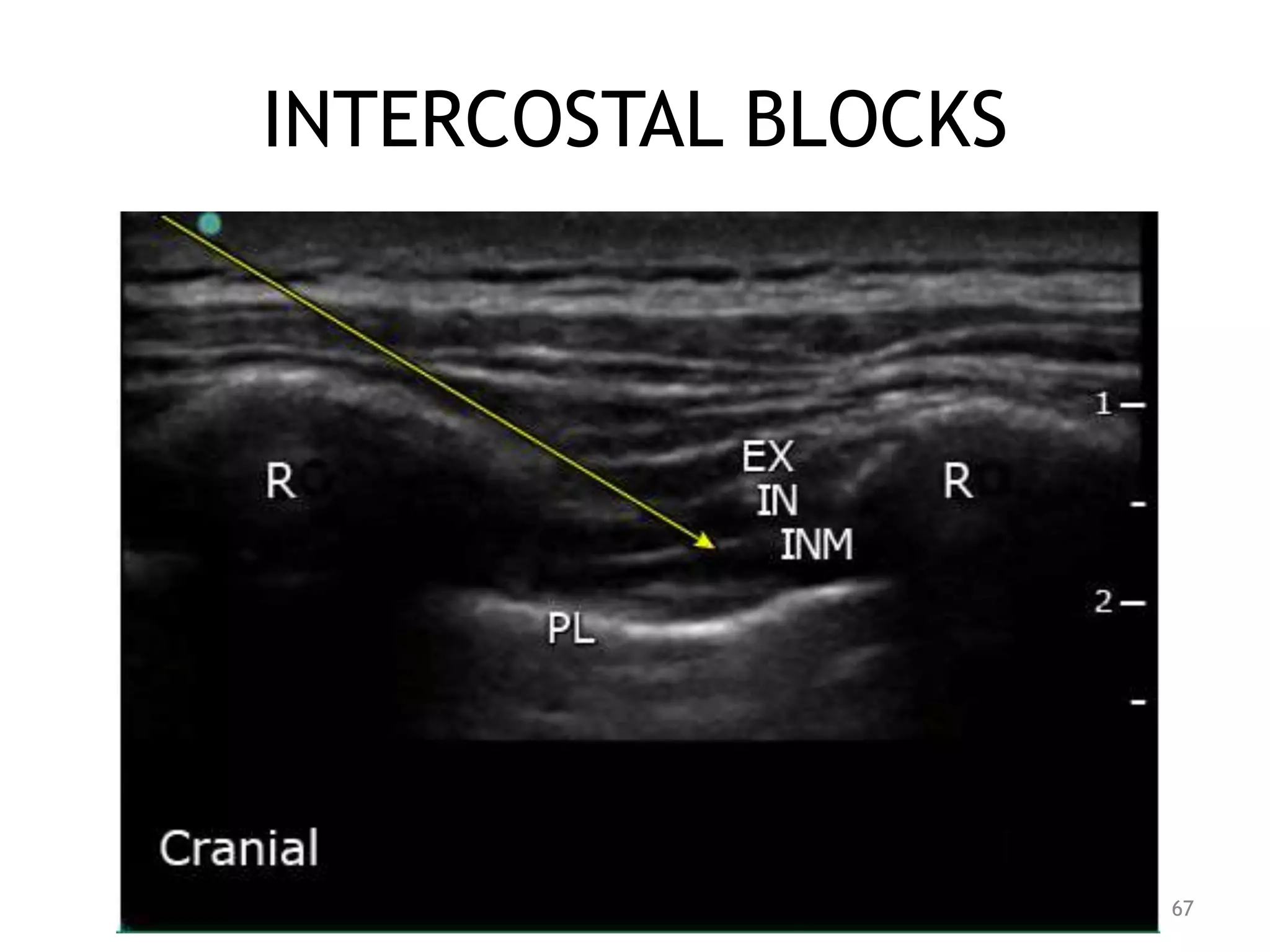
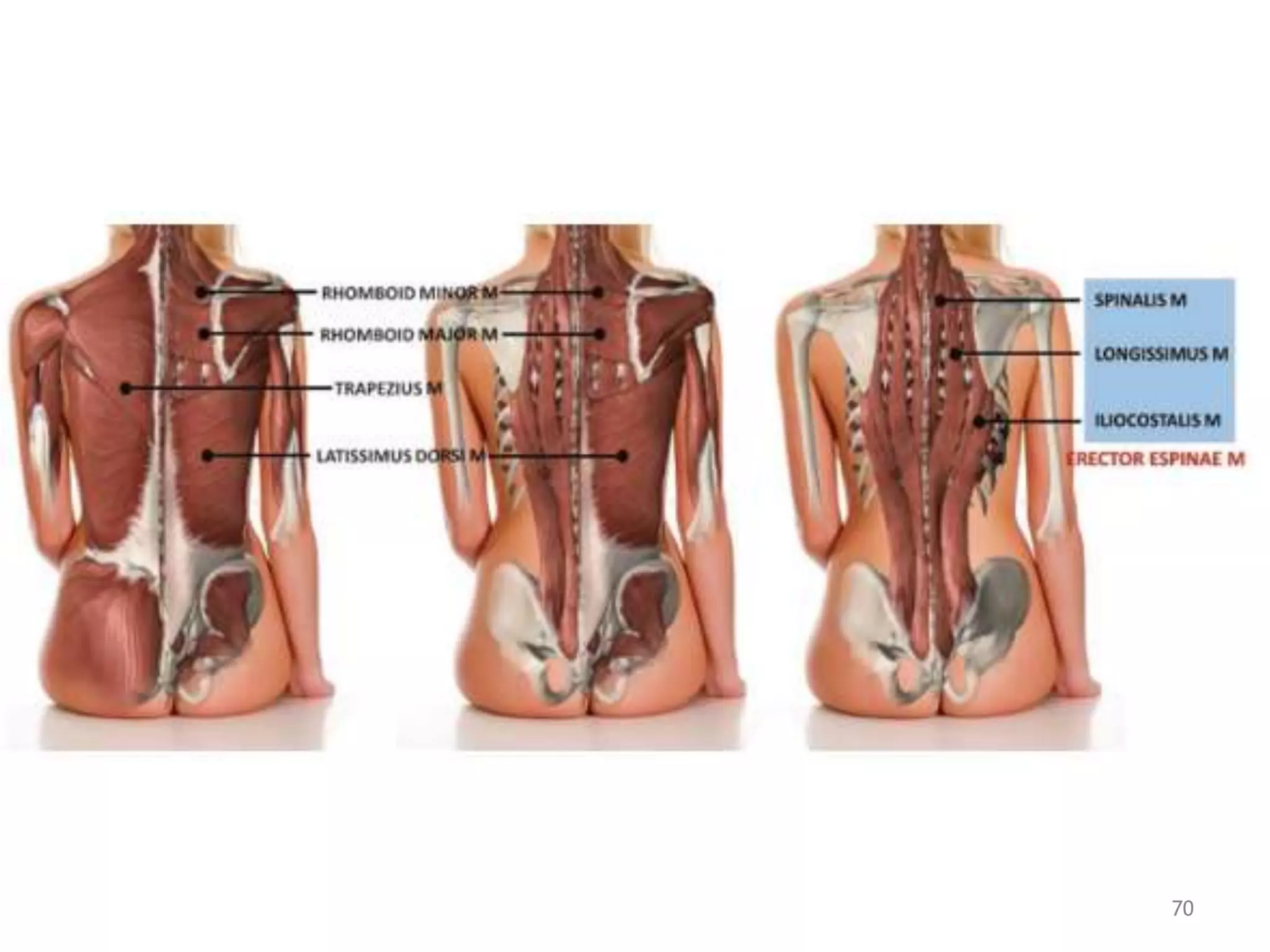
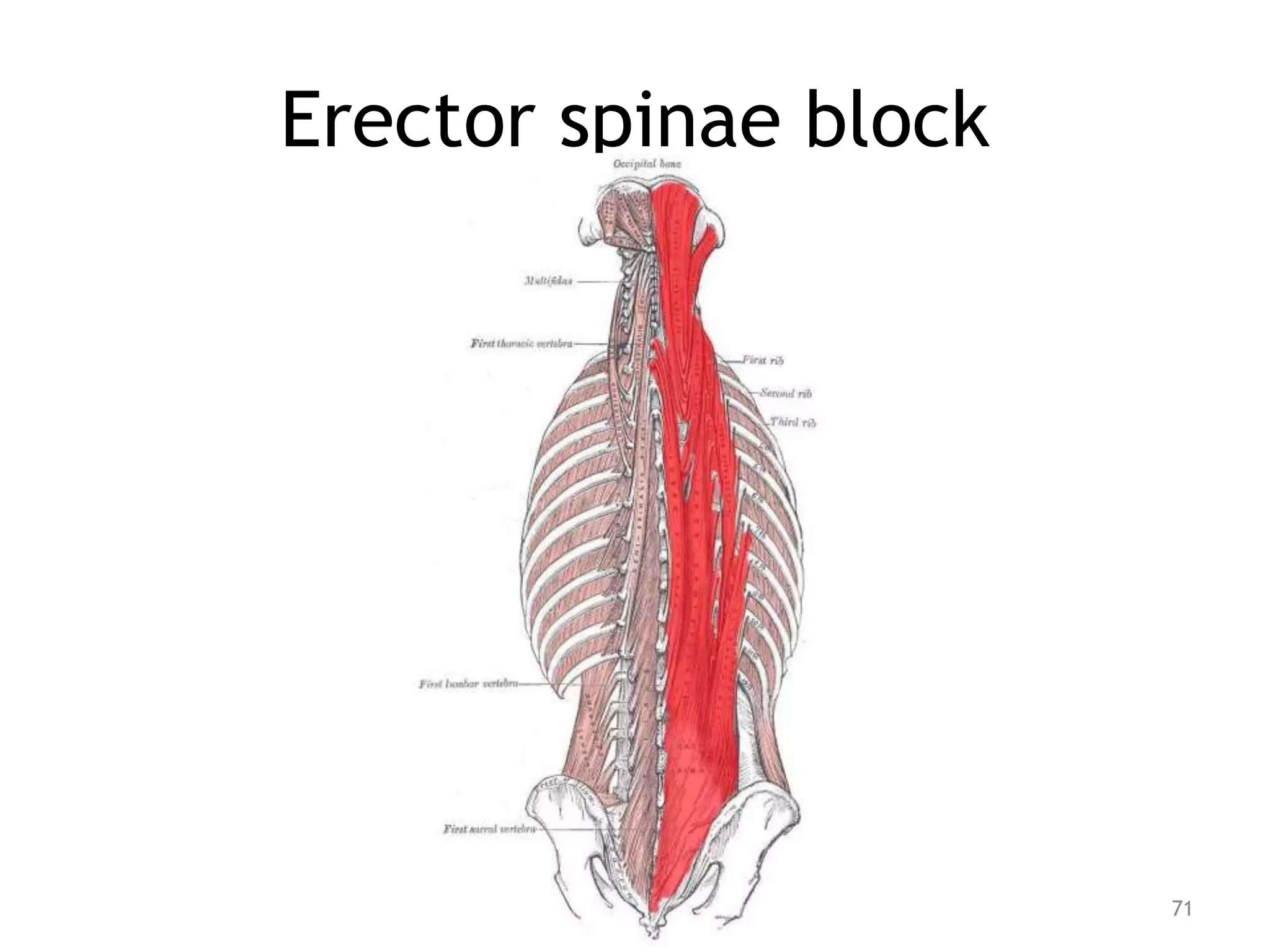
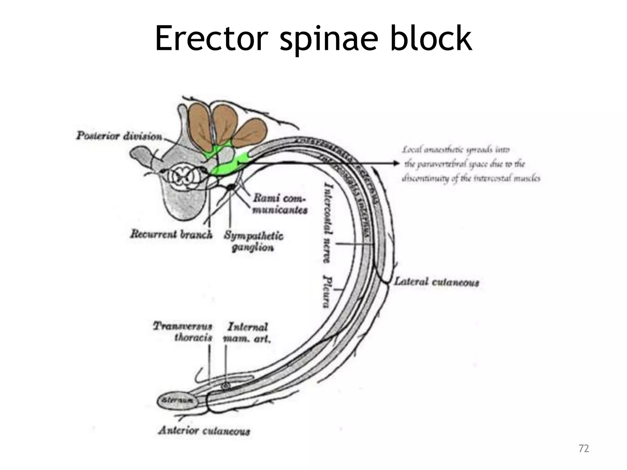
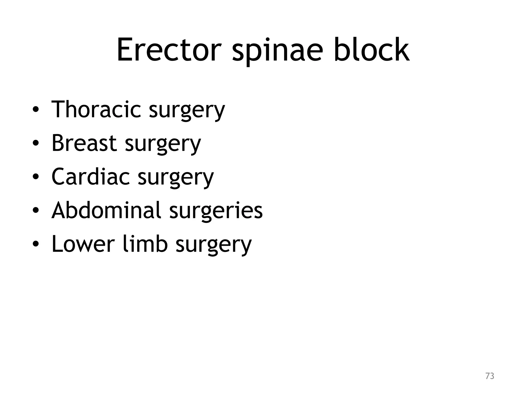
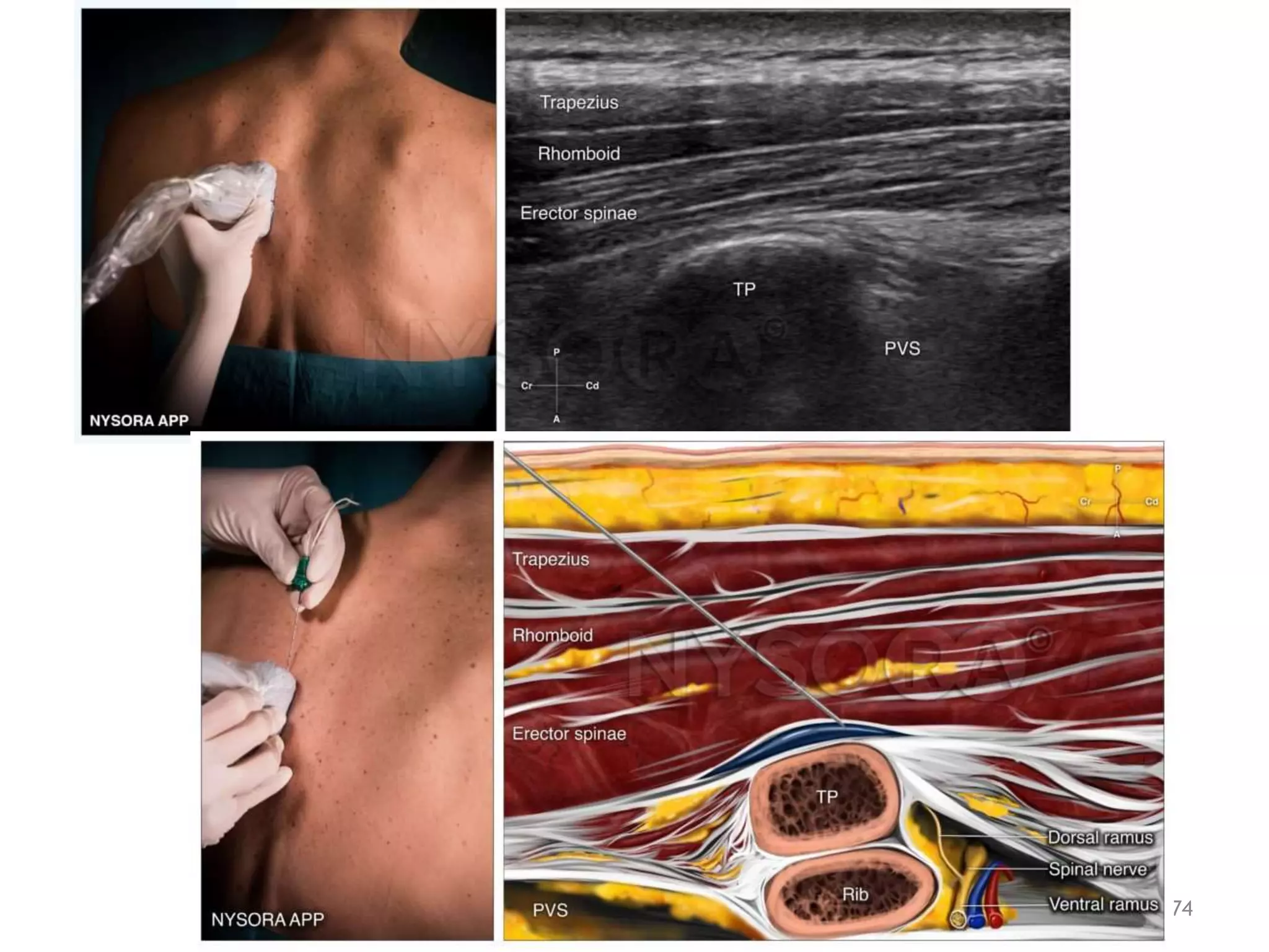
SEMINAR ON
TRUNCAL BLOCKS
Presenter: Dr Dawit G (ACCPM R1)
Moderator: Dr. Adane Getachew (Anesthesiologist)
Bahirdar University; Collage Of Medicine And Health Sciences
November 2022
OBJECTIVES
• Explain Anatomy and sonoanatomy of
abdominal wall and chest wall blocks
• Describe indications, complications and
techniques of truncal blocks
2
OUTLINES
• Introduction
• Abdominal wall blocks
• Chest wall blocks
• references
3
Introduction
• Interfascial plane blocks are the current
hot topic in regional anaesthesia
4
Abdominal wall blocks
• TAP
• Rectus sheath
• Ilioinguinal and Iieohypogastric nerve
blocks
• Quadratum lumborum block
5
Abdominal wall anatomy
6
Abdominal wall inervation
7
Abdominal wall inervation
8
TAP block
• relies on injection of LA in the
neurovascular plane between the TA and
IO muscles.
• targets dermatomes from T8 to L1.
• The subcostal TAP block is performed at
the costal margin to achieve a block as
high as T6.
9
TAP block
• part of a multimodal approach to
postoperative pain control in
– prostatectomy,
– large- and small-bowel surgery, and
– cesarean section
10
TAP block
11
TAP block
12
TAP BLOCK
13
TAP blocks comparison
14
TAP block summary
15
RECTUS SHEATH BLOCK
• involves injection of LA between the rectus
abdominus muscle and the posterior rectus
muscle sheath.
• This block results in periumbilical anesthesia of
the T9 to T11 dermatomes
• used for
– percutaneous gastrostomy surgery
– umbilical hernia and
– midline ventral hernia repairs
16
17
RECTUS SHEATH BLOCK
18
RECTUS SHEATH BLOCK
19
RECTUS SHEATH BLOCK
20
ILIOINGUINAL AND
ILIOHYPOGASTRIC NERVE BLOCKS
• blocked together with a targeted injection of LA
within the TAP.
• useful for postoperative analgesia after inguinal
hernia repair for children and adults
• employed for analgesia during inguinal hernia repair,
orchiopexy, and hydrocelectomy
• cannot be used as the only anesthetic for the
surgery because the ilioinguinal and iliohypogastric
nerves do not cover visceral pain from peritoneal
traction and manipulation of the spermatic cord.
21
LUMBAR PLEXUS
22
23
II AND IH BLOCKS
24
II AND IH BLOCKS
25
II AND IH BLOCKS
26
II AND IH BLOCKS
27
II AND IH BLOCKS
28
QUADRATUM LUMBORUM BLOCK
• more consistent method of accomplishing
somatic as well as visceral analgesia of the
abdomen than the TAP block
• may provide an extended sensory blockade
between T4 and L1.
• It can be used as an adjuvant technique for
analgesia but
• does not provide adequate blockade to be used
for anaesthesia.
29
QUADRATUM LUMBORUM BLOCK
• The QL lies between the anterior muscle
layers and the paravertebral space
• the efficacy is due to extension into
the paravertebral space
• Different approaches to QLB have been
described with no large studies to show
which is the most effective approach
30
QL BLOCK
31
QL BLOCK
32
SHAMROCK
33
SHAMROCK SIGN
34
QL BLOCK
35
QL BLOCK
36
QLB Indications
37
QUADRATUM LUMBORUM BLOCK
complications are very rare but may include the
following.
• Block Failure
• Local anaesthetic toxicity
• Sympatholysis causing hypotension
• Bowel injury
• Kidney injury
• Infection
• Vascular injury
• Unwanted femoral nerve block
38
CHEST WALL BLOCKS
• Before the advent of ultrasound-guided
regional anaesthesia, chest wall blocks were
mainly confined to
– intercostal nerve blockade,
– thoracic epidural analgesia and
– thoracic paravertebral blockade.
• development of US guided fascial plane
blocks, enabled local anaesthetic to be
injected into a tissue plane rather than
around individual nerves
CHEST WALL BLOCKS
• PEC I AND PEC II
• Seratus plane block (SPB)
• Thoracic paravertebral block (TPVB)
• Intercostal block
• Erector spina block (ESB)
40
PEC I AND PEC II BLOCK
41
PEC I BLOCK
• performed by injection of LA in the plane
between the pectoralis major and minor
muscles
• blocks the lateral and medial pectoral
nerves.
• Insert the needle in plane
• After negative aspiration, inject 10 mL of
LA
42
PEC I AND PEC II BLOCK
43
PEC I BLOCK
44
PEC II
• Also called the modified Pecs I block
• Aims to block the pectoral nerves,
intercostobrachial nerve, the intercostal nerves
3 through 6, and the long thoracic nerve.
• The Pecs I block is performed first as above,
and
• A second injection Is given in the plane
between the pectoralis minor muscle and the
serratus anterior muscle
45
SERRATUS PLANE BLOCK
• a simple, effective and safe thoracic fascial
plane block
• designed to anesthetize the thoracic intercostal
nerves in order to provide analgesia for the
lateral chest wall.
• Intercostal nerves from T2 to T9 are usually
blocked .
• The SP block is a more posterior and lateral
modification of the Pecs II block
46
SERRATUS PLANE BLOCK
• Place a linear probe in a sagittal plane under
the mid-clavicle .
• Move the probe inferolaterally, counting ribs
until the fifth rib is identified in the midaxillary
line.
• After negative aspiration, inject 20 mL of LA in
5-mL increments, aspirating between injections
47
SERRATUS PLANE BLOCK
48
SERRATUS PLANE BLOCK
49
SERRATUS PLANE BLOCK
50
SERRATUS PLANE BLOCK
Indications include
• breast surgery
• chronic pain after mastectomy,
• rib fractures
• thoracoscopy and
• thoracotomy.
51
Thoracic paravertebral block
• anesthetizes spinal nerves
as they emerge from
intervertebral foramina.
• TPVB results in somatic and
sympathetic nerve block,
52
PVB
• Compared with epidural blockade, TPVB
offers
– the possibility of unilateral block and
– is associated with a lower incidence of
• hypotension,
• urinary retention,
• respiratory problems, and
• postoperative nausea and vomiting (PONV)
53
Paramedian sagittal PVB
54
Transverse intercostal PVB
55
PVB, ANATOMIC APPROACH
56
• At a depth of 2 to 4 cm,
contact should be made
with the transverse
process;
• withdraw the needle
slightly and redirect
cranially or caudally to
walk off the transverse
process.
• The needle should be
inserted 1 cm past the
transverse process;.
• After negative aspiration,
inject 5 mL of LA.
PVB, ANATOMIC APPROACH
57
PVB, ANATOMIC APPROACH
58
59
TPVB
60
TPVB
Complications of the Paravertebral
Blockade
• vessel puncture
• hematoma
• epidural spread (via the intervertebral
foramina)
• intrathecal spread (via dural cuff),
• and pneumothorax
61
INTERCOSTAL BLOCKS
• The intercostal space is
contiguous with
paravertebral space.
• Indications are very
similar to traditional
paravertebral nerve block.
• This block is easy to
perform, though multiple
blocks are often required.
62
INTERCOSTAL BLOCKS
• useful when the placement of traditional
paravertebral blockade is contraindicated
1. if the patient is anticoagulated,
2. thrombocytopenic, or coagulopathic or
3. if there is a question of transverse process
fracture and unstable spine
63
ICB,Anatomic approach
• Palpate the rib 6 to 8
cm from the midline.
• Insert a needle at the
inferior border of the
rib, oriented
approximately 20
degrees cephalad, and
• advance 0.5 cm
underneath the rib.
• After negative
aspiration, inject 3 to 5
mL of local anesthetic
(LA) .
64
ICB,Anatomic approach
65
ICB, US GUIDED
• Place the ultrasound
transducer 4 cm from
the spinous process in
a sagittal plane
• After negative
aspiration, inject 3 to 5
mL of LA;
• as the drug is injected,
the pleura can be seen
moving away.
66
INTERCOSTAL BLOCKS
67
Erector spinae block
• An easy-to-perform regional anaesthesia
technique
• An alternative analgesic option to thoracic
epidural analgesia and paravertebral blocks,
• Has a good safety profile with very few
reported complications.
68
Erector spinae block
• uses ultrasound to deposit LA deep to the 3
columns of ES muscles
(iliocostalis, longissimus, spinalis).
• run the length of the spine from the base of
the skull to the medial crest of the sacrum.
• Overlying the ES complex are 2 further
layers of muscle: the trapezius and
rhomboid major.
69
70
Erector spinae block
71
Erector spinae block
72
Erector spinae block
• Thoracic surgery
• Breast surgery
• Cardiac surgery
• Abdominal surgeries
• Lower limb surgery
73
74
References
• The New York School Of Regional Anesthesia HADZIC’S TEXTBOOK
OF REGIONAL ANESTHESIA AND ACUTE PAIN MANAGEMENT SECOND
EDITION ; 2017
• Ultrasound for Interventional Pain Management; 2020
• Atlas of sonoanatomy for regional anesthesia and pain medicine;
2018
• Essentials of Regional Anesthesia; 2012
• Ultrasound Guidance in Regional Anaesthesia; Principles and
Practical Implementation; SECOND EDITION; 2010
• WFSA Anesthesia tutorial of the week, QLB; 2020
• Uptodate 2018
• www.nysora.com/truncalblocks
• 75
76

Truncal blocks.pptx

  • 1.
    SEMINAR ON TRUNCAL BLOCKS Presenter:Dr Dawit G (ACCPM R1) Moderator: Dr. Adane Getachew (Anesthesiologist) Bahirdar University; Collage Of Medicine And Health Sciences November 2022
  • 2.
    OBJECTIVES • Explain Anatomyand sonoanatomy of abdominal wall and chest wall blocks • Describe indications, complications and techniques of truncal blocks 2
  • 3.
    OUTLINES • Introduction • Abdominalwall blocks • Chest wall blocks • references 3
  • 4.
    Introduction • Interfascial planeblocks are the current hot topic in regional anaesthesia 4
  • 5.
    Abdominal wall blocks •TAP • Rectus sheath • Ilioinguinal and Iieohypogastric nerve blocks • Quadratum lumborum block 5
  • 6.
  • 7.
  • 8.
  • 9.
    TAP block • relieson injection of LA in the neurovascular plane between the TA and IO muscles. • targets dermatomes from T8 to L1. • The subcostal TAP block is performed at the costal margin to achieve a block as high as T6. 9
  • 10.
    TAP block • partof a multimodal approach to postoperative pain control in – prostatectomy, – large- and small-bowel surgery, and – cesarean section 10
  • 11.
  • 12.
  • 13.
  • 14.
  • 15.
  • 16.
    RECTUS SHEATH BLOCK •involves injection of LA between the rectus abdominus muscle and the posterior rectus muscle sheath. • This block results in periumbilical anesthesia of the T9 to T11 dermatomes • used for – percutaneous gastrostomy surgery – umbilical hernia and – midline ventral hernia repairs 16
  • 17.
  • 18.
  • 19.
  • 20.
  • 21.
    ILIOINGUINAL AND ILIOHYPOGASTRIC NERVEBLOCKS • blocked together with a targeted injection of LA within the TAP. • useful for postoperative analgesia after inguinal hernia repair for children and adults • employed for analgesia during inguinal hernia repair, orchiopexy, and hydrocelectomy • cannot be used as the only anesthetic for the surgery because the ilioinguinal and iliohypogastric nerves do not cover visceral pain from peritoneal traction and manipulation of the spermatic cord. 21
  • 22.
  • 23.
  • 24.
    II AND IHBLOCKS 24
  • 25.
    II AND IHBLOCKS 25
  • 26.
    II AND IHBLOCKS 26
  • 27.
    II AND IHBLOCKS 27
  • 28.
    II AND IHBLOCKS 28
  • 29.
    QUADRATUM LUMBORUM BLOCK •more consistent method of accomplishing somatic as well as visceral analgesia of the abdomen than the TAP block • may provide an extended sensory blockade between T4 and L1. • It can be used as an adjuvant technique for analgesia but • does not provide adequate blockade to be used for anaesthesia. 29
  • 30.
    QUADRATUM LUMBORUM BLOCK •The QL lies between the anterior muscle layers and the paravertebral space • the efficacy is due to extension into the paravertebral space • Different approaches to QLB have been described with no large studies to show which is the most effective approach 30
  • 31.
  • 32.
  • 33.
  • 34.
  • 35.
  • 36.
  • 37.
  • 38.
    QUADRATUM LUMBORUM BLOCK complicationsare very rare but may include the following. • Block Failure • Local anaesthetic toxicity • Sympatholysis causing hypotension • Bowel injury • Kidney injury • Infection • Vascular injury • Unwanted femoral nerve block 38
  • 39.
    CHEST WALL BLOCKS •Before the advent of ultrasound-guided regional anaesthesia, chest wall blocks were mainly confined to – intercostal nerve blockade, – thoracic epidural analgesia and – thoracic paravertebral blockade. • development of US guided fascial plane blocks, enabled local anaesthetic to be injected into a tissue plane rather than around individual nerves
  • 40.
    CHEST WALL BLOCKS •PEC I AND PEC II • Seratus plane block (SPB) • Thoracic paravertebral block (TPVB) • Intercostal block • Erector spina block (ESB) 40
  • 41.
    PEC I ANDPEC II BLOCK 41
  • 42.
    PEC I BLOCK •performed by injection of LA in the plane between the pectoralis major and minor muscles • blocks the lateral and medial pectoral nerves. • Insert the needle in plane • After negative aspiration, inject 10 mL of LA 42
  • 43.
    PEC I ANDPEC II BLOCK 43
  • 44.
  • 45.
    PEC II • Alsocalled the modified Pecs I block • Aims to block the pectoral nerves, intercostobrachial nerve, the intercostal nerves 3 through 6, and the long thoracic nerve. • The Pecs I block is performed first as above, and • A second injection Is given in the plane between the pectoralis minor muscle and the serratus anterior muscle 45
  • 46.
    SERRATUS PLANE BLOCK •a simple, effective and safe thoracic fascial plane block • designed to anesthetize the thoracic intercostal nerves in order to provide analgesia for the lateral chest wall. • Intercostal nerves from T2 to T9 are usually blocked . • The SP block is a more posterior and lateral modification of the Pecs II block 46
  • 47.
    SERRATUS PLANE BLOCK •Place a linear probe in a sagittal plane under the mid-clavicle . • Move the probe inferolaterally, counting ribs until the fifth rib is identified in the midaxillary line. • After negative aspiration, inject 20 mL of LA in 5-mL increments, aspirating between injections 47
  • 48.
  • 49.
  • 50.
  • 51.
    SERRATUS PLANE BLOCK Indicationsinclude • breast surgery • chronic pain after mastectomy, • rib fractures • thoracoscopy and • thoracotomy. 51
  • 52.
    Thoracic paravertebral block •anesthetizes spinal nerves as they emerge from intervertebral foramina. • TPVB results in somatic and sympathetic nerve block, 52
  • 53.
    PVB • Compared withepidural blockade, TPVB offers – the possibility of unilateral block and – is associated with a lower incidence of • hypotension, • urinary retention, • respiratory problems, and • postoperative nausea and vomiting (PONV) 53
  • 54.
  • 55.
  • 56.
    PVB, ANATOMIC APPROACH 56 •At a depth of 2 to 4 cm, contact should be made with the transverse process; • withdraw the needle slightly and redirect cranially or caudally to walk off the transverse process. • The needle should be inserted 1 cm past the transverse process;. • After negative aspiration, inject 5 mL of LA.
  • 57.
  • 58.
  • 59.
  • 60.
  • 61.
    TPVB Complications of theParavertebral Blockade • vessel puncture • hematoma • epidural spread (via the intervertebral foramina) • intrathecal spread (via dural cuff), • and pneumothorax 61
  • 62.
    INTERCOSTAL BLOCKS • Theintercostal space is contiguous with paravertebral space. • Indications are very similar to traditional paravertebral nerve block. • This block is easy to perform, though multiple blocks are often required. 62
  • 63.
    INTERCOSTAL BLOCKS • usefulwhen the placement of traditional paravertebral blockade is contraindicated 1. if the patient is anticoagulated, 2. thrombocytopenic, or coagulopathic or 3. if there is a question of transverse process fracture and unstable spine 63
  • 64.
    ICB,Anatomic approach • Palpatethe rib 6 to 8 cm from the midline. • Insert a needle at the inferior border of the rib, oriented approximately 20 degrees cephalad, and • advance 0.5 cm underneath the rib. • After negative aspiration, inject 3 to 5 mL of local anesthetic (LA) . 64
  • 65.
  • 66.
    ICB, US GUIDED •Place the ultrasound transducer 4 cm from the spinous process in a sagittal plane • After negative aspiration, inject 3 to 5 mL of LA; • as the drug is injected, the pleura can be seen moving away. 66
  • 67.
  • 68.
    Erector spinae block •An easy-to-perform regional anaesthesia technique • An alternative analgesic option to thoracic epidural analgesia and paravertebral blocks, • Has a good safety profile with very few reported complications. 68
  • 69.
    Erector spinae block •uses ultrasound to deposit LA deep to the 3 columns of ES muscles (iliocostalis, longissimus, spinalis). • run the length of the spine from the base of the skull to the medial crest of the sacrum. • Overlying the ES complex are 2 further layers of muscle: the trapezius and rhomboid major. 69
  • 70.
  • 71.
  • 72.
  • 73.
    Erector spinae block •Thoracic surgery • Breast surgery • Cardiac surgery • Abdominal surgeries • Lower limb surgery 73
  • 74.
  • 75.
    References • The NewYork School Of Regional Anesthesia HADZIC’S TEXTBOOK OF REGIONAL ANESTHESIA AND ACUTE PAIN MANAGEMENT SECOND EDITION ; 2017 • Ultrasound for Interventional Pain Management; 2020 • Atlas of sonoanatomy for regional anesthesia and pain medicine; 2018 • Essentials of Regional Anesthesia; 2012 • Ultrasound Guidance in Regional Anaesthesia; Principles and Practical Implementation; SECOND EDITION; 2010 • WFSA Anesthesia tutorial of the week, QLB; 2020 • Uptodate 2018 • www.nysora.com/truncalblocks • 75
  • 76.

Editor's Notes

  • #5 Fascial plane blocks??? Advantage of trucal blocks???
  • #6 ANATOMY SONOANATOMY PROBE LA COMPLICATION
  • #12 The abdominal anterolateral wall is innervated by the anterior rami of T7–L1 spinal nerves.
  • #25 NEEDLE TRAJECTORY
  • #30 Lumbar QL??
  • #36 Curvilinear 20ml on each side
  • #37 Linear probe
  • #40 The use of ultrasound in regional anaesthesia has facilitated the visualisation of anatomical structures, needle advancement and the spread of local anaesthetic.
  • #42  The pectoral nerves (lateral and medial) arise from the cords of the brachial plexus and innervate the pectoral major and minor muscles (figure 12). The lateral pectoral nerve (C5 to C7) courses along the undersurface of the pectoralis major muscle, in the fascial plane between the pectoralis major and minor muscles, and is consistently located lateral to the thoracoacromial artery. The medial pectoral nerve (C8 to T1) also runs between the pectoralis major and minor muscles [48,54]. These are small nerves that are not usually visualized using ultrasound.
  • #44 Place a linear, high-frequency ultrasound probe in the parasagittal plane below the clavicle, just medial to the coracoid process. Identify the pectoralis major and pectoralis minor muscles (image 7). Rotate the probe laterally using color-flow Doppler to identify the pectoral branch of the thoracoacromial artery between the muscles, to avoid puncture, and to more precisely deposit LA near the nerves
  • #45 The Pecs I block is performed as above. In the same Pecs I view, slide the probe caudally to identify the first rib deep to the axillary artery (image 7). Rib shadows are visualized as bright white hyperechoic structures. After identifying the first rib, move the probe in a lateral and caudal direction while counting the ribs. Slide the ultrasound probe toward the axilla until the third rib is visualized at the midaxillary line on the lateral chest wall
  • #46 After negative aspiration, inject 20 mL of LA in 5-mL increments, aspirating gently between injections.
  • #48 Identify the latissimus dorsi superficially and posteriorly, the teres major muscle superiorly, and the serratus muscle deep and inferiorly, overlying the fifth rib.
  • #49 Commence the ultrasound scan by placing the ultrasound probe in a parasagittal plane immediately inferior to the clavicle and in the deltopectoral groove adjacent to the coracoid process. Here, identify the pectoralis major and minor muscles, the axillary artery and vein as well as the underlying second rib. Move the ultrasound probe inferiorly and posteriorly with increasing coronal orientation until the fifth rib is found in the midaxillary line (Figure 3). In this position, identify a superficial and thick muscle, the latissimus dorsi, overlying the deeper serratus anterior muscle (Figure 4). Since the thoracodorsal artery is located in the fascial plane between the latissimus dorsi and the serratus anterior muscles, its identification with ultrasound imaging and use of colour Doppler is helpful to delineate the plane and decrease the risk of inadvertent accidental arterial puncture on introduction of the needle.
  • #52 Chronic pain after mastectomy Rib fracture(s) Thoracoscopy Thoracotomy
  • #53 similar to that which would be achieved with epidural blockade.
  • #54 Single-injection TPVB at T4 level is an alternative to general anesthesia for breast surgery
  • #55 Chelly et al. retrospectively reviewed 559 records for 1,318 thoracic PVT blocks and determined that the depth of PVT space at T4–T8 is shallower (3.8–5.7 cm) and more variable [8] . At T9–T12 PVT space, the depth is at 5–6 cm and appeared to be less variable. The BMI has a significant influence on the depth from the skin to paravertebral space at these levels
  • #56 Place a high-frequency (5- to 19-MHz) linear transducer 2 cm lateral to the midline at the chosen spinal level (picture 6 and picture 7). The probe can be placed either in a sagittal plane or transverse plane, and both viewing planes should be explored during the scout scan to better identify landmarks.
  • #57 Repeat the procedure at multiple levels, as needed, to effect anesthesia at multiple dermatomes.
  • #60  Cranially, the paravertebral space is in close proximity to the adipose tissue associated with the brachial plexus, phrenic nerve, and cervical sympathetic trunk. The caudal extent of the paravertebral space is debated. While one cadaver study reported that the insertion of the psoas muscle sealed the lower space [34], others reported that thoracic paravertebral injectate extended through diaphragmatic ligaments to the lumbar plexus [35,36].
  • #63 The injection can be made closer to the vertebral column (and more proximal in the course of the nerve) with ultrasound guidance because palpation of the rib is less important than with an anatomic landmark approach.
  • #65 and walked off the lower part of the rib The needle should be directed 45° cephalad and 60° medial to the sagittal plane . If the needle comes into contact with bone, "walk" the needle off the bone inferiorly. The block can be repeated at each of the levels appropriate for the surgical procedure
  • #68 For ultrasound-guided intercostal nerve block, the tip of the needle is placed between the internal intercostal muscle and the innermost intercostal muscle
  • #69 especially where these techniques are contraindicated.