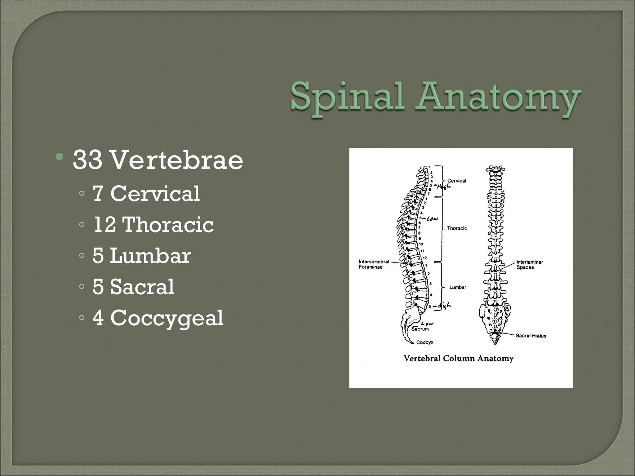
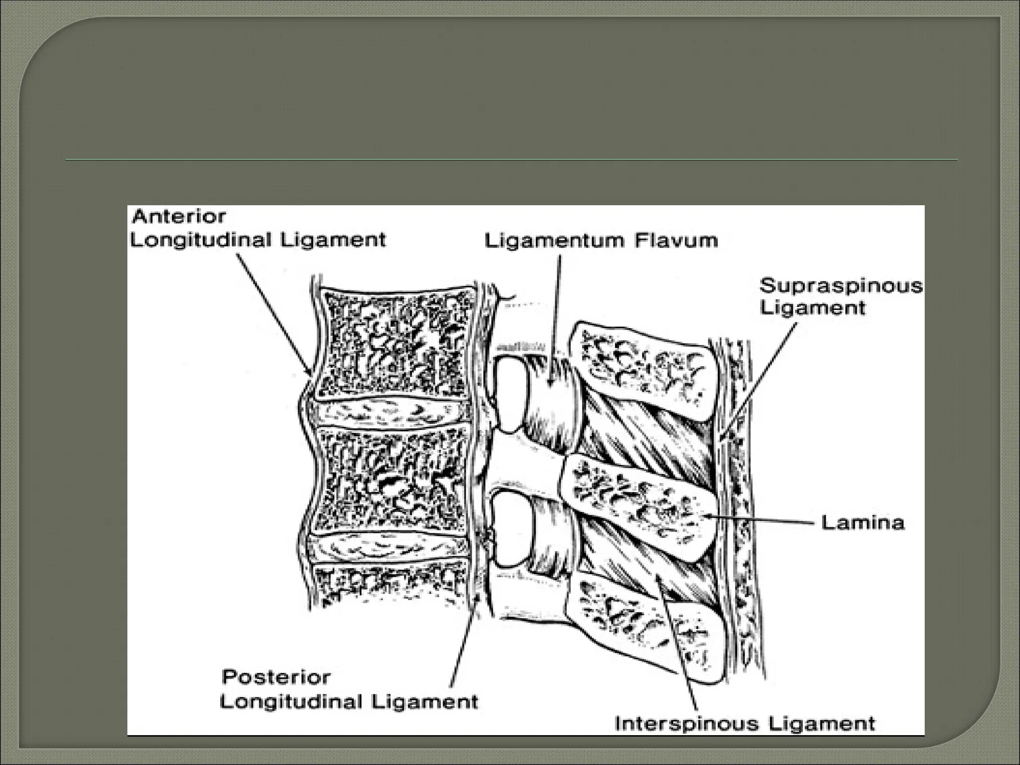
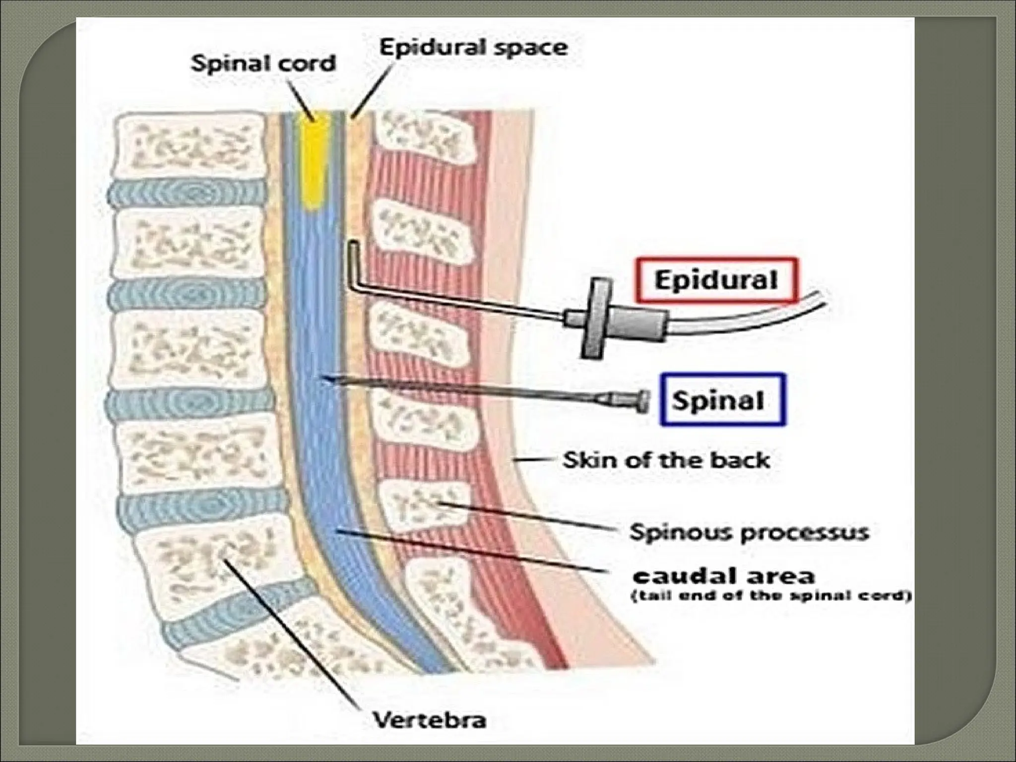
Regional Anesthesia
 Spinal, epidural, and caudal blocks are also known as
neuraxial anesthesia.
 Reduce the incidence of
Venous thrombosis and pulmonary embolism
Cardiac complications in high-risk patients
Bleeding and transfusion requirements
Vascular graft occlusion
Pneumonia and respiratory depression
following upper
abdominal or thoracic surgery in patients
with chronic lung
disease.
 33 Vertebrae
◦ 7 Cervical
◦ 12 Thoracic
◦ 5 Lumbar
◦ 5 Sacral
◦ 4 Coccygeal
 Ligaments:-The vertebral bodies are stablised by five
ligaments.These are :-
Supraspinous ligaments-This forms ligamentumnuche
above T-7 and attaches to occipital protuberance at the base
of the skull.
Ineterspinous ligaments-attaches between the
spinous process.
Ligamentum Flavum:The ligamentum flvm is thickest
in the mid line measuring 3-5 mm at the L2-L5 inter space in
adults.This is also farthest from the spinal menisges in the
midline measuring 4-6 mm at L2-L3 inter space.
Anterior and posterior longitudinal ligaments run
along the anterior and posterior surfaces of the vertebral
bodies
Meninges:-
Three protective coverings, dura mater, arachnoid
mater and pia mater cover the spinal cord.
The space before the dura mater is epidural space.
Caudially the dura mater ends approximately at S-2,
where it fuses with the final terminale.
The subarachnoid space lies between the arachnoid
mater and pia mater, containing cerebrospinal fluid.
Pia mater is adherent to the spinal cord.
The pia mater extends up to the tip of the spinal
cord where it it continues as filum terminale which
anchors the spinal cord to the sacrum.
Spinal Cord
• Adult
 Begins: Foramen Magnum
 Ends: L1
• Newborn
 Begins: Foramen Magnum
 Ends: L3
• Terminal End: Conus Medullaris
• Filum Terminale: Anchors in sacral region
• Cauda Equina: Nerve group of lower dural
sac
Space that surrounds the spinal
meninges
• Potential space
Ligamentum Flavum
• Binds epidural space posteriorly
Widest at Level L2 (5-6mm)
Narrowest at Level C5 (1-1.5mm)
 Dura Mater
◦ Outer most layer
◦ Fibrous
 Arachnoid
◦ Middle layer
◦ Non-vascular
 Pia
◦ Inner most layer
◦ Highly vascular
 Sub Arachnoid
Space
◦ Lies between the
arachnoid and pia
Vasoconstrictors
• Prolong duration of spinal block
Factors Effecting Distribution
• Site of injection
• Shape of spinal column
• Patient height
• Angulation of needle
• Volume of CSF
• Characteristics of local anesthetic
 Density
 Specific gravity
 Baracity
• Dose
• Volume
• Patient position
Anesthesia level is determined by patient
position
Uptake of local anesthetic occurs by
diffusion
Elimination determines duration of block
• Lipid solubility decreases vascular absorption
• Vasoconstriction can decrease rate of
elimination
Blockade of Sympathetic Preganglionic
Neurons
• Send signals to both arteries and veins
• Predominant action is venodilation
 Reduces:
 Venous return
 Stroke volume
 Cardiac output
 Blood pressure
• T1-T4 Blockade
 Causes unopposed vagal stimulation
 Bradycardia
 Associated with decrease venous return & cardioaccelerator fibers
blockade
 Decreased venous return to right atrium causes decreased stretch
receptor response
Treatment
• Best way to treat is physiologic not
pharmacologic
• Primary Treatment
 Increase the cardiac preload
 Large IV fluid bolus within 30 minutes prior to spinal
placement, minimum 1 liter of crystalloids
• Secondary Treatment
 Pharmacologic
 Ephedrine is more effective than Phenylephrine
Healthy Patients
• Appropriate spinal blockade has little effect
on ventilation
High Spinal
• Decrease functional residual capacity (FRC)
 Paralysis of abdominal muscles
 Intercostal muscle paralysis interferes with
coughing and clearing secretions
 Apnea is due to hypoperfusion of respiratory
center
 Preparation &
Monitoring
◦ ECG
◦ NBP
◦ Pulse Oximeter
 Patient Positioning
◦ Lateral decubitous
◦ Sitting
◦ Prone (hypobaric
technique)
Midline Approach
• Skin
• Subcutaneous tissue
• Supraspinous ligament
• Interspinous ligament
• Ligamentum flavum
• Epidural space
• Dura mater
• Arachnoid mater
Paramedian or Lateral Approach
• Same as midline excluding supraspinous &
interspinous ligaments
Advantages
• Full stomach
• Anatomic distortions of upper airway
• TURP surgery
• Obstetrical surgery (T4 Level)
• Decreased post-operative pain
• Continuous infusion
 Contraindications
• Absolute:
 Refusal
 Infection
 Coagulopathy
 Severe hypovolemia
 Increased intracranial pressure
 Severe aortic or mitral stenosis
• Relative:
Peripheral neuropathy
Mini dose heparin
Fixed output cardiac lesion
Uncooperative pt
Prolonged surgery
 Complications
 Anaphylaxis
 Backach
 Headach (PDPH)
 Urinary retention
 Spinal shock.
 Cauda equina injury
 Hypothermia
 Meningitis
 Broken needle
 Bleeding resulting in haematoma, with or without
subsequent neurological sequelae due to
compression of the spinal nerves
 Infection: immediate within six hours of the spinal
anaesthetic manifesting as meningism or
meningitis or late, at the site of injection, in the
form of pus discharge, due to improper
sterilization of the LP set.
 Test Dose: 1.5% Lido with Epi 1:200,000
◦ Tachycardia (increase >30bpm over resting HR)
◦ High blood pressure
◦ Light headedness
◦ Metallic taste in mouth
◦ Ring in ears
◦ Facial numbness
◦ Note: if beta blocked will only see increase in
BP not HR
 Bolus Dose: Preferred Local of Choice
◦ 10 milliliters for labor pain
◦ 20-30 milliliters for C-section
Distances from Skin to Epidural Space
• Average adult: 4-6cm
• Obese adult: up to 8cm
• Thin adult: 3cm
Assessment of Sensory Blockade
• Alcohol swab
 Most sensitive initial indicator to assess loss of
temperature
• Pin prick
 Most accurate assessment of overall sensory block
Hip and Knee Surgery
• Lower Extremity Vascular Surgery
• Lower Extremity Amputation
• Obstetrical – Labor & C/S
• Thoracic Surgery – Post-Op Pain
Control
• Thoracic Trauma
• Abdominal Surgery – Post-Op Pain
control
Complications
• Penetration of a blood vessel
• Hypotension (nausea & vomiting)
• Head ache
• Back pain
• Intravascular catheterization
• Wet tap
• Infection
Anatomy
• Sacrum
 Triangular bone
 5 fused sacral vertebrae
Needle Insertion
• Sacrococcygeal
membrane
• No subcutaneous bulge
or crepitous at site of
injection after 2-3ml
Post Operative Problems
• Pain at injection site is most common
• Slight risk of neurological complications
• Risk of infection
Dosages
• S5-L2: 15-20ml
• S5-T10: 25ml
 Esters
◦ Procaine
◦ Chloroprocaine
◦ Tetratcaine
◦ Cocaine
 Metabolism
◦ Hydrolysis by
psuedo-
cholinesterase
enzyme
 Amides
◦ Lidocaine
◦ Mepivacaine
◦ Bupivacaine
◦ Etidocaine
◦ Prilocaine
◦ Ropivacaine
 Metabolism
◦ Liver
Un-ionized local
anesthetic
defuses into
nerve axon & the
ionized form
binds the
receptors of the
Na channel in the
inactivated state
Thank you.

Regional_Anesthesia .powerpoint presentation

  • 1.
  • 2.
     Spinal, epidural,and caudal blocks are also known as neuraxial anesthesia.  Reduce the incidence of Venous thrombosis and pulmonary embolism Cardiac complications in high-risk patients Bleeding and transfusion requirements Vascular graft occlusion Pneumonia and respiratory depression following upper abdominal or thoracic surgery in patients with chronic lung disease.
  • 3.
     33 Vertebrae ◦7 Cervical ◦ 12 Thoracic ◦ 5 Lumbar ◦ 5 Sacral ◦ 4 Coccygeal
  • 5.
     Ligaments:-The vertebralbodies are stablised by five ligaments.These are :- Supraspinous ligaments-This forms ligamentumnuche above T-7 and attaches to occipital protuberance at the base of the skull. Ineterspinous ligaments-attaches between the spinous process. Ligamentum Flavum:The ligamentum flvm is thickest in the mid line measuring 3-5 mm at the L2-L5 inter space in adults.This is also farthest from the spinal menisges in the midline measuring 4-6 mm at L2-L3 inter space. Anterior and posterior longitudinal ligaments run along the anterior and posterior surfaces of the vertebral bodies
  • 6.
    Meninges:- Three protective coverings,dura mater, arachnoid mater and pia mater cover the spinal cord. The space before the dura mater is epidural space. Caudially the dura mater ends approximately at S-2, where it fuses with the final terminale. The subarachnoid space lies between the arachnoid mater and pia mater, containing cerebrospinal fluid. Pia mater is adherent to the spinal cord. The pia mater extends up to the tip of the spinal cord where it it continues as filum terminale which anchors the spinal cord to the sacrum.
  • 8.
    Spinal Cord • Adult Begins: Foramen Magnum  Ends: L1 • Newborn  Begins: Foramen Magnum  Ends: L3 • Terminal End: Conus Medullaris • Filum Terminale: Anchors in sacral region • Cauda Equina: Nerve group of lower dural sac
  • 9.
    Space that surroundsthe spinal meninges • Potential space Ligamentum Flavum • Binds epidural space posteriorly Widest at Level L2 (5-6mm) Narrowest at Level C5 (1-1.5mm)
  • 10.
     Dura Mater ◦Outer most layer ◦ Fibrous  Arachnoid ◦ Middle layer ◦ Non-vascular  Pia ◦ Inner most layer ◦ Highly vascular  Sub Arachnoid Space ◦ Lies between the arachnoid and pia
  • 12.
  • 13.
    Factors Effecting Distribution •Site of injection • Shape of spinal column • Patient height • Angulation of needle • Volume of CSF • Characteristics of local anesthetic  Density  Specific gravity  Baracity • Dose • Volume • Patient position
  • 14.
    Anesthesia level isdetermined by patient position Uptake of local anesthetic occurs by diffusion Elimination determines duration of block • Lipid solubility decreases vascular absorption • Vasoconstriction can decrease rate of elimination
  • 15.
    Blockade of SympatheticPreganglionic Neurons • Send signals to both arteries and veins • Predominant action is venodilation  Reduces:  Venous return  Stroke volume  Cardiac output  Blood pressure • T1-T4 Blockade  Causes unopposed vagal stimulation  Bradycardia  Associated with decrease venous return & cardioaccelerator fibers blockade  Decreased venous return to right atrium causes decreased stretch receptor response
  • 16.
    Treatment • Best wayto treat is physiologic not pharmacologic • Primary Treatment  Increase the cardiac preload  Large IV fluid bolus within 30 minutes prior to spinal placement, minimum 1 liter of crystalloids • Secondary Treatment  Pharmacologic  Ephedrine is more effective than Phenylephrine
  • 17.
    Healthy Patients • Appropriatespinal blockade has little effect on ventilation High Spinal • Decrease functional residual capacity (FRC)  Paralysis of abdominal muscles  Intercostal muscle paralysis interferes with coughing and clearing secretions  Apnea is due to hypoperfusion of respiratory center
  • 18.
     Preparation & Monitoring ◦ECG ◦ NBP ◦ Pulse Oximeter  Patient Positioning ◦ Lateral decubitous ◦ Sitting ◦ Prone (hypobaric technique)
  • 19.
    Midline Approach • Skin •Subcutaneous tissue • Supraspinous ligament • Interspinous ligament • Ligamentum flavum • Epidural space • Dura mater • Arachnoid mater Paramedian or Lateral Approach • Same as midline excluding supraspinous & interspinous ligaments
  • 20.
    Advantages • Full stomach •Anatomic distortions of upper airway • TURP surgery • Obstetrical surgery (T4 Level) • Decreased post-operative pain • Continuous infusion
  • 21.
     Contraindications • Absolute: Refusal  Infection  Coagulopathy  Severe hypovolemia  Increased intracranial pressure  Severe aortic or mitral stenosis • Relative: Peripheral neuropathy Mini dose heparin Fixed output cardiac lesion Uncooperative pt Prolonged surgery
  • 22.
     Complications  Anaphylaxis Backach  Headach (PDPH)  Urinary retention  Spinal shock.  Cauda equina injury  Hypothermia  Meningitis  Broken needle  Bleeding resulting in haematoma, with or without subsequent neurological sequelae due to compression of the spinal nerves  Infection: immediate within six hours of the spinal anaesthetic manifesting as meningism or meningitis or late, at the site of injection, in the form of pus discharge, due to improper sterilization of the LP set.
  • 26.
     Test Dose:1.5% Lido with Epi 1:200,000 ◦ Tachycardia (increase >30bpm over resting HR) ◦ High blood pressure ◦ Light headedness ◦ Metallic taste in mouth ◦ Ring in ears ◦ Facial numbness ◦ Note: if beta blocked will only see increase in BP not HR  Bolus Dose: Preferred Local of Choice ◦ 10 milliliters for labor pain ◦ 20-30 milliliters for C-section
  • 27.
    Distances from Skinto Epidural Space • Average adult: 4-6cm • Obese adult: up to 8cm • Thin adult: 3cm Assessment of Sensory Blockade • Alcohol swab  Most sensitive initial indicator to assess loss of temperature • Pin prick  Most accurate assessment of overall sensory block
  • 28.
    Hip and KneeSurgery • Lower Extremity Vascular Surgery • Lower Extremity Amputation • Obstetrical – Labor & C/S • Thoracic Surgery – Post-Op Pain Control • Thoracic Trauma • Abdominal Surgery – Post-Op Pain control
  • 29.
    Complications • Penetration ofa blood vessel • Hypotension (nausea & vomiting) • Head ache • Back pain • Intravascular catheterization • Wet tap • Infection
  • 30.
    Anatomy • Sacrum  Triangularbone  5 fused sacral vertebrae Needle Insertion • Sacrococcygeal membrane • No subcutaneous bulge or crepitous at site of injection after 2-3ml
  • 31.
    Post Operative Problems •Pain at injection site is most common • Slight risk of neurological complications • Risk of infection Dosages • S5-L2: 15-20ml • S5-T10: 25ml
  • 32.
     Esters ◦ Procaine ◦Chloroprocaine ◦ Tetratcaine ◦ Cocaine  Metabolism ◦ Hydrolysis by psuedo- cholinesterase enzyme  Amides ◦ Lidocaine ◦ Mepivacaine ◦ Bupivacaine ◦ Etidocaine ◦ Prilocaine ◦ Ropivacaine  Metabolism ◦ Liver
  • 33.
    Un-ionized local anesthetic defuses into nerveaxon & the ionized form binds the receptors of the Na channel in the inactivated state
  • 35.