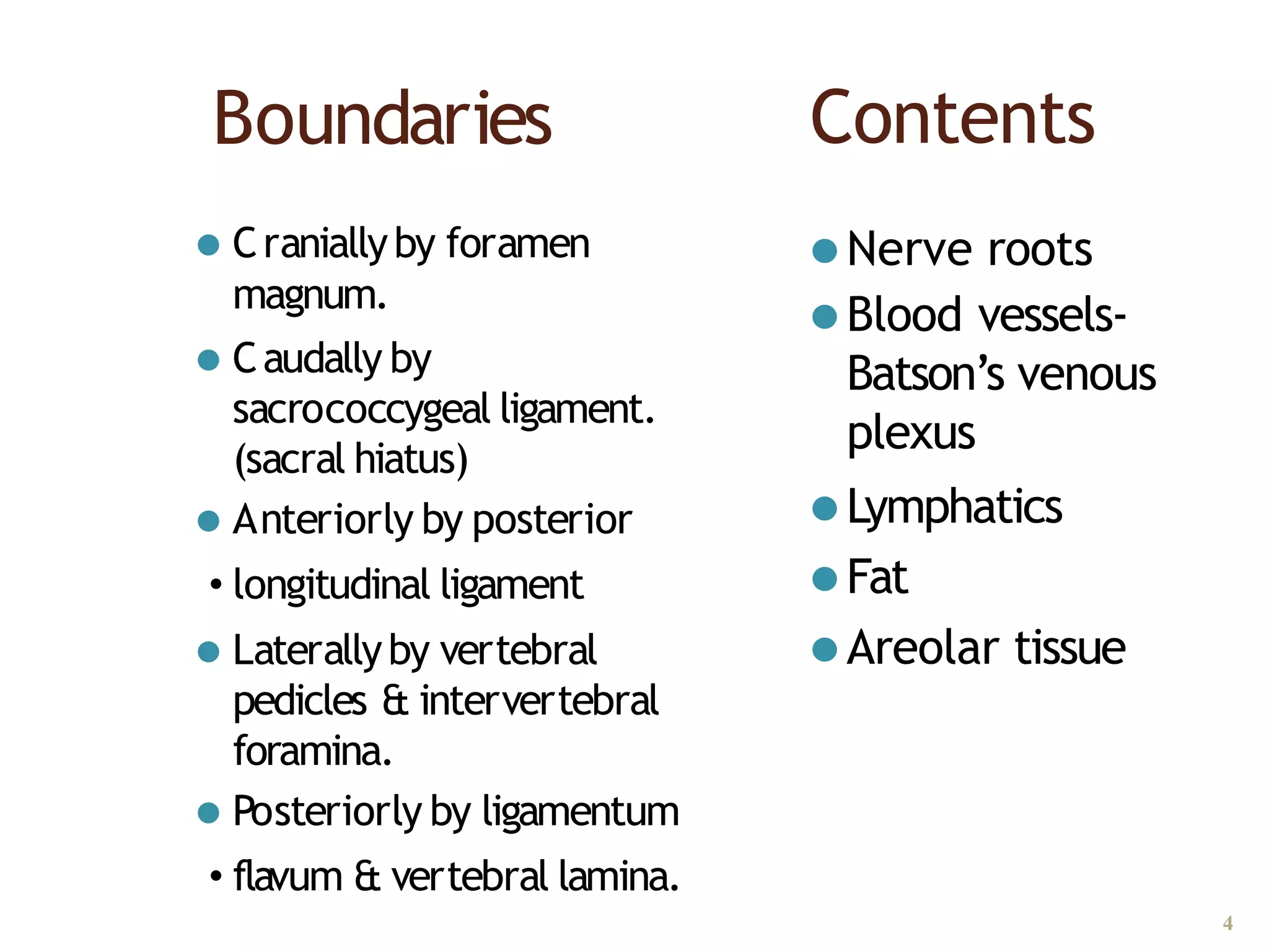
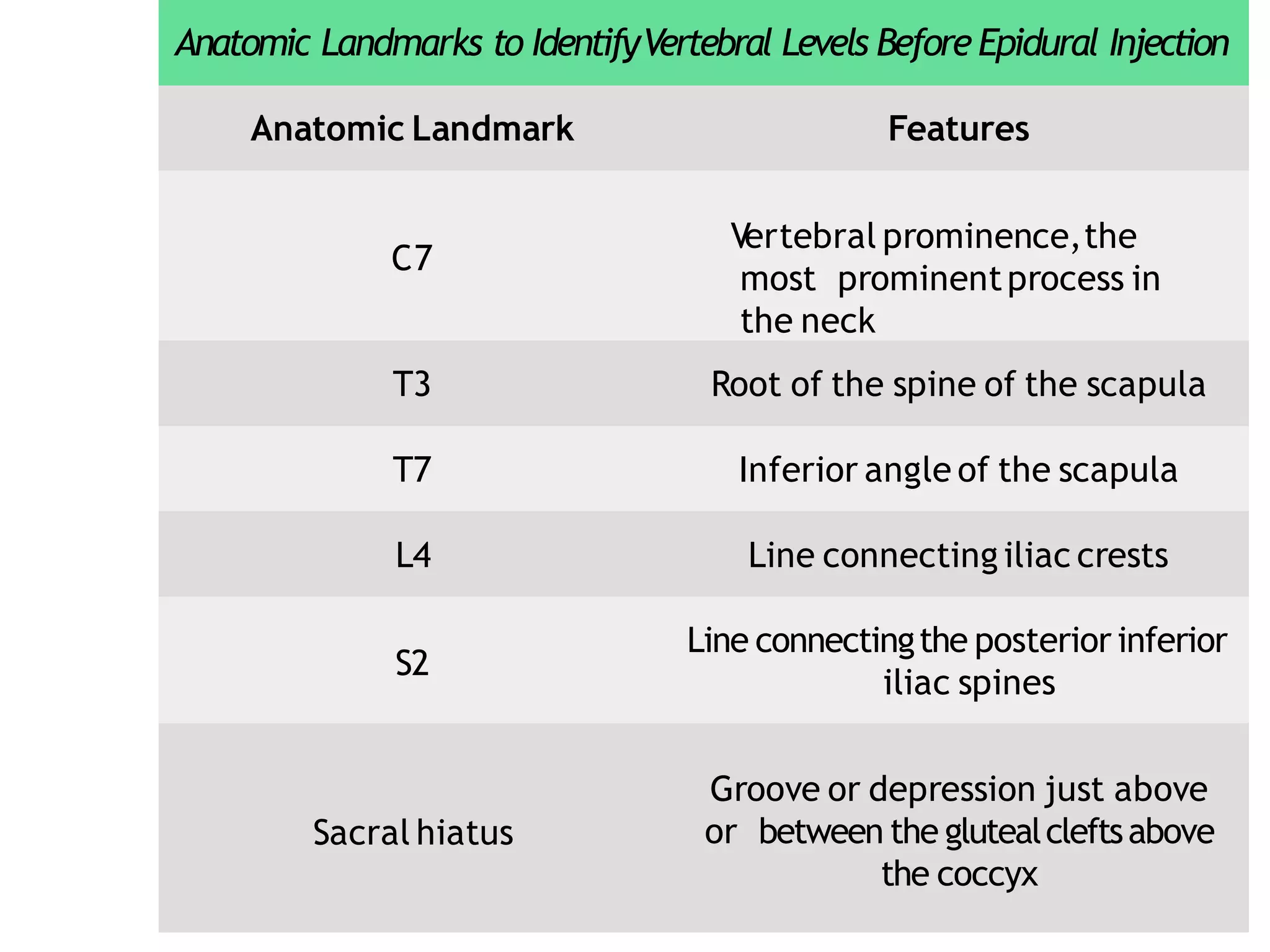
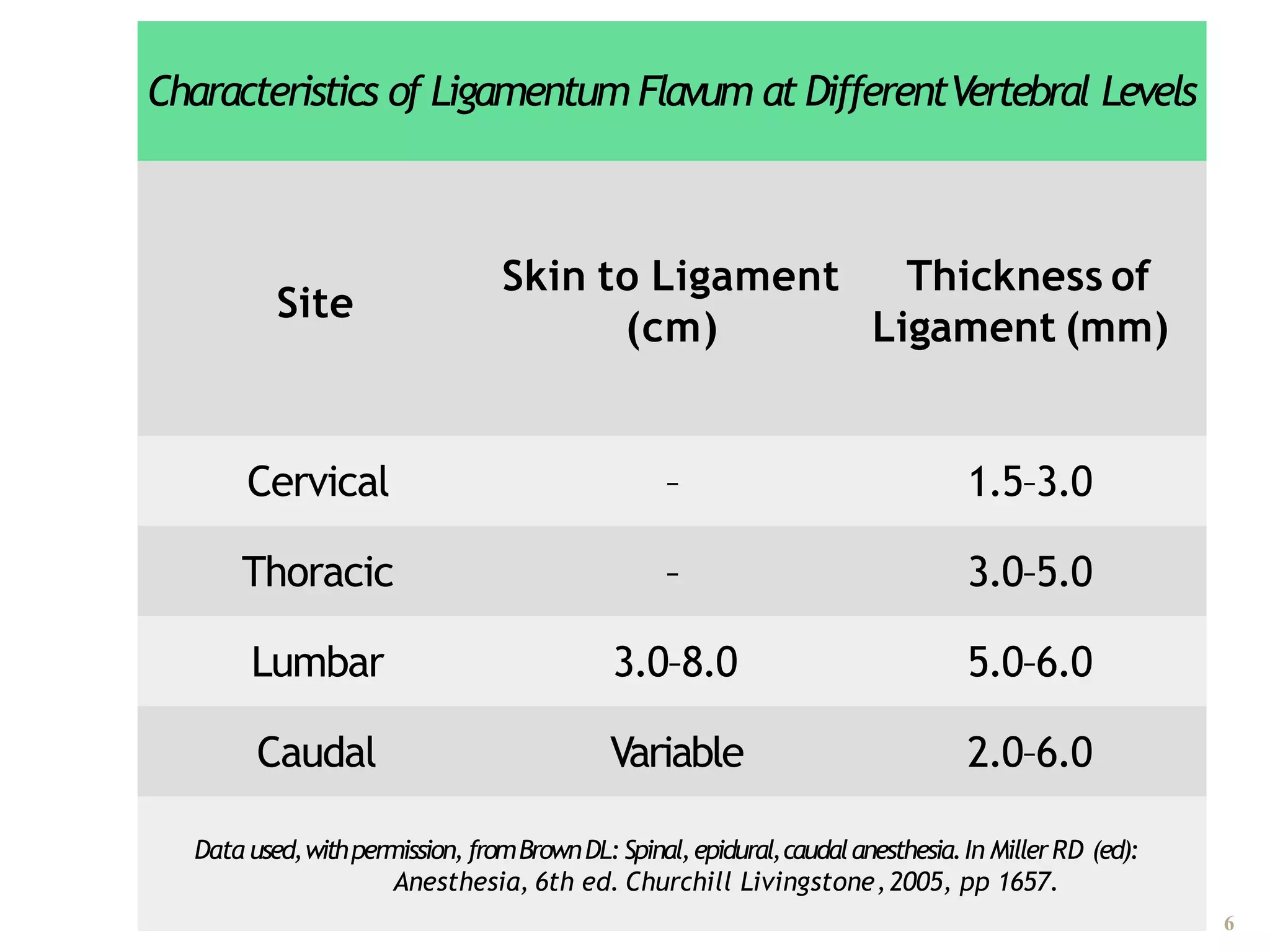
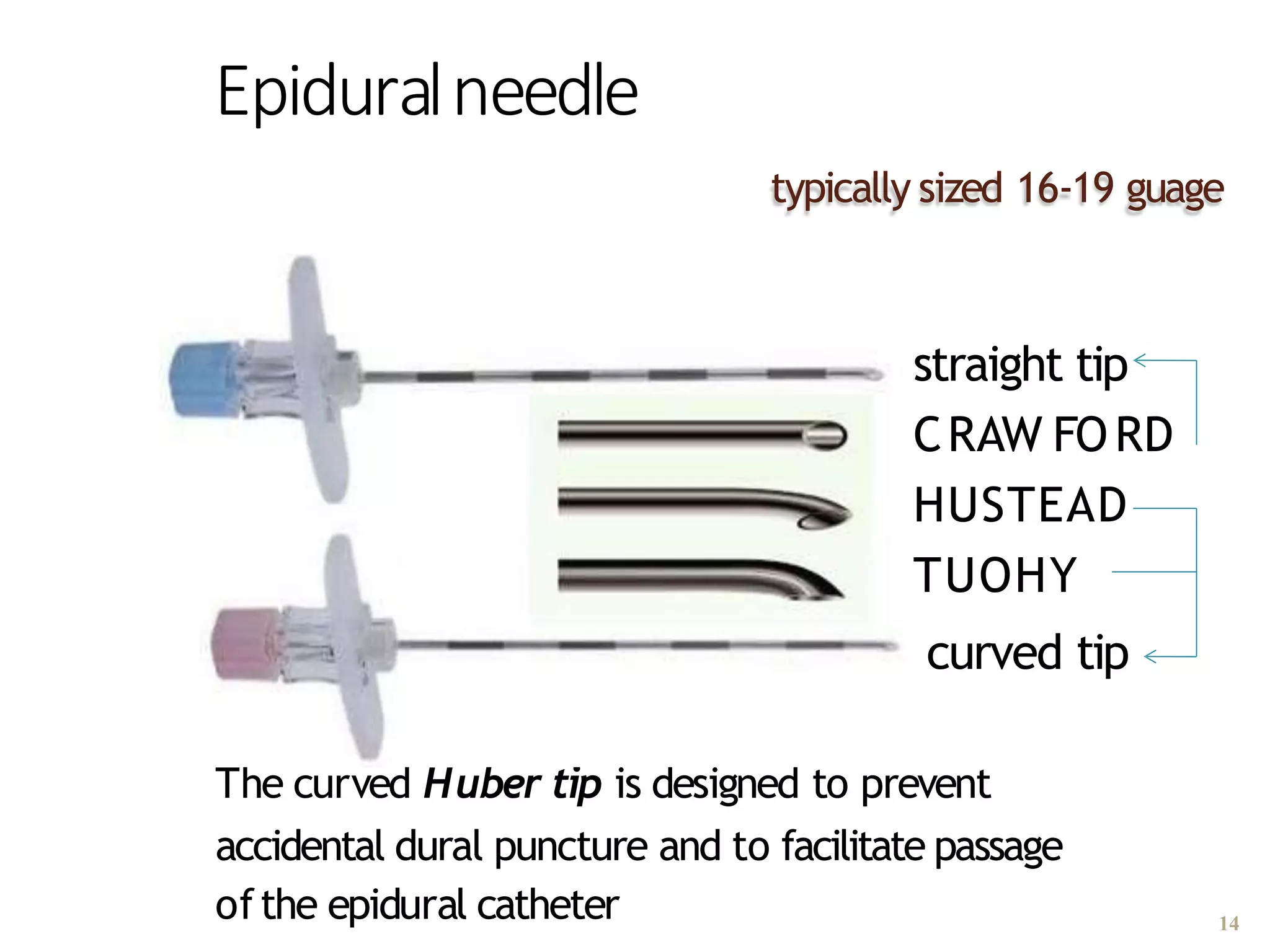
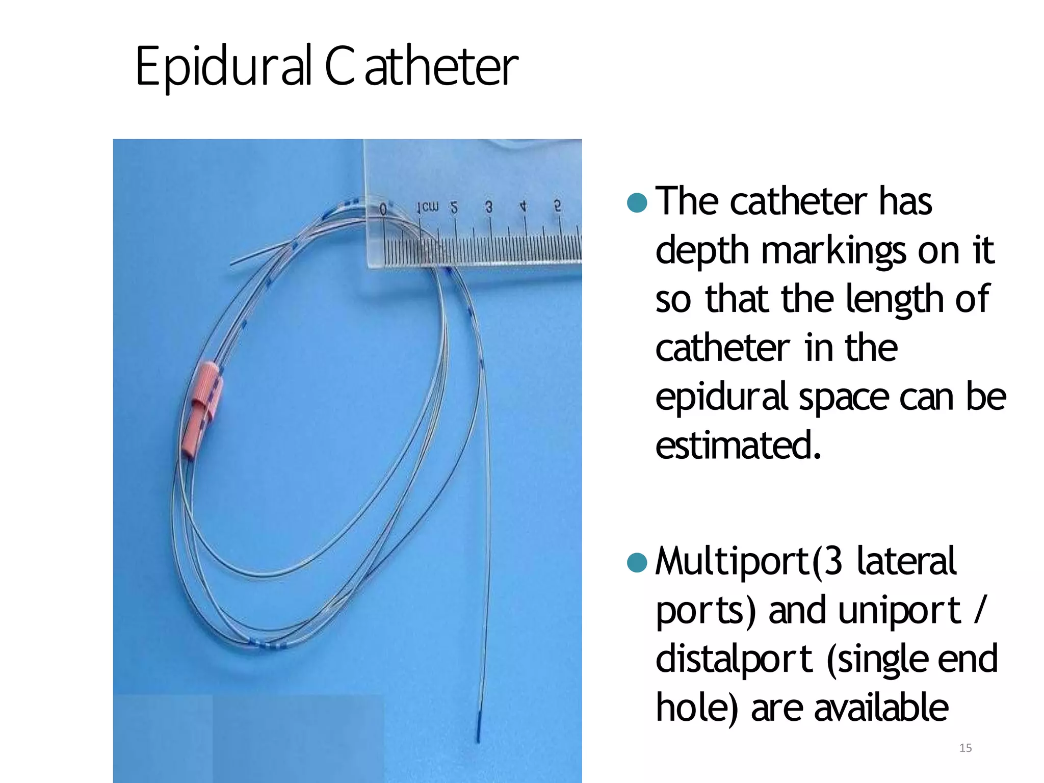
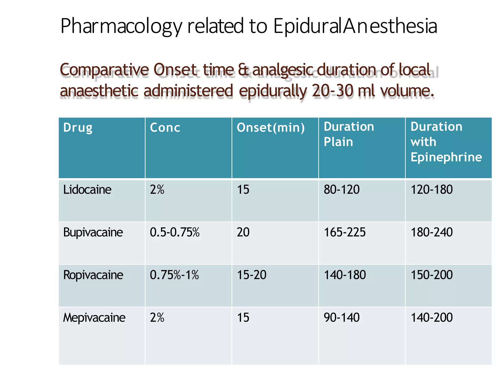
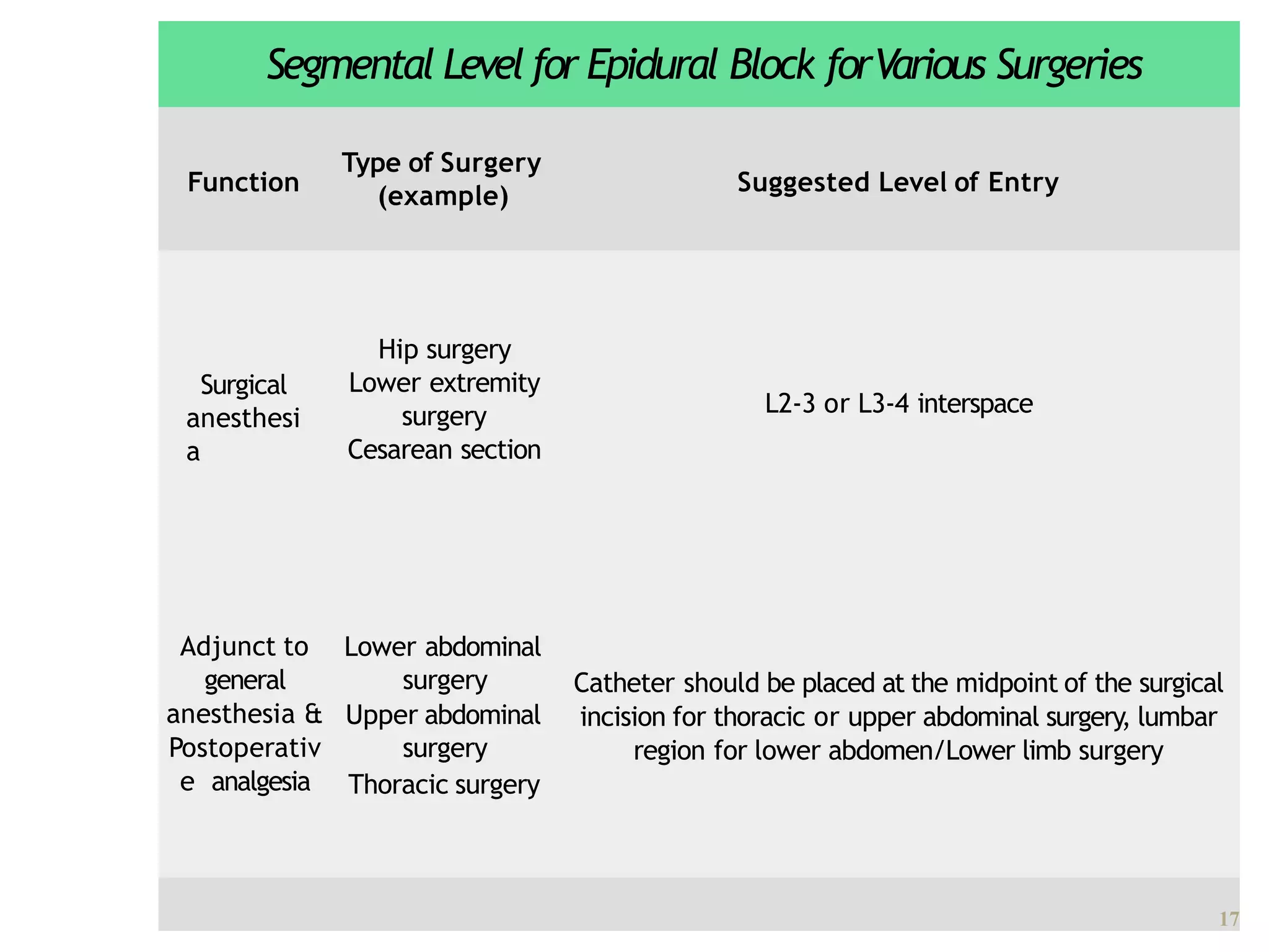
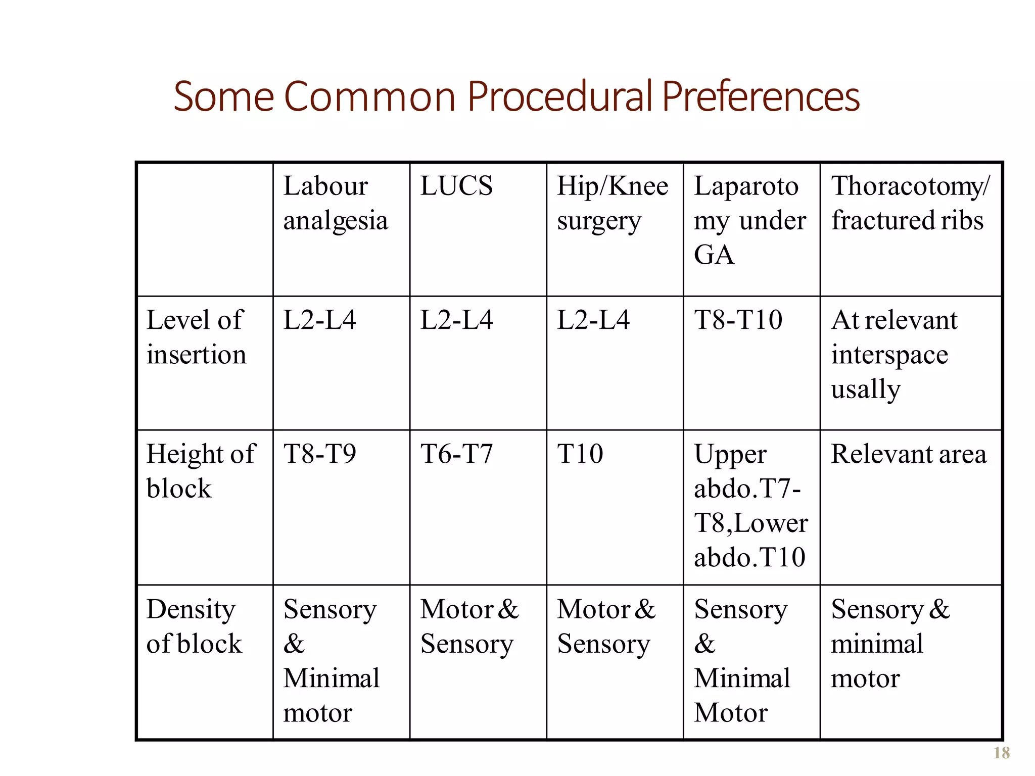
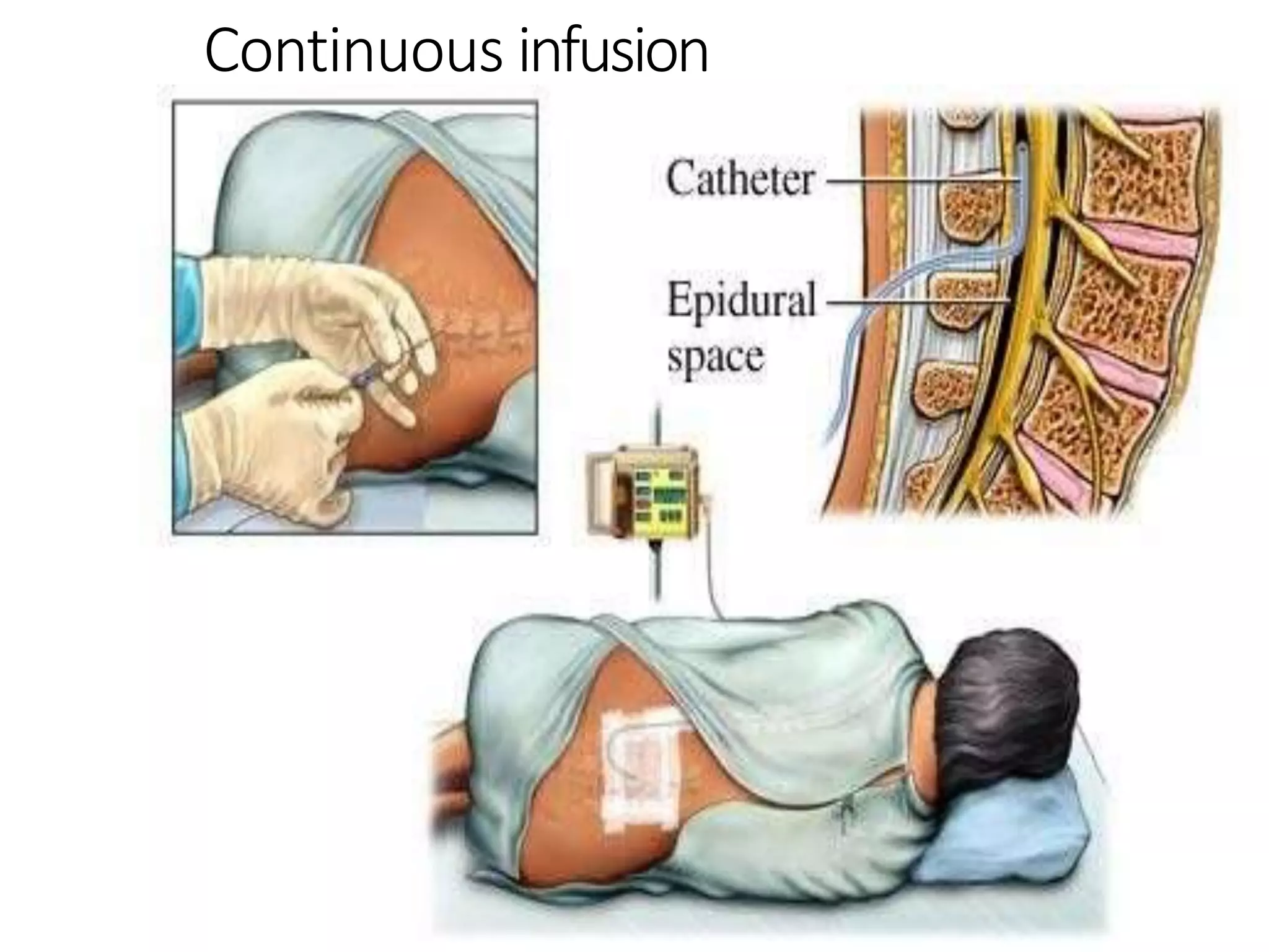
This document discusses epidural, caudal, and paravertebral block techniques. It begins with a brief history of epidural anesthesia and describes relevant anatomy like the epidural space and its boundaries. It then covers the indications, contraindications, advantages, and disadvantages of epidural anesthesia. The document discusses epidural needle and catheter types and placement techniques. It also provides details on pharmacology, dosing, and procedures related to epidural anesthesia.
The transversus abdominis plane, more commonly referred to as the TAP block,
Places local anesthetic in the lateral abdominal wall in a plane between the internal oblique and the transversus abdominis muscles.
Here, the local anesthetic block can block many of the abdominal nerves as they pass to the abdominal structures.
A basic overview on the management of intra-operative bronchospasm: the risk factors, triggers, diagnosis, prevention and management. Includes a case scenario – discussion.
A case report of open reduction, internal fixation and platting of clavicle f...iosrjce
IOSR Journal of Dental and Medical Sciences is one of the speciality Journal in Dental Science and Medical Science published by International Organization of Scientific Research (IOSR). The Journal publishes papers of the highest scientific merit and widest possible scope work in all areas related to medical and dental science. The Journal welcome review articles, leading medical and clinical research articles, technical notes, case reports and others.
Ozempic: Preoperative Management of Patients on GLP-1 Receptor Agonists Saeid Safari
Preoperative Management of Patients on GLP-1 Receptor Agonists like Ozempic and Semiglutide
ASA GUIDELINE
NYSORA Guideline
2 Case Reports of Gastric Ultrasound
Lung Cancer: Artificial Intelligence, Synergetics, Complex System Analysis, S...Oleg Kshivets
RESULTS: Overall life span (LS) was 2252.1±1742.5 days and cumulative 5-year survival (5YS) reached 73.2%, 10 years – 64.8%, 20 years – 42.5%. 513 LCP lived more than 5 years (LS=3124.6±1525.6 days), 148 LCP – more than 10 years (LS=5054.4±1504.1 days).199 LCP died because of LC (LS=562.7±374.5 days). 5YS of LCP after bi/lobectomies was significantly superior in comparison with LCP after pneumonectomies (78.1% vs.63.7%, P=0.00001 by log-rank test). AT significantly improved 5YS (66.3% vs. 34.8%) (P=0.00000 by log-rank test) only for LCP with N1-2. Cox modeling displayed that 5YS of LCP significantly depended on: phase transition (PT) early-invasive LC in terms of synergetics, PT N0—N12, cell ratio factors (ratio between cancer cells- CC and blood cells subpopulations), G1-3, histology, glucose, AT, blood cell circuit, prothrombin index, heparin tolerance, recalcification time (P=0.000-0.038). Neural networks, genetic algorithm selection and bootstrap simulation revealed relationships between 5YS and PT early-invasive LC (rank=1), PT N0—N12 (rank=2), thrombocytes/CC (3), erythrocytes/CC (4), eosinophils/CC (5), healthy cells/CC (6), lymphocytes/CC (7), segmented neutrophils/CC (8), stick neutrophils/CC (9), monocytes/CC (10); leucocytes/CC (11). Correct prediction of 5YS was 100% by neural networks computing (area under ROC curve=1.0; error=0.0).
CONCLUSIONS: 5YS of LCP after radical procedures significantly depended on: 1) PT early-invasive cancer; 2) PT N0--N12; 3) cell ratio factors; 4) blood cell circuit; 5) biochemical factors; 6) hemostasis system; 7) AT; 8) LC characteristics; 9) LC cell dynamics; 10) surgery type: lobectomy/pneumonectomy; 11) anthropometric data. Optimal diagnosis and treatment strategies for LC are: 1) screening and early detection of LC; 2) availability of experienced thoracic surgeons because of complexity of radical procedures; 3) aggressive en block surgery and adequate lymph node dissection for completeness; 4) precise prediction; 5) adjuvant chemoimmunoradiotherapy for LCP with unfavorable prognosis.
Couples presenting to the infertility clinic- Do they really have infertility...Sujoy Dasgupta
Dr Sujoy Dasgupta presented the study on "Couples presenting to the infertility clinic- Do they really have infertility? – The unexplored stories of non-consummation" in the 13th Congress of the Asia Pacific Initiative on Reproduction (ASPIRE 2024) at Manila on 24 May, 2024.
Report Back from SGO 2024: What’s the Latest in Cervical Cancer?bkling
Are you curious about what’s new in cervical cancer research or unsure what the findings mean? Join Dr. Emily Ko, a gynecologic oncologist at Penn Medicine, to learn about the latest updates from the Society of Gynecologic Oncology (SGO) 2024 Annual Meeting on Women’s Cancer. Dr. Ko will discuss what the research presented at the conference means for you and answer your questions about the new developments.
New Drug Discovery and Development .....NEHA GUPTA
The "New Drug Discovery and Development" process involves the identification, design, testing, and manufacturing of novel pharmaceutical compounds with the aim of introducing new and improved treatments for various medical conditions. This comprehensive endeavor encompasses various stages, including target identification, preclinical studies, clinical trials, regulatory approval, and post-market surveillance. It involves multidisciplinary collaboration among scientists, researchers, clinicians, regulatory experts, and pharmaceutical companies to bring innovative therapies to market and address unmet medical needs.
Prix Galien International 2024 Forum ProgramLevi Shapiro
June 20, 2024, Prix Galien International and Jerusalem Ethics Forum in ROME. Detailed agenda including panels:
- ADVANCES IN CARDIOLOGY: A NEW PARADIGM IS COMING
- WOMEN’S HEALTH: FERTILITY PRESERVATION
- WHAT’S NEW IN THE TREATMENT OF INFECTIOUS,
ONCOLOGICAL AND INFLAMMATORY SKIN DISEASES?
- ARTIFICIAL INTELLIGENCE AND ETHICS
- GENE THERAPY
- BEYOND BORDERS: GLOBAL INITIATIVES FOR DEMOCRATIZING LIFE SCIENCE TECHNOLOGIES AND PROMOTING ACCESS TO HEALTHCARE
- ETHICAL CHALLENGES IN LIFE SCIENCES
- Prix Galien International Awards Ceremony
ARTIFICIAL INTELLIGENCE IN HEALTHCARE.pdfAnujkumaranit
Artificial intelligence (AI) refers to the simulation of human intelligence processes by machines, especially computer systems. It encompasses tasks such as learning, reasoning, problem-solving, perception, and language understanding. AI technologies are revolutionizing various fields, from healthcare to finance, by enabling machines to perform tasks that typically require human intelligence.
These lecture slides, by Dr Sidra Arshad, offer a quick overview of physiological basis of a normal electrocardiogram.
Learning objectives:
1. Define an electrocardiogram (ECG) and electrocardiography
2. Describe how dipoles generated by the heart produce the waveforms of the ECG
3. Describe the components of a normal electrocardiogram of a typical bipolar leads (limb II)
4. Differentiate between intervals and segments
5. Enlist some common indications for obtaining an ECG
Study Resources:
1. Chapter 11, Guyton and Hall Textbook of Medical Physiology, 14th edition
2. Chapter 9, Human Physiology - From Cells to Systems, Lauralee Sherwood, 9th edition
3. Chapter 29, Ganong’s Review of Medical Physiology, 26th edition
4. Electrocardiogram, StatPearls - https://www.ncbi.nlm.nih.gov/books/NBK549803/
5. ECG in Medical Practice by ABM Abdullah, 4th edition
6. ECG Basics, http://www.nataliescasebook.com/tag/e-c-g-basics
Ethanol (CH3CH2OH), or beverage alcohol, is a two-carbon alcohol
that is rapidly distributed in the body and brain. Ethanol alters many
neurochemical systems and has rewarding and addictive properties. It
is the oldest recreational drug and likely contributes to more morbidity,
mortality, and public health costs than all illicit drugs combined. The
5th edition of the Diagnostic and Statistical Manual of Mental Disorders
(DSM-5) integrates alcohol abuse and alcohol dependence into a single
disorder called alcohol use disorder (AUD), with mild, moderate,
and severe subclassifications (American Psychiatric Association, 2013).
In the DSM-5, all types of substance abuse and dependence have been
combined into a single substance use disorder (SUD) on a continuum
from mild to severe. A diagnosis of AUD requires that at least two of
the 11 DSM-5 behaviors be present within a 12-month period (mild
AUD: 2–3 criteria; moderate AUD: 4–5 criteria; severe AUD: 6–11 criteria).
The four main behavioral effects of AUD are impaired control over
drinking, negative social consequences, risky use, and altered physiological
effects (tolerance, withdrawal). This chapter presents an overview
of the prevalence and harmful consequences of AUD in the U.S.,
the systemic nature of the disease, neurocircuitry and stages of AUD,
comorbidities, fetal alcohol spectrum disorders, genetic risk factors, and
pharmacotherapies for AUD.
New Directions in Targeted Therapeutic Approaches for Older Adults With Mantl...i3 Health
i3 Health is pleased to make the speaker slides from this activity available for use as a non-accredited self-study or teaching resource.
This slide deck presented by Dr. Kami Maddocks, Professor-Clinical in the Division of Hematology and
Associate Division Director for Ambulatory Operations
The Ohio State University Comprehensive Cancer Center, will provide insight into new directions in targeted therapeutic approaches for older adults with mantle cell lymphoma.
STATEMENT OF NEED
Mantle cell lymphoma (MCL) is a rare, aggressive B-cell non-Hodgkin lymphoma (NHL) accounting for 5% to 7% of all lymphomas. Its prognosis ranges from indolent disease that does not require treatment for years to very aggressive disease, which is associated with poor survival (Silkenstedt et al, 2021). Typically, MCL is diagnosed at advanced stage and in older patients who cannot tolerate intensive therapy (NCCN, 2022). Although recent advances have slightly increased remission rates, recurrence and relapse remain very common, leading to a median overall survival between 3 and 6 years (LLS, 2021). Though there are several effective options, progress is still needed towards establishing an accepted frontline approach for MCL (Castellino et al, 2022). Treatment selection and management of MCL are complicated by the heterogeneity of prognosis, advanced age and comorbidities of patients, and lack of an established standard approach for treatment, making it vital that clinicians be familiar with the latest research and advances in this area. In this activity chaired by Michael Wang, MD, Professor in the Department of Lymphoma & Myeloma at MD Anderson Cancer Center, expert faculty will discuss prognostic factors informing treatment, the promising results of recent trials in new therapeutic approaches, and the implications of treatment resistance in therapeutic selection for MCL.
Target Audience
Hematology/oncology fellows, attending faculty, and other health care professionals involved in the treatment of patients with mantle cell lymphoma (MCL).
Learning Objectives
1.) Identify clinical and biological prognostic factors that can guide treatment decision making for older adults with MCL
2.) Evaluate emerging data on targeted therapeutic approaches for treatment-naive and relapsed/refractory MCL and their applicability to older adults
3.) Assess mechanisms of resistance to targeted therapies for MCL and their implications for treatment selection
Title: Sense of Smell
Presenter: Dr. Faiza, Assistant Professor of Physiology
Qualifications:
MBBS (Best Graduate, AIMC Lahore)
FCPS Physiology
ICMT, CHPE, DHPE (STMU)
MPH (GC University, Faisalabad)
MBA (Virtual University of Pakistan)
Learning Objectives:
Describe the primary categories of smells and the concept of odor blindness.
Explain the structure and location of the olfactory membrane and mucosa, including the types and roles of cells involved in olfaction.
Describe the pathway and mechanisms of olfactory signal transmission from the olfactory receptors to the brain.
Illustrate the biochemical cascade triggered by odorant binding to olfactory receptors, including the role of G-proteins and second messengers in generating an action potential.
Identify different types of olfactory disorders such as anosmia, hyposmia, hyperosmia, and dysosmia, including their potential causes.
Key Topics:
Olfactory Genes:
3% of the human genome accounts for olfactory genes.
400 genes for odorant receptors.
Olfactory Membrane:
Located in the superior part of the nasal cavity.
Medially: Folds downward along the superior septum.
Laterally: Folds over the superior turbinate and upper surface of the middle turbinate.
Total surface area: 5-10 square centimeters.
Olfactory Mucosa:
Olfactory Cells: Bipolar nerve cells derived from the CNS (100 million), with 4-25 olfactory cilia per cell.
Sustentacular Cells: Produce mucus and maintain ionic and molecular environment.
Basal Cells: Replace worn-out olfactory cells with an average lifespan of 1-2 months.
Bowman’s Gland: Secretes mucus.
Stimulation of Olfactory Cells:
Odorant dissolves in mucus and attaches to receptors on olfactory cilia.
Involves a cascade effect through G-proteins and second messengers, leading to depolarization and action potential generation in the olfactory nerve.
Quality of a Good Odorant:
Small (3-20 Carbon atoms), volatile, water-soluble, and lipid-soluble.
Facilitated by odorant-binding proteins in mucus.
Membrane Potential and Action Potential:
Resting membrane potential: -55mV.
Action potential frequency in the olfactory nerve increases with odorant strength.
Adaptation Towards the Sense of Smell:
Rapid adaptation within the first second, with further slow adaptation.
Psychological adaptation greater than receptor adaptation, involving feedback inhibition from the central nervous system.
Primary Sensations of Smell:
Camphoraceous, Musky, Floral, Pepperminty, Ethereal, Pungent, Putrid.
Odor Detection Threshold:
Examples: Hydrogen sulfide (0.0005 ppm), Methyl-mercaptan (0.002 ppm).
Some toxic substances are odorless at lethal concentrations.
Characteristics of Smell:
Odor blindness for single substances due to lack of appropriate receptor protein.
Behavioral and emotional influences of smell.
Transmission of Olfactory Signals:
From olfactory cells to glomeruli in the olfactory bulb, involving lateral inhibition.
Primitive, less old, and new olfactory systems with different path
Recomendações da OMS sobre cuidados maternos e neonatais para uma experiência pós-natal positiva.
Em consonância com os ODS – Objetivos do Desenvolvimento Sustentável e a Estratégia Global para a Saúde das Mulheres, Crianças e Adolescentes, e aplicando uma abordagem baseada nos direitos humanos, os esforços de cuidados pós-natais devem expandir-se para além da cobertura e da simples sobrevivência, de modo a incluir cuidados de qualidade.
Estas diretrizes visam melhorar a qualidade dos cuidados pós-natais essenciais e de rotina prestados às mulheres e aos recém-nascidos, com o objetivo final de melhorar a saúde e o bem-estar materno e neonatal.
Uma “experiência pós-natal positiva” é um resultado importante para todas as mulheres que dão à luz e para os seus recém-nascidos, estabelecendo as bases para a melhoria da saúde e do bem-estar a curto e longo prazo. Uma experiência pós-natal positiva é definida como aquela em que as mulheres, pessoas que gestam, os recém-nascidos, os casais, os pais, os cuidadores e as famílias recebem informação consistente, garantia e apoio de profissionais de saúde motivados; e onde um sistema de saúde flexível e com recursos reconheça as necessidades das mulheres e dos bebês e respeite o seu contexto cultural.
Estas diretrizes consolidadas apresentam algumas recomendações novas e já bem fundamentadas sobre cuidados pós-natais de rotina para mulheres e neonatos que recebem cuidados no pós-parto em unidades de saúde ou na comunidade, independentemente dos recursos disponíveis.
É fornecido um conjunto abrangente de recomendações para cuidados durante o período puerperal, com ênfase nos cuidados essenciais que todas as mulheres e recém-nascidos devem receber, e com a devida atenção à qualidade dos cuidados; isto é, a entrega e a experiência do cuidado recebido. Estas diretrizes atualizam e ampliam as recomendações da OMS de 2014 sobre cuidados pós-natais da mãe e do recém-nascido e complementam as atuais diretrizes da OMS sobre a gestão de complicações pós-natais.
O estabelecimento da amamentação e o manejo das principais intercorrências é contemplada.
Recomendamos muito.
Vamos discutir essas recomendações no nosso curso de pós-graduação em Aleitamento no Instituto Ciclos.
Esta publicação só está disponível em inglês até o momento.
Prof. Marcus Renato de Carvalho
www.agostodourado.com
2. History ofEpidural Anaesthesia
2
⚫ Fidel Pages first used Epidural anaesthesia in human in
1921.
⚫ T
uohy introduced the needle used in Epidural
Anaesthesia in 1945 .
⚫ Manuel Martinez used the T
uohy needle with a silk
ureteral catheter to provide continuous segmental
lumbar “peridural”anesthesia.
3. Anatomy
⚫ The epidural space is the
potential space between
ligamentum flavum and dura
inside the bony spinal canal
⚫ It extends from the base of
the skull to the sacral hiatus,
and surrounds the dura
mater anteriorly
,laterally
,and
posteriorly.
⚫ It comprises of anterior and
posterior epidural spaces.
3
5. 5
Anatomic Landmarks to IdentifyVertebral Levels Before Epidural Injection
Anatomic Landmark Features
C7
Vertebral prominence,the
most prominentprocess in
the neck
T3 Root of the spine of the scapula
T7 Inferior angle of the scapula
L4 Line connecting iliac crests
S2
Line connectingthe posterior inferior
iliac spines
Sacral hiatus
Groove or depression just above
or between the glutealcleftsabove
the coccyx
6. 6
Characteristics of Ligamentum Flavum at DifferentVertebral Levels
Site
Skin to Ligament
(cm)
Thickness of
Ligament (mm)
Cervical – 1.5–3.0
Thoracic – 3.0–5.0
Lumbar 3.0–8.0 5.0–6.0
Caudal Variable 2.0–6.0
Data used,withpermission, fromBrownDL:Spinal,epidural,caudalanesthesia.In MillerRD (ed):
Anesthesia, 6th ed. Churchill Livingstone,2005, pp 1657.
7. Epidural anesthesia
7
⚫Form of regional anesthesiainvolving
injection ofdrugs into the epidural space.
⚫It can be performed as a single shot
techniqueor a catheter can be placed for
either bolus dosing or continuous infusion.
8. Indications
8
⚫ Epidural anaesthesiawith or without sedation has been
used as the sole anaesthetic or as an adjunct to general
anaesthesia (reduces patient’srequirement for opioid
analgesics)
⚫ Orthopaedic surgery :Major hip/kneesurgery,pelvic
fractures
⚫ Obstetrics:Caesarean section,labour analgesia
⚫ Gynaecologic surgery :Procedures involvingfemale
pelvicorgans
⚫ Urologic surgery :Prostate,bladder procedures
⚫ General surgery :Upper and lower abdominal
procedures
⚫ Paediatric procedures :Penile procedures,inguinalhernia
repair
, analsurgery, orthopaedic procedures on the feet;
supplementto GA,postoperative pain relief.
9. ⚫ Vascular surgery :Vascular reconstruction of the
lower limb vessels, amputations involving the lower
extremities.
⚫ Thoracic surgery :Postoperative analgesia,
combinationwith GA to reduce GA requirements.
⚫ Diagnosis and managementof chronic pain :
Chronic benign pain- Cervical & lumbar
radiculopathy
,vertebralcompression fracture ,
degenerativedisc disease,peripheralneuropathy
,
low back pain,pelvic pain syndrome.
⚫ Cancer related pain- pain secondary to face,neck,
shoulder, genital, pelvic, perineal etc .malignancy
.&
chemotherapyrelated peripheral neuropathy.
9
10. Contraindications
10
⚫Patient refusal
⚫Infection at site
⚫Raised ICP
⚫SevereAS, severe MS
⚫Allergy to LA drugs
⚫Severe hypovolaemia/shock
⚫Coagulation disorder
⚫Pre-existing neurological disease
⚫Demyelinating disease(Multiple sclerosis)
⚫Abnormalities of spine
⚫Uncooperative patient
11. Advantages
11
Epidural blockade is becoming one of the most useful and
versatile procedures in modern anesthesiology.It is uniquein
that it can be placedat virtually any levelof the spine,
allowingmore flexibilityin its applicationto clinicalpractice.
Minimal effectof surgery on Cardiopulmonaryreserve
compared to GA
In pt.with compromised Respiratorysystem
Morbid obesity
COPD
Elderly
Earlier mobilization
Decreased chancesof DVT
It can be used to supplementGA, decreasingthe need for
deep levels of GA,therefore providinga more
hemodynamicallystable operative course.
12. Thoracic epidural analgesia has been shown to
decrease the incidence of myocardial infarction,
postoperative pulmonary complications and to
promote the return of gastrointestinal motility
without compromising fresh suture lines in the GI
tract.
Effective postoperative analgesia without taking
systemic opioids (analgesics are given through
epidural catheter)-rapid recovery
Neuroendocrine stress response to surgery is
decreased.
Blood loss is less.
12
13. Disadvantages
13
⚫Risk of block failure /patchy block
⚫Onset is slower
⚫Risk of introducing infection
⚫Epidural hematoma
⚫Continuous epidural catheter should not
be used in the ward ifmonitoring is not
proper.
⚫Risk ofdural puncture with 18-guage
needle
14. Epiduralneedle
typically sized 16-19 guage
straight tip
CRAW FORD
HUSTEAD
TUOHY
curved tip
The curved Huber tip is designed to prevent
accidental dural puncture and to facilitate passage
ofthe epidural catheter 14
15. EpiduralCatheter
15
⚫The catheter has
depth markings on it
so that the length of
catheter in the
epidural space can be
estimated.
⚫Multiport(3 lateral
ports) and uniport /
distalport (single end
hole) are available
16. Pharmacology related to EpiduralAnesthesia
16
Comparative Onset time & analgesic duration of local
anaesthetic administered epidurally 20-30 ml volume.
Drug Conc Onset(min) Duration
Plain
Duration
with
Epinephrine
Lidocaine 2% 15 80-120 120-180
Bupivacaine 0.5-0.75% 20 165-225 180-240
Ropivacaine 0.75%-1% 15-20 140-180 150-200
Mepivacaine 2% 15 90-140 140-200
17. Segmental Levelfor Epidural Block forVarious Surgeries
Function
Type of Surgery
(example)
Suggested Level of Entry
Surgical
anesthesi
a
Hip surgery
Lower extremity
surgery
Cesarean section
L2-3 or L3-4 interspace
Adjunct to
general
anesthesia &
Postoperativ
e analgesia
Lower abdominal
surgery
Upper abdominal
surgery
Thoracic surgery
Catheter should be placed at the midpoint of the surgical
incision for thoracic or upper abdominal surgery, lumbar
region for lower abdomen/Lower limb surgery
17
18. SomeCommon ProceduralPreferences
Labour
analgesia
LUCS Hip/Knee
surgery
Laparoto
my under
GA
Thoracotomy/
fractured ribs
Level of
insertion
L2-L4 L2-L4 L2-L4 T8-T10 At relevant
interspace
usally
Height of
block
T8-T9 T6-T7 T10 Upper
abdo.T7-
T8,Lower
abdo.T10
Relevant area
Density
of block
Sensory
&
Minimal
motor
Motor&
Sensory
Motor&
Sensory
Sensory
&
Minimal
Motor
Sensory&
minimal
motor
18
19. Labour
Analgesia
LSCS Hip/Knee
surgery
Laparotomy
under GA
Thoracotomy
/
Fractured ribs
Choice of
local
anaesthetic
0.1%-0.25%
Bupivacaine
Lignocaine2
% +
Bupivacaine
0.5%
Bupivacaine
0.5%
0.25%-
0.5%
Bupivacain
in theatre
0.25%-0.5%
Bupivacaine
in theatre or
establish
block
Infusion Bupivacaine.
1%+
Fentanyl
2mcg/ml
Post op
Bupivacaine
0.166%+Dia
morphine
.1mg/ml
Not usually
necessary
Post op
Bupivacain
0.166%+Di
amorphine
0.1mg/ml
Post op
Bupivacaine
0.166%+Dia
morphine
.1mg/ml
Rate of
infusion
0-12 ml/hr 0-8mls/hr -- 0-12mls/hr 0-8mls/hr
19
20. Pre operative preparation
20
⚫ Intravenous access with a catheter large enough to
administer fluids or emergency drugs should be in
place (i.e.,18- to 20-gauge).
⚫ Reversible conditions such as severe hypovolemia
should be managed prior to block placement.
⚫ Drugs and equipmentfor life support,including
airway management,must be readily available
⚫ Sedatingthe patientwith a benzodiazepineor a
narcotic allows the clinician to safely place the
epiduralblock.(Exception:pregnantmothers for
labour and delivery or caesarian section).
⚫ Monitoring blood pressure and pulse oximetry are
a minimumrequirement.
21. Performing theprocedure
21
Position of patient- Careful attention to the patient’
s
position is essential to successful placement ofthe
epidural needle and catheter
.
Depending on the patient’
s medical status,weight,and
ability to cooperate,the sitting or lateral decubitus
position can be used.
Easier in sitting position.
Approach - Four common approaches to the epidural
space are possible:
1. Midline,
2. Paramedian,
3. T
aylor (modified paramedian),
4. Caudal
22. ⚫ Needle angulation requiredtoaccomplishepiduralblockadein
the high thoracic/low thoracic/lum
bar regions.
A: High thoracic region.B: Low thoracic region.C: Lumbar region.
22
23. LocatingtheEpiduralspace
23
⚫All aseptic precaution is taken.
⚫Skin is infiltrated with local anaesthetic in to
desired space(identified).
⚫Needle is advanced slowly,feel of increase
resistance.
⚫ 3 methods are used to identify Epidural space-
Loss of resistance (to with air or fluid)
Hanging drop method
Ultrasonography /Fluoroscopy
⚫As needle reaches Epidural space Loss of
Resistance is felt /Hanging drop is sucked in.
24. Loss of resistance technique
24
Glass syringe (Luer-Lok syringe) is used
27. Feedingthecatheter
27
⚫Catheter is threaded through needle after
placing in space.
⚫Needle is withdrawn over the catheter
.
⚫4-6 cms catheter remain in epidural space.
Threading more catheter may increase the
likelihood of catheter malposition.
⚫Catheter is firmly secured to skin with
surgical tape.
30. EpiduralDosing
30
⚫ As a general guideline,
1. 1–2 mL per segment in a lumbar epidural,
2. 0.7 mL per segment in a thoracic epidural,and
3. 3 mL per segment for a sacral/caudal epidural
is used as an initial loading dose.
⚫ T
est Dose
⚫ Incremental Dosing
⚫ Aspiration to check for blood or CSF before each
dose.
⚫ After the initial loading dose, one quarter to one third
ofthe amount can be administered 10–15 min later to
intensify the sensory block.The overall level ofthe
block will not be significantly increased with this
method.
31. T
estDose
⚫ The purpose of the“test dose” is to make sure that the
catheter is not in the subarachnoid, intravascular
, or
subduralspace.
⚫ The classic test dose combines 3 mL of1.5% lidocainewith
15 mcg ofepinephrine.
⚫ The intrathecal injection of 45 mg of lidocaine will
producea significantmotor block consistentwith spinal
anaesthesia.
⚫ A change in heart rate of 20% or greater is an indication
ofintravascular injection warrantingthe removal and
replacementofthe catheter
.
⚫ If the heart rate does not increase by 20% or greater
, or if
a significant motor block does not develop within 5 min of
administeringthe test dose,it is considerednegative.
⚫ False-ve ifpt is on β blocker
,false +ve in pregnancyif
coincideswith labour pain. 31
32. Incremental Dosing
32
⚫Its purposeis to avoid excessively high
anaesthetic levels.
⚫The loading dose should be given in 5-mL
aliquots through the catheter
,repeated at
3- to 5-min intervals,giving the clinician
time to assess the patient’
s response to
dosing.
⚫If at any time the patient demonstrates an
exaggerated response,further
incremental doses should be withheld and
the patient reassessed.
33. Repeat dose of LA:
3
Doses are administered before the block regresses to the
point where the patient experiences pain,the “time to
two-segment regression.” Defined as the time it takes
for the sensory block to regress by two dermatome levels.
At this point, one-third to one-half of the initial loading
dose can safely be administered to maintain the block.
Depends on the duration ofaction ofthe drug
3
Table 8. ClinicalEffectsof EpidurallyInjectedLocalAnesthetics
Drug (Concentration%)
Time toT
wo-segment
Regression (min)
RecommendedTime for "T
op-Up"
Dose fromInitialActivationof Dose
(min)
Chloroprocaine(3) 45–75 45
Lidocaine (2) 60–140 60
Mepivacaine (2) 90–160 90
Bupivacaine(0.5) 180–260 120
Ropivacaine (0.5–0.75) 180–260
120
34. Thoracic Epidurals - salient features
⚫ The paramedian approach is easier especiallyin the
midthoracic region.
⚫ Expectmore frequentfalse loss of resistance,especiallyif the
midlineapproach is used.
⚫ The test dose not only identifiesintravascularinjection,but
also serves as a means of identifyingplacementas a band of
anesthesiashould developin the segmentwhere the local
anestheticwas injected.
⚫ Because of the proximity to cardiac accelerator fibers,smaller
bolus doses oflocal anestheticshould be used and response
checked carefully before redosing to prevent large drops in
heart rate or blood pressure.
⚫ Remember that hypotension can occur in nearly allpatients
with a high thoracic epidural blockade.In fact,it has been said
that ifthere is no hypotension after an initial bolus in the high
thoracic epidural space,it is likelythat the epidural catheter is
not in the epidural space. 34
35. Factors affecting EpiduralAnaesthesia
Site of injection-
Lumbar- spread cranially more than caudally
Thoracic- spread evenly from site of injection
Upper thoracic & lower cervical fibres are
comparatively resistant d/
t larger size of nerve
roots-requires larger dose of LA.
Thoracic epidural space is smaller
,require lower
volume of drug.
Dose- 1-2 ml /segment.
Depends on volume & concentration of drug.
Higher conc. produces a profound motor and
sensory block,whereas low conc.a selective
sensory block. 35
36. Age - as pt age increases
intervertebral foramina
reduced size of
decreased epidural
space size and compliance.Decreased epidural fat
necessitates decrease of dose in elderly.
Weight - There is little correlation between
the spread of analgesia and the weight of the
patient.
In morbidly obese patients,there may be
compression of the epidural space secondarily to
increased intraabdominal pressure,creating a
higher block for a given dose of local anesthetic.
36
37. H eight - The correlation with height is usually
not clinically significant.
Ht.<5 ft 2 inch,reduce the dose to 1 mL per
segment to be blocked.
Bromage dosing regime - Increasing the dose of
local anaesthetic by 0.1 mL per segment for
each 2 in.over 5 ft of height.
Addition of Vasoconstrictors -
Epinephrine 5 mcg/
ml (1:200000) is most
commonly added.
Prolongs duration of action by reducing the
vascular absorption of drug.
37
38. Posture-
Block Ht. - Whether the patient is sitting or in
the lateral position, there is no significant
difference in block height.This is explained by the
fact that gravity and soln.baricity are not
intimately related to block spread.
Onset,Duration & Density - slightly faster on the
dependent side when the epidural in placed with
the patient in the lateral position
Pregnancy- Increased sensitivity to regional
anesthetics leads to faster onset time.
Engorgement of Epidural veins from caval
compression leads to increased incidence of
blood vessel puncture during procedure. 38
39. Physiologic EffectsofEpiduralBlock
39
⚫Most physiologic effects of epidural block stem
from Autonomic Blockade due to action of LA
on autonomic nerve fibres of the spinal cord.
⚫The actions mostly pertain to either Blockage
of Sympathetic outflow or Unopposed
dominance of Parasympathetic outflow.
40. Cardiovascular system
Block belowT5
⚫ Sympatheticblockade
venous return CO
venodilationin blocked segments
hypotension
⚫ The compensatory mechanismfor the decrease in mean
arterial pressurecauses
1)reflex vasoconstriction above the levelof the block
2)release in catecholaminesfrom the adrenal medulla.
Block aboveT4 (cardiac sym fibresT1-T4)
In addition to profound hypotensionand bradycardia,high
levelofsympatheticblockadecauses
⚫ Increased CVP without an increase in stroke volume
⚫ Vasoconstriction in the head,neck,and upper limbs
⚫ Splanchnicnerve blockade with blockadeof medullary
secretion of catecholamines
⚫ Blockade of vasoconstrictiveeffecton the capacitance
vessels ofthe lower limbs 41
41. Respiratory system-
Minimal impact on Pulmonary function in normal
& healthy patient,even in case of high thoracic
block
There is concern regarding the use of epidural
blockade in patients with severe chronic lung
disease dependent on accessory muscle function
to maintain adequate ventilation, because paralysis
of respiratory muscles and changes in bronchial
tone from epidural analgesia can occur
.
41
42. Effects ofThoracic EpiduralAnalgesia on ventilatory
mechanisms in pts with severe lungdisease (Gruber etal)
42
Variable BeforeTEA AfterTEA ‘p’ value
Peak
inspiratory
flow rate
(L/sec)
0.48± 0.17 0.55 ± 0.14 0.02
Peak
expiratory
flow rate
(L/sec)
0.38 ± 0.17 0.40 ± 0.09 0.78
PEEP 4.8 ± 3.6 4.7 ± 3.9 0.67
Work of
breathing
(J/Ltr)
1.5 ± 0.5 1.5 ± 0.6 0.79
Maximum
inspirator
y
pressure
81.7 ± 25.5 76.8 ± 32 0.52
43. Gastrointestinal system
Lumbar segments (T5-L1) for major abdominal
surgeries
vagal dominance leads to increased secretions,inc
peristalsis and an contracted gut
Stable visceral perfusion prevents decrease in
intramucosal pH, post-op period epidural analgesia has a
protective effect on gastric mucosa
Thoracic segments (T1-T5) as adjunct to GA in thoracic
/cardiac /abdominal surgeries
Segmental sympatholysis creating an increase of
sympathetic activity in segments below the block
leading to impaired splanchnic blood flow has been a
concern.
Nausea is a common problem (20%) – treat with
atropine 44
44. Renal system-
Block both Sympathetic & Parasympathetic
fibres
Loss of control of bladder function
Urinary retention
Endocrine system-
⚫ Abolishes stress response to surgery.
⚫ Decreased release of catecholamines,GH,
cortisol, TSH, ADH, vasopressin, renin,
angiotensin,norepinephrine
44
45. Complications
45
Drug Related Complications:-
⚫ When an excessive dose of local anaesthetics is injected
into the epidural space or when a moderate dose is
accidentally injected into an epidural vein, systemic
toxicity can occur
.
CNS toxicity : Symptoms – Light headedness, tinnitus,
circumoral numbness and tingling, numbness of the
tongue, and blurred vision. Signs - Muscle twitching,
confusion,tremors of the facial muscles and extremities,
and shivering.
CVS toxicity :Range from mild changes in blood
pressure and pulse to complete cardiovascular collapse.
Treatment :Supportive,directed toward maintaining the
airway
,supporting ventilation, and cardiopulmonary
resuscitation ifnecessary
.
46. ProcedureRelatedComplications
46
⚫Minor Back Pain :Incidence between 20 and 30%.
Self-limiting.T
reated by NSAIDS,paracetamol.
⚫Postdural Puncture Headache:Due to
inadvertent dural puncture.
TOC – Epidural blood patch.
Rapid recovery in between 90 to 95% of patients after
blood patch.
Using sterile techniques, aneedle is inserted into the
epidural space at or one interspace below the prior
level of dural puncture.15 to 20 mL ofthe patient’
s
blood (drawn aseptically) is slowly injected into the
space.Rare complication – DuralAbcess after blood
patch.
47. ⚫Subarachnoid Injection/High or Total
Spinal :Due to inadvertent injection of epidural drug
dose in sub arachnoid space.Profound hypotension,
bradycardia,and apnea will occur
.
Unconsciousness follows as a result of the effect of
local anesthetic action on the brainstem.
Treatment includes airway support and intubation, 100%
oxygen,intravenous fluids and vasopressors to maintain
hemodynamic stability.Epinephrine should be used early
and in large enough doses.
⚫Major SubduralInjection:
Subdural space extends intracranially.
A small dose of local anesthetic can have a profound
effect.
Delayed onset by 10 to 15 min compared with a high
spinal.Treatment is similar to that of a high spinal. 47
48. N euraxialblockin settingofanticoagulant and
antiplateletdrugs
48
(recommended byAmerican Society ofRegional
Anesthesia)
⚫ Neuraxialblock and indwellingcatheters are safe in
patientson aspirin ,NSAID’s & cox-2 inhibitors
⚫ Discontinueclopidogrel for 7 days ,ticlopidinefor
14 days ,abciximab for 24-48 hrs ,tirofiban &
eptifibatidefor 4-8 hrs before technique.
⚫ Wait at least 12 hrs before last thromboprophylaxis
dose of LMWH and 24 hrs after last full dose
⚫ W hen LMW H is begun post-op first dose should
be withheldfor at least 24 hrs if using a twice daily
dosing regimen and 6-8 hrs ifusing once daily
dosing regimen
49. ⚫ An indwelling epidural catheter should not be removed
until 12 hrs after the last prophylaxis dose of LMWH,
and the next dose should be administered no sooner
than 2 hrs after catheter removal
⚫ If a single daily thromboprophylaxis dose of LMWH is
administered,then indwelling catheters may be
maintained postoperatively. But the concurrent use of
twice daily or therapeutic LMWH and an indwelling
epidural catheter is not recommended.
⚫ The LMWH dose is delayed for 24 hr if the patient
experienced excessive traumaduring attempted
epidural or spinal anesthesia.
⚫ Neuraxial blocks should not be performed in patients
chronically taking warfarin unless the warfarin is
stopped and the INR is <1.5
⚫ Neuraxial catheters should be removed only when the
INR is <1.5 49
50. Caudal space anatomy
50
⚫ The sacrum is a large triangularly shaped bone
formed by the fusion of the five sacral vertebrae.
⚫ The sacral canal contains the cauda equina
(including the filum terminale) and the spinal
meninges.
⚫ Sacral hiatus (a bony defect) is identified in the
posterior wall of the sacral canal,due to the
failure of fusion of the laminae of S5 and partially
S4
⚫ The caudal opening of the Sacral canal is the
sacral hiatus roofed by the firm elastic membrane,
the sacrococcygeal ligament,which is an
extension of the ligamentum flavum.
52. CaudalEpiduralAnaesthesia
52
⚫ Common regional technique in infants ,neonates and
pediatric pts
⚫ For lower abdominal and genitourological procedures
⚫ In adults,it is usually reserved for procedures requiring
blockage of the sacral and lumbar nerves,epidurography,
for lysis of adhesions in patients with low back pain
with radiculopathy after spinal surgery,cancer pain-bony
mets in pelvis or chemotherapy related peripheral
neuropathy.
⚫ Positions :
Lateraldecubitus- mainly paediatric
Prone- mainly in adults
Knee-chest position
53. ⚫ Point ofEntry is the Sacral Hiatus.
⚫ T
wo ways oflocating sacral hiatus:
1.Locate the posterior superior iliac spines.A line drawn
between them becomes one side of a equilateral triangle. At
the apex of the triangleis the sacral hiatus.
2.With firm pressure, identify the coccyx with the index finger
.
As the finger moves cephalad,the first pair of bony
protuberances are the cornu,which surrounds the hiatus.
53
54. Procedure for Caudal block
54
⚫ Prep and drape the skin in sterilefashion.
⚫ Patientis placed in a lateral or prone position (pillow
under pelvis ifprone).
⚫ Either asmaller gauge IV catheter (18- to 23-gauge) or a
20-gaugeepiduralneedleis advanced at a 45-degreeangle
from the back with the bevelup (to avoid penetratingthe
anterior sacral wall).
⚫ A distinct“pop” or “snap”is feltwhen the needlepierces
the sacrococcygealmembrane.
⚫ The needleangle is lowered to 160 degrees (almost flat)
toward the back. It is advanced not more than 1.5 cm
(usually between5 and 7 mm) in adults and not more than
0.5 cm in children.
⚫ Aspirate for blood or CSF before injectinglocal
anesthetic.
56. PARAVERTEBRAL BLOCK
•HISTORY
• Paravertebral block(PVB) was first performed in 1905 and
became a popular technique for the provision of analgesia in
the early part of twentieth century. However their use
declined over the years until a publication by EASON and
WYATT in 1979 began a renaissance.
• Since then a considerable number of good quality studies
have been published on PVB and it is now an established
regional anaesthesia technique.
57. ANATOMY OF THORACIC PARAVERTEBRAL SPACE
• The thoracic paravertebral space begins at T1 and extends caudally to
terminate at T12.Although PVBs can be performed in the cervical and
lumber regions,there is no direct communication between adjacent
levels in these areas.Most PVBs are therefore performed at the
thoracic level.
•BOUNDARIES
• Medial wall is formed by bodies of the vertebrae,
intervertebral disc and intervertebral foraminae.
• Anterolaterally the space is bounded by the parietal pleura
and the innermost intercostal membrane.
• Posteriorly it is bounded by the transverse processes(TPs) of
the thoracic vertebrae,head of the ribs, and the superior
costotransverse ligament.
58. CONTENTS OF PARAVERTEBRAL SPACE
•Spinal nerves
•White rami communicantes
•Grey rami communicantes
•The sympathetic chain
•Intercostal vessels
•Fat
59. INDICATIONS FOR PVB
Unilateral surgeries in
thoracoabdominal region
• Breast surgery
• Thoracic surgery
• Cholecystectomy
• Renal surgery
• Appendicectomy
• Inguinal hernia repair
Relief of acute pain
• Fractured ribs
• Liver capsule pain(trauma or
ruptured cysts)
Relief of chronic pain
• Neuropathic chest or abdominal
pain
• Complex regional pain syndrome
• Refractory angina pectoris
• Relief of cancer pain
60. CONTRAINDICATIONS
Absolute contraindications
• Patient refusal
• Local sepsis
• Tumours in the paravertebral
space at the level of injections
• Allergy to local anaesthetic
drugs
Relative contraindications
• Severe coagulopathy
• Severe respiratory
disease(where the patient
depends on intercostal
muscle function for
ventilation)
• Ipsilateral diaphragmatic
paresis.
• Severe spinal
deformities(kyphosis or
scoliosis)
61. PROCEDURE FOR PVB
POSITIONING
• If awake, the patient should be seated with the neck and back flexed.
• If performed under sedation or general anaesthesia,the patient is
turned to the lateral position with the operated side uppermost.
• A bag of saline or pillow can be placed between the patient and the
operating table surface at the level of the intended block,to open up
the spaces between adjacent TPs.
CHOOSING THE LEVEL
• If only one to four dermatomes need to be blocked, a single level PVB at
or bellow the mid-dermatomal level is usually significant.(e.g. for simple
mastectomy;T3 or T4 is an appropriate level.For open cholecystectomy
T6 or T7 should be selected)
• If spread greater than four dermatomes is required,then multiple
injections will block the area more reliably;(e.g. for mastectomy and
axillary dissection, a block from atleast T1-T6 will be required. Therefore
block should be performed at each level or at T1,T3 and T5)
62. ADVANTAGES OF PVB
• PVB is easier to learn and perform than thoracic epidural
anaesthesia.
• Analgesia is comparable with that provided by a thoracic
epidural, in terms of success rate and analgesic efficacy.
• PVB can be performed safely in fully anaesthetized patients.
• There is less risk of neurological complications than with
most other regional anaesthetic techniques.
• Pronounced hypotension is unusual because sympathetic
block is rarely bilateral.
• Urinary retention does not occur,unlike neuraxial technique.
• Compared with interpleural blocks,PVB analgesia is more
intense and longer lasting.Serum levels of local anaesthetic
are lower.
63. COMPLICATIONS
PVB is safe and complications are rare.Although some reportrd
complications include
• Hypotension
• Vascular puncture
• Pleural puncture
• Pneumothorax