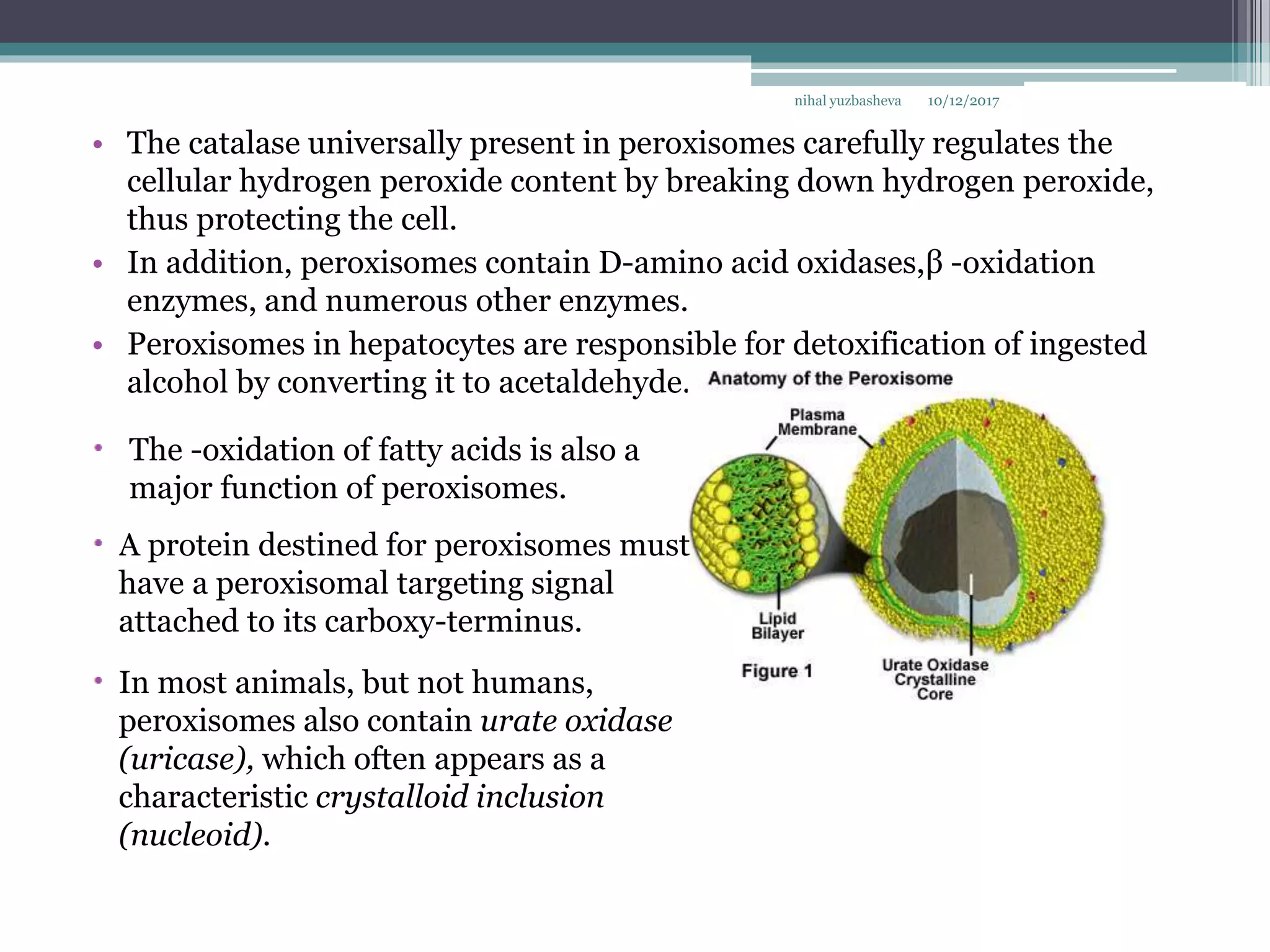
SlideShare a Scribd company logo
Endosomes.Peroxisomes. Proteasomes
Cytoplasmic inclusions
Structure.Functions.Their role in
protection of cell
10/12/2017nihal yuzbasheva
Endosomes
• Endosomes are divided into two compartments: early endosomes, near the
periphery of the cell, and late endosomes, situated deeper within the
cytoplasm near nucleus and Golgi apparatus.
• Early endosomes, are restricted to a portion of the cytoplasm near the cell
membrane where vesicles originating from the cell membrane fuse.
Early endosomes in live HeLa
cells identified after a 10-minute
incubation with green
10/12/2017nihal yuzbasheva
• From here, many vesicles return to the plasma membrane. However, large
numbers of vesicles originating in early endosomes travel to deeper
structures in the cytoplasm called late endosomes. The latter typically
mature into lysosomes.
Electron micrograph of an early endosome.
This deep-etch electron micrograph shows the structure of an
early endosome in Dictyostelium. Early endosomes are located
near the plasma membrane and, as in many other sorting
compartments, have a typical tubulovesicle structure. The tubular
portions contain the majority of integral membrane proteins
destined for membrane recycling, whereas the luminal portions
collect secretory cargo proteins. The lumen of the endosome is
subdivided into multiple compartments, or cisternae, by the
invagination of its membrane and undergoes frequent changes in
shape. 15,000. (Courtesy of Dr. John E. Heuser, Washington
University School of Medicine.)
Endosomes can be viewed either as stable cytoplasmic organelles or as
transient structures formed as the result of endocytosis.
10/12/2017nihal yuzbasheva
• Recent experimental observations of endocytotic pathwaysconducted in
vitro and in vivo suggest two different models that explain the origin and
formation of the endosomal compartments in the cell:
• The stable compartment model describes early and late endosomes as
stable cellular organelles connected by vesicular transport with the external
environment of the cell and with the Golgi apparatus. Coated vesicles
formed at the plasma membrane fuse only with early endosomes because of
their expression of specific surface receptors. The receptor remains a
resident component of the early endosomal membrane.
• In the maturation model, early endosomes are formed de novo from
endocytotic vesicles originating from the plasma membrane. Therefore, the
composition of the early endosomal membrane changes progressively as
some components are recycled between the cell surface and the Golgi
apparatus. This maturation process leads to formation of late endosomes
and then to lysosomes. Specific receptors present on early endosomes (e.g.,
for coated vesicles) are removed by recycling, degradation, or inactivation
as this compartment matures.
10/12/2017nihal yuzbasheva
• Endosomes destined to become lysosomes receive newly synthesized
lysosomal enzymes that are targeted via the mannose-6-phosphate
receptor.
• Some endosomes also communicate with the vesicular transport system of
the rER. This pathway provides constant delivery of newly synthesized
lysosomal enzymes, or hydrolases.
• A hydrolase is synthesized in the rER as an enzymatically inactive precursor
called a prohydrolase. This heavily glycosylated protein then folds in a
specific way so that a signal patch is formed and exposed on its surface.
• The signal patch on a protein destined for a lysosome is then modified by
several enzymes that attach mannose-6-phosphate (M-6-P) to the
prohydrolase surface. M-6-P acts as a target for proteins possessing an M-
6-P receptor.
10/12/2017nihal yuzbasheva
The Mannose-6-Phosphate Pathway
10/12/2017nihal yuzbasheva
• Early and late endosomes differ in their cellular localization,
morphology, and state of acidification and function.
An early endosome has a tubulovesicular
structure: The lumen is subdivided into
cisternae that are separated by
invagination of its membrane. It exhibits
only a slightly more acidic environment
(pH 6.2 to 6.5) than the cytoplasm of the
cell.
In contrast, late endosomes have a more
complex structure and often exhibit
onionlike internal membranes. Their pH is
more acidic, averaging 5.5.
• TEM studies reveal specific vesicles that transport substances between early
and late endosomes. These vesicles, called multivesicular bodies (MVBs),
are highly selective transporters.
• Because late endosomes mature into lysosomes, they are also called
prelysosomes
10/12/2017nihal yuzbasheva
Pathways for delivery of newly synthesized
lysosomal enzymes. Lysosomal enzymes (such as
lysosomal hydrolases)
are synthesized and glycosylated within the rough endoplasmic
reticulum (rER). The enzymes then fold in a specific
way so that a signal patch is formed, which allows for further
modification by the addition of M-6-P, which allows the enzyme to
be targeted to specific proteins that possess M-6-P receptor
activity.
M-6-P receptors are present in the TGN of the Golgi apparatus,
where the lysosomal enzymes are sorted and packaged
into vesicles later transported to the early or late endosomes.
10/12/2017nihal yuzbasheva
• The major function of early endosomes is to sort and recycle proteins
internalized by endocytotic pathways.
• The morphologic shape and geometry of the tubules and vesicles emerging
from the early endosome create an environment in which localized changes
in pH constitute the basis of the sorting mechanism.
• This mechanism includes dissociation of ligands from their receptor
protein; thus, in the past, early endosomes were referred to as
compartments of uncoupling receptors and ligands (CURLs).
Following endocytosis, ligand–drug
conjugates may be trafficked through
different intracellular compartments,
depending on the receptor that is exploited
for the internalization of the conjugate.
Some of the more common compartments
that are encountered during intracellular
trafficking include: early endosomes;
compartments for uncoupling of receptor
and ligand (CURLs), where dissociation of
the conjugate from the receptor may occur;
recycling endosomes, which can deliver the
internalized receptor back to the cell surface;
and lysosomes, where the receptor and the
conjugate can be degraded.
10/12/2017nihal yuzbasheva
• In addition, the narrow diameter of the tubules and vesicles may also aid in
the sorting of large molecules, which can be mechanically prevented from
entering specific sorting compartments. After sorting, most of the protein is
rapidly recycled, and the excess membrane is returned to the plasma
membrane.
• The fate of the internalized ligand–receptor complex depends on the
sorting and recycling ability of the early endosome.
• The following pathways for processing internalized ligand–receptor
complexes are present in the cell:The receptor is recycled and the ligand is
degraded, Both receptor and ligand are recycled,Both receptor and ligand
are degraded, Both receptor and ligand are transported through the cell.
• The receptor is recycled and the ligand is degraded.Surface receptors allow the
cell to bring in substances selectively through the process of endocytosis. This
pathway occurs most often in the cell; it is important because it allows surface
receptors to be recycled. Most ligand–receptor complexes dissociate in the
acidic pH of the early endosome. The receptor, most likely an integral
membrane protein , is recycled to the surface via vesicles that bud off the ends of
narrow-diameter tubules of the early endosome. Ligands are usually
sequestered in the spherical vacuolar part of the endosome that will later form
MVBs, which will transport the ligand to late endosomes for further degradation
in the lysosome . This pathway is described for the low-density lipoprotein
(LDL)–receptor complex, insulin–glucose transporter (GLUT) receptor
complex, and a variety of peptide hormones and their receptors.
• Both receptor and ligand are recycled. Ligand–receptor complex dissociation
does not always accompany receptor recycling. For example, the low pH of the
endosome dissociates iron from the iron-carrier protein transferrin, but
transferrin remains associated with its receptor. Once the transferrin–receptor
complex returns to the cell surface, however, transferrin is released. At neutral
extracellular pH, transferrin must again bind iron to be recognized by and
bound to its receptor. A similar pathway is recognized for major
histocompatibility complex (MHC) I and II molecules, which are recycled to the
cell surface with a foreign antigen protein attached to them.
10/12/2017nihal yuzbasheva
• Both receptor and ligand are degraded. This pathway has been identified for
epidermal growth factor (EGF) and its receptor. Like many other proteins, EGF binds
to its receptor on the cell surface. The complex is internalized and carried to the early
endosomes. Here EGF dissociates from its receptor, and both are sorted, packaged in
separate MVBs, and transferred to the late endosome. From there, both ligand and
receptor are transferred to lysosomes, where they are degraded
• Both receptor and ligand are transported through the cell. This pathway is used for
secretion of immunoglobulins (secretory IgA) into the saliva and human milk. During
this process, commonly referred to as transcytosis, substances can be altered as they
are transported across the epithelial cell. Transport of maternal IgG across the
placental barrier into the fetus also follows a similar pathway.
10/12/2017nihal yuzbasheva
Peroxisomes (Microbodies)
• Peroxisomes (microbodies) are small (0.5 m in diameter), membrane-
limited spherical organelles that contain oxidative enzymes, particularly
catalase and other peroxidases. Virtually all oxidative enzymes produce
hydrogen peroxide (H2O2) as a product of the oxidation reaction.
10/12/2017nihal yuzbasheva
• The catalase universally present in peroxisomes carefully regulates the
cellular hydrogen peroxide content by breaking down hydrogen peroxide,
thus protecting the cell.
• In addition, peroxisomes contain D-amino acid oxidases,β -oxidation
enzymes, and numerous other enzymes.
• Peroxisomes in hepatocytes are responsible for detoxification of ingested
alcohol by converting it to acetaldehyde.
10/12/2017nihal yuzbasheva
The -oxidation of fatty acids is also a
major function of peroxisomes.
A protein destined for peroxisomes must
have a peroxisomal targeting signal
attached to its carboxy-terminus.
In most animals, but not humans,
peroxisomes also contain urate oxidase
(uricase), which often appears as a
characteristic crystalloid inclusion
(nucleoid).
10/12/2017nihal yuzbasheva
• In the most common inherited disease related to nonfunctional
peroxisomes, Zellweger syndrome, which leads to early death, peroxisomes
lose their ability to function because of a lack of necessary enzymes. The
disorder is caused by a mutation in the gene encoding the receptor for the
peroxisome targeting signal that does not recognize the signal Ser-Lys-Leu
at the carboxyterminus of enzymes directed to peroxisomes.
Contribution of Fetal MR
Imaging in the Prenatal
Diagnosis of Zellweger
Syndrome
Proteasomes
• Proteasomes are small organelles composed of protein complexes that are
responsible for proteolysis of malformed and ubiquitin-tagged proteins.
• The protein population of a cell is in a constant flux as a result of the
continuous synthesis, export, and degradation of these macromolecules.
10/12/2017nihal yuzbasheva
Frequently, proteins, such as those that act in
metabolic regulation, have to be degraded to
ensure that the metabolic response to a single
stimulus is not prolonged.
• Additionally, proteins that have been denatured, damaged, or malformed
have to be eliminated; moreover, antigenic proteins that have been
endocytosed by antigen-presenting cells (APCs) have to be cleaved into
small polypeptide fragments (epitopes) so that they can be presented to T
lymphocytes for recognition and the mounting of an immune response.
• The process of cytosolic proteolysis is carefully controlled by the cell, and it
requires that the protein be recognized as a potential candidate for
degradation. This recognition involves ubiquination, a process whereby
several ubiquitin molecules (a 76-amino acid long polypeptide chain) are
attached to a lysine residue of the candidate protein to form a
polyubiquinated protein.
10/12/2017nihal yuzbasheva
• Once a protein has been thus tagged, it is degraded by proteasomes,
multisubunit protein complexes that have a molecular weight in excess of 2
million daltons.
• During proteolysis, the ubiquitin molecules are released and reenter the
cytosolic pool. The mechanism of ubiquitination requires:
1.The cooperation of a series of enzymes, including ubiquitin-activating
enzyme
2.A family of ubiquitin-conjugating enzymes
3.A number of ubiquitin ligases each of which recognizes one or more
substrate proteins
INCLUSIONS
• Inclusions are cytoplasmic or nuclear structures with characteristic staining
properties that are formed from the metabolic products of cell. They are
considered nonmoving and nonliving components of the cell.
• Some of them, such as pigment granules, are surrounded by a plasma
membrane; others (e.g., lipid droplets or glycogen) instead reside within the
cytoplasmic or nuclear matrix.
10/12/2017nihal yuzbasheva
• Lipofuscin is a brownish-gold pigment visible in routine H&E preparation.
It is easily seen in nondividing cells such as neurons and skeletal and
cardiac muscle cells.
• Lipofuscin accumulates during the years in most eukaryotic cells as a result
of cellular senescence (aging); thus, it is often called the “wear-and-tear”
pigment.
• Lipofuscin is a conglomerate of oxidized lipids, phospholipids, metals, and
organic molecules that accumulate within the cells as a result of oxidative
degradation of mitochondria and lysosomal digestion.
10/12/2017nihal yuzbasheva
Phagocytotic cells such as
macrophages may also contain
lipofuscin, which accumulates from
the digestion of bacteria, foreign
particles, dead cells, and their own
organelles. Recent experiments
indicate that lipofuscin
accumulation may be an accurate
indicator of cellular stress.
• Hemosiderin is an iron-storage complex found within the cytoplasm of
many cells. It is most likely formed by the indigestible residues of
hemoglobin, and its presence is related to phagocytosis of red blood cells.
• Hemosiderin is most easily demonstrated in the spleen, where aged
erythrocytes are phagocytosed, but it can also be found in alveolar
macrophages in the lung tissue, especially after pulmonary infection
accompanied by small hemorrhage into the alveoli.
10/12/2017nihal yuzbasheva
Hemosiderin-laden macrophages in the lung.
KU Collection
It is visible in light microscopy as
a deep brown granule, more or
less indistinguishable from
lipofuscin.
Hemosiderin granules can be
differentially stained
using histochemical methods for
iron detection.
• Glycogen is a highly branched polymer used as a storage material for
glucose. It is not stained in the routine H&E preparation. However, it may
be seen in the light microscope with special fixation and staining
procedures (such as toluidine blue or the PAS method).
• Liver and striated muscle cells, which usually contain large amounts of
glycogen, may display unstained regions where glycogen is located.
Glycogen appears in EM as granules 25 to 30 nm in diameter or as clusters
of granules that often occupy significant portions of the cytoplasm
10/12/2017nihal yuzbasheva
• Lipid inclusions (fat droplets) are usually nutritive inclusions that provide
energy for cellular metabolism. The lipid droplets may appear in a cell for a
brief time (e.g., in intestinal absorptive cells) or may reside for a long period
(e.g., in adipocytes)
• In adipocytes, lipid inclusions often constitute most of the cytoplasmic
volume, compressing the other formed organelles into a thin rim at the
margin of the cell. Lipid droplets are usually extracted by the organic solvents
used to prepare tissues for both light and electron microscopy. What is seen as
a fat droplet in light microscopy is actually a hole in the cytoplasm that
represents the site from which the lipid was extracted.
10/12/2017nihal yuzbasheva
In individuals with genetic
defects
of enzymes involved in lipid
metabolism, lipid droplets
may accumulate in abnormal
locations or in abnormal
amounts. Such diseases are
classified as lipid storage
diseases.
• Crystalline inclusions contained in certain cells are recognized in the light
microscope. In humans, such inclusions are found in the Sertoli
(sustentacular) and Leydig (interstitial) cells of the testis.
• With the TEM, crystalline inclusions have been found in many cell types
and in virtually all parts of the cell, including the nucleus and most
cytoplasmic organelles.
• Although some of these inclusions contain viral proteins, storage material,
or cellular metabolites, the significance of others is not clear.
10/12/2017nihal yuzbasheva
Schaumann bodies with crystalline inclusions,
polarized
Multinucleate plasma cell with intracytoplasmic
crystalline inclusions, (Geimsa stain, ×1000)

More Related Content

What's hot

cell_junctions
 cell_junctions cell_junctions
cell_junctions
MedicTalks
 
Membrane Dynamics:Properties of biological membrane (plasma membrane)
Membrane Dynamics:Properties of biological membrane (plasma membrane)Membrane Dynamics:Properties of biological membrane (plasma membrane)
Membrane Dynamics:Properties of biological membrane (plasma membrane)
Manju Chhetri
 
Lysosomes and peroxisomes
Lysosomes and peroxisomesLysosomes and peroxisomes
Lysosomes and peroxisomes
Sara Hassan
 
Structure and functions of endoplasmic reticulum
Structure and functions of endoplasmic reticulumStructure and functions of endoplasmic reticulum
Structure and functions of endoplasmic reticulum
ICHHA PURAK
 
Endoplasmic reticulum- cell Organelle
Endoplasmic reticulum- cell OrganelleEndoplasmic reticulum- cell Organelle
Endoplasmic reticulum- cell Organelle
AshishNain
 
Cell adhesion molecules
Cell adhesion moleculesCell adhesion molecules
Cell adhesion molecules
Mital Chandegara
 
Structure and functions of Mitochondria
Structure and functions of MitochondriaStructure and functions of Mitochondria
Structure and functions of Mitochondria
ICHHA PURAK
 
Peroxisomes
PeroxisomesPeroxisomes
Peroxisomes
Sam Shaikh
 
Biological membrane & its chemical composition
Biological membrane & its chemical compositionBiological membrane & its chemical composition
Biological membrane & its chemical composition
Pradeep Singh Narwat
 
Membrane proteins
Membrane proteinsMembrane proteins
Membrane proteins
Lovnish Thakur
 
Cell adhesion molecules and matrix proteins
Cell adhesion    molecules and matrix proteinsCell adhesion    molecules and matrix proteins
Cell adhesion molecules and matrix proteinsUSmile Ï Ṩṃïlệ
 
Plasma membrane
Plasma membranePlasma membrane
Plasma membrane
Saranraj P
 
Cytoskeleton
CytoskeletonCytoskeleton
Cytoskeleton
Dilip Pandya
 
Peroxisome
PeroxisomePeroxisome
Peroxisome
Dilip Pandya
 
microtubules and microfilaments
microtubules and microfilamentsmicrotubules and microfilaments
microtubules and microfilaments
karthika05
 
Endoplasmic reticulum
Endoplasmic reticulumEndoplasmic reticulum
Endoplasmic reticulum
Dilip Pandya
 
Cell Surface Membrane
Cell Surface MembraneCell Surface Membrane
Cell Surface Membrane
Hanna Elise
 
Membrane Proteins & its types
Membrane Proteins & its typesMembrane Proteins & its types
Membrane Proteins & its types
Sabahat Ali
 

What's hot (20)

cell_junctions
 cell_junctions cell_junctions
cell_junctions
 
Membrane Dynamics:Properties of biological membrane (plasma membrane)
Membrane Dynamics:Properties of biological membrane (plasma membrane)Membrane Dynamics:Properties of biological membrane (plasma membrane)
Membrane Dynamics:Properties of biological membrane (plasma membrane)
 
Lysosomes and peroxisomes
Lysosomes and peroxisomesLysosomes and peroxisomes
Lysosomes and peroxisomes
 
Structure and functions of endoplasmic reticulum
Structure and functions of endoplasmic reticulumStructure and functions of endoplasmic reticulum
Structure and functions of endoplasmic reticulum
 
Endoplasmic reticulum- cell Organelle
Endoplasmic reticulum- cell OrganelleEndoplasmic reticulum- cell Organelle
Endoplasmic reticulum- cell Organelle
 
Cell adhesion molecules
Cell adhesion moleculesCell adhesion molecules
Cell adhesion molecules
 
Structure and functions of Mitochondria
Structure and functions of MitochondriaStructure and functions of Mitochondria
Structure and functions of Mitochondria
 
Peroxisomes
PeroxisomesPeroxisomes
Peroxisomes
 
Biological membrane & its chemical composition
Biological membrane & its chemical compositionBiological membrane & its chemical composition
Biological membrane & its chemical composition
 
Membrane proteins
Membrane proteinsMembrane proteins
Membrane proteins
 
Cell adhesion molecules and matrix proteins
Cell adhesion    molecules and matrix proteinsCell adhesion    molecules and matrix proteins
Cell adhesion molecules and matrix proteins
 
Mitochondria
MitochondriaMitochondria
Mitochondria
 
Lysosomes
LysosomesLysosomes
Lysosomes
 
Plasma membrane
Plasma membranePlasma membrane
Plasma membrane
 
Cytoskeleton
CytoskeletonCytoskeleton
Cytoskeleton
 
Peroxisome
PeroxisomePeroxisome
Peroxisome
 
microtubules and microfilaments
microtubules and microfilamentsmicrotubules and microfilaments
microtubules and microfilaments
 
Endoplasmic reticulum
Endoplasmic reticulumEndoplasmic reticulum
Endoplasmic reticulum
 
Cell Surface Membrane
Cell Surface MembraneCell Surface Membrane
Cell Surface Membrane
 
Membrane Proteins & its types
Membrane Proteins & its typesMembrane Proteins & its types
Membrane Proteins & its types
 

Similar to Endosomes.Peroxisomes. Proteasomes Cytoplasmic inclusions

endocytosis and exocytosis vvvvvvvvvvvvv
endocytosis and exocytosis vvvvvvvvvvvvvendocytosis and exocytosis vvvvvvvvvvvvv
endocytosis and exocytosis vvvvvvvvvvvvv
Aakashdeep669682
 
Cell structure, Ola Elgaddar
Cell structure, Ola ElgaddarCell structure, Ola Elgaddar
Cell structure, Ola Elgaddar
Ola Elgaddar
 
Cell Biology class 1(2).pptx
Cell Biology class 1(2).pptxCell Biology class 1(2).pptx
Cell Biology class 1(2).pptx
ANJUHARIDAS5
 
Protein trafficking in lysosomes
Protein trafficking in lysosomesProtein trafficking in lysosomes
Protein trafficking in lysosomes
Sheryl Bhatnagar
 
Interactions among organelles
Interactions among organellesInteractions among organelles
Interactions among organelles
Nawfal Aldujaily
 
Lecture1 cytology
Lecture1 cytologyLecture1 cytology
Lecture1 cytologyReach Na
 
Plasma membrane : cell biology
Plasma membrane : cell biologyPlasma membrane : cell biology
Plasma membrane : cell biology
Gauri Haval
 
Role of lysosomes as hyrolaytic activity of an enzymes
 Role of lysosomes as hyrolaytic activity of an enzymes Role of lysosomes as hyrolaytic activity of an enzymes
Role of lysosomes as hyrolaytic activity of an enzymes
Eram Rashid
 
THE HUMAN CELL ANATOMY & PHYSIOLOGY / TRANSPORT ACROSS CELL MEMBRANE /INTER C...
THE HUMAN CELL ANATOMY & PHYSIOLOGY / TRANSPORT ACROSS CELL MEMBRANE /INTER C...THE HUMAN CELL ANATOMY & PHYSIOLOGY / TRANSPORT ACROSS CELL MEMBRANE /INTER C...
THE HUMAN CELL ANATOMY & PHYSIOLOGY / TRANSPORT ACROSS CELL MEMBRANE /INTER C...
Kameshwaran Sugavanam
 
Cell Membrane Structure-1.pptx cell outer layer
Cell Membrane Structure-1.pptx cell outer layerCell Membrane Structure-1.pptx cell outer layer
Cell Membrane Structure-1.pptx cell outer layer
SalsabeelArif
 
Endoplasmic reticulum
Endoplasmic reticulumEndoplasmic reticulum
Endoplasmic reticulum
bikashsingh2510
 
Lecture 3.pptx
Lecture 3.pptxLecture 3.pptx
Lecture 3.pptx
backup007
 
Role of endoplasmic reticulum in protein systhesis and1
Role of endoplasmic reticulum in protein systhesis and1Role of endoplasmic reticulum in protein systhesis and1
Role of endoplasmic reticulum in protein systhesis and1
Monalisa Behera
 
Plasma membrane or cell membrane (2).pptx
Plasma membrane or cell membrane (2).pptxPlasma membrane or cell membrane (2).pptx
Plasma membrane or cell membrane (2).pptx
aminagulshan526
 
Cell Biology Lec 1.ppt
Cell Biology Lec 1.pptCell Biology Lec 1.ppt
Cell Biology Lec 1.ppt
MohamedTharwat970652
 
cell structure and cell membrain // cell
cell structure and cell membrain // cellcell structure and cell membrain // cell
cell structure and cell membrain // cell
StarletMG
 
Plasma membrane
Plasma membranePlasma membrane
Plasma membrane
Subramaniya Sharma
 

Similar to Endosomes.Peroxisomes. Proteasomes Cytoplasmic inclusions (20)

endocytosis and exocytosis vvvvvvvvvvvvv
endocytosis and exocytosis vvvvvvvvvvvvvendocytosis and exocytosis vvvvvvvvvvvvv
endocytosis and exocytosis vvvvvvvvvvvvv
 
Cell structure, Ola Elgaddar
Cell structure, Ola ElgaddarCell structure, Ola Elgaddar
Cell structure, Ola Elgaddar
 
Cell Biology class 1(2).pptx
Cell Biology class 1(2).pptxCell Biology class 1(2).pptx
Cell Biology class 1(2).pptx
 
Cell
CellCell
Cell
 
1. cell
1. cell1. cell
1. cell
 
Protein trafficking in lysosomes
Protein trafficking in lysosomesProtein trafficking in lysosomes
Protein trafficking in lysosomes
 
Interactions among organelles
Interactions among organellesInteractions among organelles
Interactions among organelles
 
cytology
cytologycytology
cytology
 
Lecture1 cytology
Lecture1 cytologyLecture1 cytology
Lecture1 cytology
 
Plasma membrane : cell biology
Plasma membrane : cell biologyPlasma membrane : cell biology
Plasma membrane : cell biology
 
Role of lysosomes as hyrolaytic activity of an enzymes
 Role of lysosomes as hyrolaytic activity of an enzymes Role of lysosomes as hyrolaytic activity of an enzymes
Role of lysosomes as hyrolaytic activity of an enzymes
 
THE HUMAN CELL ANATOMY & PHYSIOLOGY / TRANSPORT ACROSS CELL MEMBRANE /INTER C...
THE HUMAN CELL ANATOMY & PHYSIOLOGY / TRANSPORT ACROSS CELL MEMBRANE /INTER C...THE HUMAN CELL ANATOMY & PHYSIOLOGY / TRANSPORT ACROSS CELL MEMBRANE /INTER C...
THE HUMAN CELL ANATOMY & PHYSIOLOGY / TRANSPORT ACROSS CELL MEMBRANE /INTER C...
 
Cell Membrane Structure-1.pptx cell outer layer
Cell Membrane Structure-1.pptx cell outer layerCell Membrane Structure-1.pptx cell outer layer
Cell Membrane Structure-1.pptx cell outer layer
 
Endoplasmic reticulum
Endoplasmic reticulumEndoplasmic reticulum
Endoplasmic reticulum
 
Lecture 3.pptx
Lecture 3.pptxLecture 3.pptx
Lecture 3.pptx
 
Role of endoplasmic reticulum in protein systhesis and1
Role of endoplasmic reticulum in protein systhesis and1Role of endoplasmic reticulum in protein systhesis and1
Role of endoplasmic reticulum in protein systhesis and1
 
Plasma membrane or cell membrane (2).pptx
Plasma membrane or cell membrane (2).pptxPlasma membrane or cell membrane (2).pptx
Plasma membrane or cell membrane (2).pptx
 
Cell Biology Lec 1.ppt
Cell Biology Lec 1.pptCell Biology Lec 1.ppt
Cell Biology Lec 1.ppt
 
cell structure and cell membrain // cell
cell structure and cell membrain // cellcell structure and cell membrain // cell
cell structure and cell membrain // cell
 
Plasma membrane
Plasma membranePlasma membrane
Plasma membrane
 

More from Nihal Yuzbasheva

Stomach Cancer
Stomach CancerStomach Cancer
Stomach Cancer
Nihal Yuzbasheva
 
Juvenile Idiopathic Arthritis
Juvenile Idiopathic ArthritisJuvenile Idiopathic Arthritis
Juvenile Idiopathic Arthritis
Nihal Yuzbasheva
 
Ocular Manifestations of Tuberculosis
Ocular Manifestations of TuberculosisOcular Manifestations of Tuberculosis
Ocular Manifestations of Tuberculosis
Nihal Yuzbasheva
 
Prostate Cancer (Urology)
Prostate Cancer (Urology)Prostate Cancer (Urology)
Prostate Cancer (Urology)
Nihal Yuzbasheva
 
Botulism
BotulismBotulism
Genital Warts(HPV), Genital Herpes
Genital Warts(HPV), Genital HerpesGenital Warts(HPV), Genital Herpes
Genital Warts(HPV), Genital Herpes
Nihal Yuzbasheva
 
Diabetic Retinopathy
Diabetic RetinopathyDiabetic Retinopathy
Diabetic Retinopathy
Nihal Yuzbasheva
 
Meninges of the Brain
Meninges of the BrainMeninges of the Brain
Meninges of the Brain
Nihal Yuzbasheva
 
Features of Skeletal System in Children
Features of Skeletal System in ChildrenFeatures of Skeletal System in Children
Features of Skeletal System in Children
Nihal Yuzbasheva
 
Infectious Mononucleosis - Pathology
Infectious Mononucleosis - PathologyInfectious Mononucleosis - Pathology
Infectious Mononucleosis - Pathology
Nihal Yuzbasheva
 
Diabetes Mellitus
Diabetes MellitusDiabetes Mellitus
Diabetes Mellitus
Nihal Yuzbasheva
 
Scleroderma
SclerodermaScleroderma
Scleroderma
Nihal Yuzbasheva
 
Leprosy - Dermatology
Leprosy - DermatologyLeprosy - Dermatology
Leprosy - Dermatology
Nihal Yuzbasheva
 
Respiratory Diseases - Pediatrics
Respiratory Diseases - PediatricsRespiratory Diseases - Pediatrics
Respiratory Diseases - Pediatrics
Nihal Yuzbasheva
 
Cardiomyopathy - clinical findings
Cardiomyopathy - clinical findingsCardiomyopathy - clinical findings
Cardiomyopathy - clinical findings
Nihal Yuzbasheva
 
Local anesthesia (General Surgery)
Local anesthesia (General Surgery)Local anesthesia (General Surgery)
Local anesthesia (General Surgery)
Nihal Yuzbasheva
 
Ovarian Tumors (Ovarian Cancers)
Ovarian Tumors (Ovarian Cancers)Ovarian Tumors (Ovarian Cancers)
Ovarian Tumors (Ovarian Cancers)
Nihal Yuzbasheva
 
Metaplasia & Dysplasia
Metaplasia & DysplasiaMetaplasia & Dysplasia
Metaplasia & Dysplasia
Nihal Yuzbasheva
 
Immunology of Bronchial Asthma
Immunology of Bronchial AsthmaImmunology of Bronchial Asthma
Immunology of Bronchial Asthma
Nihal Yuzbasheva
 
Reflexes of Newborns
Reflexes of NewbornsReflexes of Newborns
Reflexes of Newborns
Nihal Yuzbasheva
 

More from Nihal Yuzbasheva (20)

Stomach Cancer
Stomach CancerStomach Cancer
Stomach Cancer
 
Juvenile Idiopathic Arthritis
Juvenile Idiopathic ArthritisJuvenile Idiopathic Arthritis
Juvenile Idiopathic Arthritis
 
Ocular Manifestations of Tuberculosis
Ocular Manifestations of TuberculosisOcular Manifestations of Tuberculosis
Ocular Manifestations of Tuberculosis
 
Prostate Cancer (Urology)
Prostate Cancer (Urology)Prostate Cancer (Urology)
Prostate Cancer (Urology)
 
Botulism
BotulismBotulism
Botulism
 
Genital Warts(HPV), Genital Herpes
Genital Warts(HPV), Genital HerpesGenital Warts(HPV), Genital Herpes
Genital Warts(HPV), Genital Herpes
 
Diabetic Retinopathy
Diabetic RetinopathyDiabetic Retinopathy
Diabetic Retinopathy
 
Meninges of the Brain
Meninges of the BrainMeninges of the Brain
Meninges of the Brain
 
Features of Skeletal System in Children
Features of Skeletal System in ChildrenFeatures of Skeletal System in Children
Features of Skeletal System in Children
 
Infectious Mononucleosis - Pathology
Infectious Mononucleosis - PathologyInfectious Mononucleosis - Pathology
Infectious Mononucleosis - Pathology
 
Diabetes Mellitus
Diabetes MellitusDiabetes Mellitus
Diabetes Mellitus
 
Scleroderma
SclerodermaScleroderma
Scleroderma
 
Leprosy - Dermatology
Leprosy - DermatologyLeprosy - Dermatology
Leprosy - Dermatology
 
Respiratory Diseases - Pediatrics
Respiratory Diseases - PediatricsRespiratory Diseases - Pediatrics
Respiratory Diseases - Pediatrics
 
Cardiomyopathy - clinical findings
Cardiomyopathy - clinical findingsCardiomyopathy - clinical findings
Cardiomyopathy - clinical findings
 
Local anesthesia (General Surgery)
Local anesthesia (General Surgery)Local anesthesia (General Surgery)
Local anesthesia (General Surgery)
 
Ovarian Tumors (Ovarian Cancers)
Ovarian Tumors (Ovarian Cancers)Ovarian Tumors (Ovarian Cancers)
Ovarian Tumors (Ovarian Cancers)
 
Metaplasia & Dysplasia
Metaplasia & DysplasiaMetaplasia & Dysplasia
Metaplasia & Dysplasia
 
Immunology of Bronchial Asthma
Immunology of Bronchial AsthmaImmunology of Bronchial Asthma
Immunology of Bronchial Asthma
 
Reflexes of Newborns
Reflexes of NewbornsReflexes of Newborns
Reflexes of Newborns
 

Recently uploaded

KDIGO 2024 guidelines for diabetologists
KDIGO 2024 guidelines for diabetologistsKDIGO 2024 guidelines for diabetologists
KDIGO 2024 guidelines for diabetologists
د.محمود نجيب
 
POST OPERATIVE OLIGURIA and its management
POST OPERATIVE OLIGURIA and its managementPOST OPERATIVE OLIGURIA and its management
POST OPERATIVE OLIGURIA and its management
touseefaziz1
 
Flu Vaccine Alert in Bangalore Karnataka
Flu Vaccine Alert in Bangalore KarnatakaFlu Vaccine Alert in Bangalore Karnataka
Flu Vaccine Alert in Bangalore Karnataka
addon Scans
 
Lung Cancer: Artificial Intelligence, Synergetics, Complex System Analysis, S...
Lung Cancer: Artificial Intelligence, Synergetics, Complex System Analysis, S...Lung Cancer: Artificial Intelligence, Synergetics, Complex System Analysis, S...
Lung Cancer: Artificial Intelligence, Synergetics, Complex System Analysis, S...
Oleg Kshivets
 
Ozempic: Preoperative Management of Patients on GLP-1 Receptor Agonists
Ozempic: Preoperative Management of Patients on GLP-1 Receptor Agonists  Ozempic: Preoperative Management of Patients on GLP-1 Receptor Agonists
Ozempic: Preoperative Management of Patients on GLP-1 Receptor Agonists
Saeid Safari
 
How STIs Influence the Development of Pelvic Inflammatory Disease.pptx
How STIs Influence the Development of Pelvic Inflammatory Disease.pptxHow STIs Influence the Development of Pelvic Inflammatory Disease.pptx
How STIs Influence the Development of Pelvic Inflammatory Disease.pptx
FFragrant
 
BRACHYTHERAPY OVERVIEW AND APPLICATORS
BRACHYTHERAPY OVERVIEW  AND  APPLICATORSBRACHYTHERAPY OVERVIEW  AND  APPLICATORS
BRACHYTHERAPY OVERVIEW AND APPLICATORS
Krishan Murari
 
NVBDCP.pptx Nation vector borne disease control program
NVBDCP.pptx Nation vector borne disease control programNVBDCP.pptx Nation vector borne disease control program
NVBDCP.pptx Nation vector borne disease control program
Sapna Thakur
 
Non-respiratory Functions of the Lungs.pdf
Non-respiratory Functions of the Lungs.pdfNon-respiratory Functions of the Lungs.pdf
Non-respiratory Functions of the Lungs.pdf
MedicoseAcademics
 
New Drug Discovery and Development .....
New Drug Discovery and Development .....New Drug Discovery and Development .....
New Drug Discovery and Development .....
NEHA GUPTA
 
Pharynx and Clinical Correlations BY Dr.Rabia Inam Gandapore.pptx
Pharynx and Clinical Correlations BY Dr.Rabia Inam Gandapore.pptxPharynx and Clinical Correlations BY Dr.Rabia Inam Gandapore.pptx
Pharynx and Clinical Correlations BY Dr.Rabia Inam Gandapore.pptx
Dr. Rabia Inam Gandapore
 
Maxilla, Mandible & Hyoid Bone & Clinical Correlations by Dr. RIG.pptx
Maxilla, Mandible & Hyoid Bone & Clinical Correlations by Dr. RIG.pptxMaxilla, Mandible & Hyoid Bone & Clinical Correlations by Dr. RIG.pptx
Maxilla, Mandible & Hyoid Bone & Clinical Correlations by Dr. RIG.pptx
Dr. Rabia Inam Gandapore
 
New Directions in Targeted Therapeutic Approaches for Older Adults With Mantl...
New Directions in Targeted Therapeutic Approaches for Older Adults With Mantl...New Directions in Targeted Therapeutic Approaches for Older Adults With Mantl...
New Directions in Targeted Therapeutic Approaches for Older Adults With Mantl...
i3 Health
 
24 Upakrama.pptx class ppt useful in all
24 Upakrama.pptx class ppt useful in all24 Upakrama.pptx class ppt useful in all
24 Upakrama.pptx class ppt useful in all
DrSathishMS1
 
Alcohol_Dr. Jeenal Mistry MD Pharmacology.pdf
Alcohol_Dr. Jeenal Mistry MD Pharmacology.pdfAlcohol_Dr. Jeenal Mistry MD Pharmacology.pdf
Alcohol_Dr. Jeenal Mistry MD Pharmacology.pdf
Dr Jeenal Mistry
 
Couples presenting to the infertility clinic- Do they really have infertility...
Couples presenting to the infertility clinic- Do they really have infertility...Couples presenting to the infertility clinic- Do they really have infertility...
Couples presenting to the infertility clinic- Do they really have infertility...
Sujoy Dasgupta
 
Prix Galien International 2024 Forum Program
Prix Galien International 2024 Forum ProgramPrix Galien International 2024 Forum Program
Prix Galien International 2024 Forum Program
Levi Shapiro
 
Novas diretrizes da OMS para os cuidados perinatais de mais qualidade
Novas diretrizes da OMS para os cuidados perinatais de mais qualidadeNovas diretrizes da OMS para os cuidados perinatais de mais qualidade
Novas diretrizes da OMS para os cuidados perinatais de mais qualidade
Prof. Marcus Renato de Carvalho
 
Evaluation of antidepressant activity of clitoris ternatea in animals
Evaluation of antidepressant activity of clitoris ternatea in animalsEvaluation of antidepressant activity of clitoris ternatea in animals
Evaluation of antidepressant activity of clitoris ternatea in animals
Shweta
 
Report Back from SGO 2024: What’s the Latest in Cervical Cancer?
Report Back from SGO 2024: What’s the Latest in Cervical Cancer?Report Back from SGO 2024: What’s the Latest in Cervical Cancer?
Report Back from SGO 2024: What’s the Latest in Cervical Cancer?
bkling
 

Recently uploaded (20)

KDIGO 2024 guidelines for diabetologists
KDIGO 2024 guidelines for diabetologistsKDIGO 2024 guidelines for diabetologists
KDIGO 2024 guidelines for diabetologists
 
POST OPERATIVE OLIGURIA and its management
POST OPERATIVE OLIGURIA and its managementPOST OPERATIVE OLIGURIA and its management
POST OPERATIVE OLIGURIA and its management
 
Flu Vaccine Alert in Bangalore Karnataka
Flu Vaccine Alert in Bangalore KarnatakaFlu Vaccine Alert in Bangalore Karnataka
Flu Vaccine Alert in Bangalore Karnataka
 
Lung Cancer: Artificial Intelligence, Synergetics, Complex System Analysis, S...
Lung Cancer: Artificial Intelligence, Synergetics, Complex System Analysis, S...Lung Cancer: Artificial Intelligence, Synergetics, Complex System Analysis, S...
Lung Cancer: Artificial Intelligence, Synergetics, Complex System Analysis, S...
 
Ozempic: Preoperative Management of Patients on GLP-1 Receptor Agonists
Ozempic: Preoperative Management of Patients on GLP-1 Receptor Agonists  Ozempic: Preoperative Management of Patients on GLP-1 Receptor Agonists
Ozempic: Preoperative Management of Patients on GLP-1 Receptor Agonists
 
How STIs Influence the Development of Pelvic Inflammatory Disease.pptx
How STIs Influence the Development of Pelvic Inflammatory Disease.pptxHow STIs Influence the Development of Pelvic Inflammatory Disease.pptx
How STIs Influence the Development of Pelvic Inflammatory Disease.pptx
 
BRACHYTHERAPY OVERVIEW AND APPLICATORS
BRACHYTHERAPY OVERVIEW  AND  APPLICATORSBRACHYTHERAPY OVERVIEW  AND  APPLICATORS
BRACHYTHERAPY OVERVIEW AND APPLICATORS
 
NVBDCP.pptx Nation vector borne disease control program
NVBDCP.pptx Nation vector borne disease control programNVBDCP.pptx Nation vector borne disease control program
NVBDCP.pptx Nation vector borne disease control program
 
Non-respiratory Functions of the Lungs.pdf
Non-respiratory Functions of the Lungs.pdfNon-respiratory Functions of the Lungs.pdf
Non-respiratory Functions of the Lungs.pdf
 
New Drug Discovery and Development .....
New Drug Discovery and Development .....New Drug Discovery and Development .....
New Drug Discovery and Development .....
 
Pharynx and Clinical Correlations BY Dr.Rabia Inam Gandapore.pptx
Pharynx and Clinical Correlations BY Dr.Rabia Inam Gandapore.pptxPharynx and Clinical Correlations BY Dr.Rabia Inam Gandapore.pptx
Pharynx and Clinical Correlations BY Dr.Rabia Inam Gandapore.pptx
 
Maxilla, Mandible & Hyoid Bone & Clinical Correlations by Dr. RIG.pptx
Maxilla, Mandible & Hyoid Bone & Clinical Correlations by Dr. RIG.pptxMaxilla, Mandible & Hyoid Bone & Clinical Correlations by Dr. RIG.pptx
Maxilla, Mandible & Hyoid Bone & Clinical Correlations by Dr. RIG.pptx
 
New Directions in Targeted Therapeutic Approaches for Older Adults With Mantl...
New Directions in Targeted Therapeutic Approaches for Older Adults With Mantl...New Directions in Targeted Therapeutic Approaches for Older Adults With Mantl...
New Directions in Targeted Therapeutic Approaches for Older Adults With Mantl...
 
24 Upakrama.pptx class ppt useful in all
24 Upakrama.pptx class ppt useful in all24 Upakrama.pptx class ppt useful in all
24 Upakrama.pptx class ppt useful in all
 
Alcohol_Dr. Jeenal Mistry MD Pharmacology.pdf
Alcohol_Dr. Jeenal Mistry MD Pharmacology.pdfAlcohol_Dr. Jeenal Mistry MD Pharmacology.pdf
Alcohol_Dr. Jeenal Mistry MD Pharmacology.pdf
 
Couples presenting to the infertility clinic- Do they really have infertility...
Couples presenting to the infertility clinic- Do they really have infertility...Couples presenting to the infertility clinic- Do they really have infertility...
Couples presenting to the infertility clinic- Do they really have infertility...
 
Prix Galien International 2024 Forum Program
Prix Galien International 2024 Forum ProgramPrix Galien International 2024 Forum Program
Prix Galien International 2024 Forum Program
 
Novas diretrizes da OMS para os cuidados perinatais de mais qualidade
Novas diretrizes da OMS para os cuidados perinatais de mais qualidadeNovas diretrizes da OMS para os cuidados perinatais de mais qualidade
Novas diretrizes da OMS para os cuidados perinatais de mais qualidade
 
Evaluation of antidepressant activity of clitoris ternatea in animals
Evaluation of antidepressant activity of clitoris ternatea in animalsEvaluation of antidepressant activity of clitoris ternatea in animals
Evaluation of antidepressant activity of clitoris ternatea in animals
 
Report Back from SGO 2024: What’s the Latest in Cervical Cancer?
Report Back from SGO 2024: What’s the Latest in Cervical Cancer?Report Back from SGO 2024: What’s the Latest in Cervical Cancer?
Report Back from SGO 2024: What’s the Latest in Cervical Cancer?
 

Endosomes.Peroxisomes. Proteasomes Cytoplasmic inclusions

  • 1. Endosomes.Peroxisomes. Proteasomes Cytoplasmic inclusions Structure.Functions.Their role in protection of cell 10/12/2017nihal yuzbasheva
  • 2. Endosomes • Endosomes are divided into two compartments: early endosomes, near the periphery of the cell, and late endosomes, situated deeper within the cytoplasm near nucleus and Golgi apparatus. • Early endosomes, are restricted to a portion of the cytoplasm near the cell membrane where vesicles originating from the cell membrane fuse. Early endosomes in live HeLa cells identified after a 10-minute incubation with green 10/12/2017nihal yuzbasheva
  • 3. • From here, many vesicles return to the plasma membrane. However, large numbers of vesicles originating in early endosomes travel to deeper structures in the cytoplasm called late endosomes. The latter typically mature into lysosomes. Electron micrograph of an early endosome. This deep-etch electron micrograph shows the structure of an early endosome in Dictyostelium. Early endosomes are located near the plasma membrane and, as in many other sorting compartments, have a typical tubulovesicle structure. The tubular portions contain the majority of integral membrane proteins destined for membrane recycling, whereas the luminal portions collect secretory cargo proteins. The lumen of the endosome is subdivided into multiple compartments, or cisternae, by the invagination of its membrane and undergoes frequent changes in shape. 15,000. (Courtesy of Dr. John E. Heuser, Washington University School of Medicine.) Endosomes can be viewed either as stable cytoplasmic organelles or as transient structures formed as the result of endocytosis. 10/12/2017nihal yuzbasheva
  • 4. • Recent experimental observations of endocytotic pathwaysconducted in vitro and in vivo suggest two different models that explain the origin and formation of the endosomal compartments in the cell: • The stable compartment model describes early and late endosomes as stable cellular organelles connected by vesicular transport with the external environment of the cell and with the Golgi apparatus. Coated vesicles formed at the plasma membrane fuse only with early endosomes because of their expression of specific surface receptors. The receptor remains a resident component of the early endosomal membrane. • In the maturation model, early endosomes are formed de novo from endocytotic vesicles originating from the plasma membrane. Therefore, the composition of the early endosomal membrane changes progressively as some components are recycled between the cell surface and the Golgi apparatus. This maturation process leads to formation of late endosomes and then to lysosomes. Specific receptors present on early endosomes (e.g., for coated vesicles) are removed by recycling, degradation, or inactivation as this compartment matures. 10/12/2017nihal yuzbasheva
  • 5. • Endosomes destined to become lysosomes receive newly synthesized lysosomal enzymes that are targeted via the mannose-6-phosphate receptor. • Some endosomes also communicate with the vesicular transport system of the rER. This pathway provides constant delivery of newly synthesized lysosomal enzymes, or hydrolases. • A hydrolase is synthesized in the rER as an enzymatically inactive precursor called a prohydrolase. This heavily glycosylated protein then folds in a specific way so that a signal patch is formed and exposed on its surface. • The signal patch on a protein destined for a lysosome is then modified by several enzymes that attach mannose-6-phosphate (M-6-P) to the prohydrolase surface. M-6-P acts as a target for proteins possessing an M- 6-P receptor. 10/12/2017nihal yuzbasheva
  • 7. 10/12/2017nihal yuzbasheva • Early and late endosomes differ in their cellular localization, morphology, and state of acidification and function. An early endosome has a tubulovesicular structure: The lumen is subdivided into cisternae that are separated by invagination of its membrane. It exhibits only a slightly more acidic environment (pH 6.2 to 6.5) than the cytoplasm of the cell. In contrast, late endosomes have a more complex structure and often exhibit onionlike internal membranes. Their pH is more acidic, averaging 5.5.
  • 8. • TEM studies reveal specific vesicles that transport substances between early and late endosomes. These vesicles, called multivesicular bodies (MVBs), are highly selective transporters. • Because late endosomes mature into lysosomes, they are also called prelysosomes 10/12/2017nihal yuzbasheva Pathways for delivery of newly synthesized lysosomal enzymes. Lysosomal enzymes (such as lysosomal hydrolases) are synthesized and glycosylated within the rough endoplasmic reticulum (rER). The enzymes then fold in a specific way so that a signal patch is formed, which allows for further modification by the addition of M-6-P, which allows the enzyme to be targeted to specific proteins that possess M-6-P receptor activity. M-6-P receptors are present in the TGN of the Golgi apparatus, where the lysosomal enzymes are sorted and packaged into vesicles later transported to the early or late endosomes.
  • 9. 10/12/2017nihal yuzbasheva • The major function of early endosomes is to sort and recycle proteins internalized by endocytotic pathways. • The morphologic shape and geometry of the tubules and vesicles emerging from the early endosome create an environment in which localized changes in pH constitute the basis of the sorting mechanism. • This mechanism includes dissociation of ligands from their receptor protein; thus, in the past, early endosomes were referred to as compartments of uncoupling receptors and ligands (CURLs). Following endocytosis, ligand–drug conjugates may be trafficked through different intracellular compartments, depending on the receptor that is exploited for the internalization of the conjugate. Some of the more common compartments that are encountered during intracellular trafficking include: early endosomes; compartments for uncoupling of receptor and ligand (CURLs), where dissociation of the conjugate from the receptor may occur; recycling endosomes, which can deliver the internalized receptor back to the cell surface; and lysosomes, where the receptor and the conjugate can be degraded.
  • 10. 10/12/2017nihal yuzbasheva • In addition, the narrow diameter of the tubules and vesicles may also aid in the sorting of large molecules, which can be mechanically prevented from entering specific sorting compartments. After sorting, most of the protein is rapidly recycled, and the excess membrane is returned to the plasma membrane. • The fate of the internalized ligand–receptor complex depends on the sorting and recycling ability of the early endosome. • The following pathways for processing internalized ligand–receptor complexes are present in the cell:The receptor is recycled and the ligand is degraded, Both receptor and ligand are recycled,Both receptor and ligand are degraded, Both receptor and ligand are transported through the cell.
  • 11. • The receptor is recycled and the ligand is degraded.Surface receptors allow the cell to bring in substances selectively through the process of endocytosis. This pathway occurs most often in the cell; it is important because it allows surface receptors to be recycled. Most ligand–receptor complexes dissociate in the acidic pH of the early endosome. The receptor, most likely an integral membrane protein , is recycled to the surface via vesicles that bud off the ends of narrow-diameter tubules of the early endosome. Ligands are usually sequestered in the spherical vacuolar part of the endosome that will later form MVBs, which will transport the ligand to late endosomes for further degradation in the lysosome . This pathway is described for the low-density lipoprotein (LDL)–receptor complex, insulin–glucose transporter (GLUT) receptor complex, and a variety of peptide hormones and their receptors. • Both receptor and ligand are recycled. Ligand–receptor complex dissociation does not always accompany receptor recycling. For example, the low pH of the endosome dissociates iron from the iron-carrier protein transferrin, but transferrin remains associated with its receptor. Once the transferrin–receptor complex returns to the cell surface, however, transferrin is released. At neutral extracellular pH, transferrin must again bind iron to be recognized by and bound to its receptor. A similar pathway is recognized for major histocompatibility complex (MHC) I and II molecules, which are recycled to the cell surface with a foreign antigen protein attached to them. 10/12/2017nihal yuzbasheva
  • 12. • Both receptor and ligand are degraded. This pathway has been identified for epidermal growth factor (EGF) and its receptor. Like many other proteins, EGF binds to its receptor on the cell surface. The complex is internalized and carried to the early endosomes. Here EGF dissociates from its receptor, and both are sorted, packaged in separate MVBs, and transferred to the late endosome. From there, both ligand and receptor are transferred to lysosomes, where they are degraded • Both receptor and ligand are transported through the cell. This pathway is used for secretion of immunoglobulins (secretory IgA) into the saliva and human milk. During this process, commonly referred to as transcytosis, substances can be altered as they are transported across the epithelial cell. Transport of maternal IgG across the placental barrier into the fetus also follows a similar pathway. 10/12/2017nihal yuzbasheva
  • 13. Peroxisomes (Microbodies) • Peroxisomes (microbodies) are small (0.5 m in diameter), membrane- limited spherical organelles that contain oxidative enzymes, particularly catalase and other peroxidases. Virtually all oxidative enzymes produce hydrogen peroxide (H2O2) as a product of the oxidation reaction. 10/12/2017nihal yuzbasheva
  • 14. • The catalase universally present in peroxisomes carefully regulates the cellular hydrogen peroxide content by breaking down hydrogen peroxide, thus protecting the cell. • In addition, peroxisomes contain D-amino acid oxidases,β -oxidation enzymes, and numerous other enzymes. • Peroxisomes in hepatocytes are responsible for detoxification of ingested alcohol by converting it to acetaldehyde. 10/12/2017nihal yuzbasheva The -oxidation of fatty acids is also a major function of peroxisomes. A protein destined for peroxisomes must have a peroxisomal targeting signal attached to its carboxy-terminus. In most animals, but not humans, peroxisomes also contain urate oxidase (uricase), which often appears as a characteristic crystalloid inclusion (nucleoid).
  • 15. 10/12/2017nihal yuzbasheva • In the most common inherited disease related to nonfunctional peroxisomes, Zellweger syndrome, which leads to early death, peroxisomes lose their ability to function because of a lack of necessary enzymes. The disorder is caused by a mutation in the gene encoding the receptor for the peroxisome targeting signal that does not recognize the signal Ser-Lys-Leu at the carboxyterminus of enzymes directed to peroxisomes. Contribution of Fetal MR Imaging in the Prenatal Diagnosis of Zellweger Syndrome
  • 16. Proteasomes • Proteasomes are small organelles composed of protein complexes that are responsible for proteolysis of malformed and ubiquitin-tagged proteins. • The protein population of a cell is in a constant flux as a result of the continuous synthesis, export, and degradation of these macromolecules. 10/12/2017nihal yuzbasheva Frequently, proteins, such as those that act in metabolic regulation, have to be degraded to ensure that the metabolic response to a single stimulus is not prolonged.
  • 17. • Additionally, proteins that have been denatured, damaged, or malformed have to be eliminated; moreover, antigenic proteins that have been endocytosed by antigen-presenting cells (APCs) have to be cleaved into small polypeptide fragments (epitopes) so that they can be presented to T lymphocytes for recognition and the mounting of an immune response. • The process of cytosolic proteolysis is carefully controlled by the cell, and it requires that the protein be recognized as a potential candidate for degradation. This recognition involves ubiquination, a process whereby several ubiquitin molecules (a 76-amino acid long polypeptide chain) are attached to a lysine residue of the candidate protein to form a polyubiquinated protein. 10/12/2017nihal yuzbasheva
  • 18. 10/12/2017nihal yuzbasheva • Once a protein has been thus tagged, it is degraded by proteasomes, multisubunit protein complexes that have a molecular weight in excess of 2 million daltons. • During proteolysis, the ubiquitin molecules are released and reenter the cytosolic pool. The mechanism of ubiquitination requires: 1.The cooperation of a series of enzymes, including ubiquitin-activating enzyme 2.A family of ubiquitin-conjugating enzymes 3.A number of ubiquitin ligases each of which recognizes one or more substrate proteins
  • 19. INCLUSIONS • Inclusions are cytoplasmic or nuclear structures with characteristic staining properties that are formed from the metabolic products of cell. They are considered nonmoving and nonliving components of the cell. • Some of them, such as pigment granules, are surrounded by a plasma membrane; others (e.g., lipid droplets or glycogen) instead reside within the cytoplasmic or nuclear matrix. 10/12/2017nihal yuzbasheva
  • 20. • Lipofuscin is a brownish-gold pigment visible in routine H&E preparation. It is easily seen in nondividing cells such as neurons and skeletal and cardiac muscle cells. • Lipofuscin accumulates during the years in most eukaryotic cells as a result of cellular senescence (aging); thus, it is often called the “wear-and-tear” pigment. • Lipofuscin is a conglomerate of oxidized lipids, phospholipids, metals, and organic molecules that accumulate within the cells as a result of oxidative degradation of mitochondria and lysosomal digestion. 10/12/2017nihal yuzbasheva Phagocytotic cells such as macrophages may also contain lipofuscin, which accumulates from the digestion of bacteria, foreign particles, dead cells, and their own organelles. Recent experiments indicate that lipofuscin accumulation may be an accurate indicator of cellular stress.
  • 21. • Hemosiderin is an iron-storage complex found within the cytoplasm of many cells. It is most likely formed by the indigestible residues of hemoglobin, and its presence is related to phagocytosis of red blood cells. • Hemosiderin is most easily demonstrated in the spleen, where aged erythrocytes are phagocytosed, but it can also be found in alveolar macrophages in the lung tissue, especially after pulmonary infection accompanied by small hemorrhage into the alveoli. 10/12/2017nihal yuzbasheva Hemosiderin-laden macrophages in the lung. KU Collection It is visible in light microscopy as a deep brown granule, more or less indistinguishable from lipofuscin. Hemosiderin granules can be differentially stained using histochemical methods for iron detection.
  • 22. • Glycogen is a highly branched polymer used as a storage material for glucose. It is not stained in the routine H&E preparation. However, it may be seen in the light microscope with special fixation and staining procedures (such as toluidine blue or the PAS method). • Liver and striated muscle cells, which usually contain large amounts of glycogen, may display unstained regions where glycogen is located. Glycogen appears in EM as granules 25 to 30 nm in diameter or as clusters of granules that often occupy significant portions of the cytoplasm 10/12/2017nihal yuzbasheva
  • 23. • Lipid inclusions (fat droplets) are usually nutritive inclusions that provide energy for cellular metabolism. The lipid droplets may appear in a cell for a brief time (e.g., in intestinal absorptive cells) or may reside for a long period (e.g., in adipocytes) • In adipocytes, lipid inclusions often constitute most of the cytoplasmic volume, compressing the other formed organelles into a thin rim at the margin of the cell. Lipid droplets are usually extracted by the organic solvents used to prepare tissues for both light and electron microscopy. What is seen as a fat droplet in light microscopy is actually a hole in the cytoplasm that represents the site from which the lipid was extracted. 10/12/2017nihal yuzbasheva In individuals with genetic defects of enzymes involved in lipid metabolism, lipid droplets may accumulate in abnormal locations or in abnormal amounts. Such diseases are classified as lipid storage diseases.
  • 24. • Crystalline inclusions contained in certain cells are recognized in the light microscope. In humans, such inclusions are found in the Sertoli (sustentacular) and Leydig (interstitial) cells of the testis. • With the TEM, crystalline inclusions have been found in many cell types and in virtually all parts of the cell, including the nucleus and most cytoplasmic organelles. • Although some of these inclusions contain viral proteins, storage material, or cellular metabolites, the significance of others is not clear. 10/12/2017nihal yuzbasheva Schaumann bodies with crystalline inclusions, polarized Multinucleate plasma cell with intracytoplasmic crystalline inclusions, (Geimsa stain, ×1000)