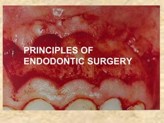
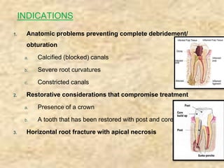
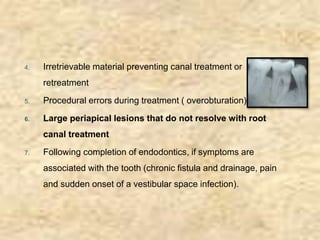
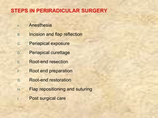
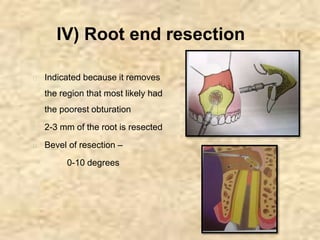
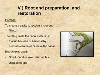
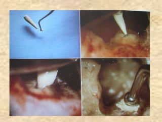
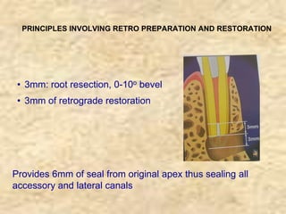
PRINCIPLES OF
ENDODONTIC SURGERY
DEFINITION:
Endodontic surgery is the management or prevention of
periradicular pathosis by a surgical approach.
� It includes:
● Abscess drainage
● Periapical surgery
● Corrective surgery
● Root removal
RATIONALE:
� To remove the causative agents of periradicular
pathology.
� To restore the periodontium to a state of biologic and
functional health.
OBJECTIVE:
� To ensure the placement of a proper seal between the
periodontium and the root canal foramina.
PERIAPICAL SURGERY
1. Anatomic problems preventing complete debridement/
obturation
a. Calcified (blocked) canals
b. Severe root curvatures
c. Constricted canals
2. Restorative considerations that compromise treatment
a. Presence of a crown
b. A tooth that has been restored with post and core
3. Horizontal root fracture with apical necrosis
INDICATIONS
4. Irretrievable material preventing canal treatment or
retreatment
5. Procedural errors during treatment ( overobturation)
6. Large periapical lesions that do not resolve with root
canal treatment
7. Following completion of endodontics, if symptoms are
associated with the tooth (chronic fistula and drainage, pain
and sudden onset of a vestibular space infection).
Contraindications (or cautions) for periapical
surgery
1. Unidentified cause of root canal treatment failure
2. When conventional root canal treatment is possible
3. When retreatment of a treatment failure is possible
4. Simultaneous root canal treatment and apical surgery
5. Anatomic structures (e.g., adjacent nerves and vessels) are in
close proximity
6. Structures interfere with access and visibility
7. Poor crown/root ratio
8. Systemic complications (e.g., bleeding disorders)
STEPS IN PERIRADICULAR SURGERY
A. Anesthesia
B. Incision and flap reflection
C. Periapical exposure
D. Periapical curettage
E. Root-end resection
F. Root end preparation
G. Root-end restoration
H. Flap repositioning and suturing
I. Post surgical care
I) FLAP DESIGN
▪ A properly designed and carefully reflected flap
results in good access and uncomplicated
healing
▪ The incisions most commonly used are:
a. Submarginal curved (semilunar)
b. Submarginal ,and
c. Full mucoperiosteal ( sulcular)
a. Semilunar incision
� Slightly curved half-moon horizontal
incision in the alveolar mucosa
� Advantage:
● Easy access and quick access to the
periradicular structures
● Gingival attachment is not disturbed
� Disadvantage:
● Full root surface not seen
● Root resection difficult
● Slow healing
● Excessive hemorrhage
● Scarring
� Hence, contraindicated for most
endodontic surgery
b. Submarginal incision (LUEBKE OCHSENBEIN) FLAP
� The horizontal component of the
submarginal incision is in attached
gingiva with vertical relieving
incisions
� The incision is scalloped in the
horizontal line, with obtuse angles
at the corners
� Indications
● Maxillary anteriors
● Maxillary premolars
� Advantages:
● Esthetics (less incidence of gingival recession)
● Less risk of incising over a bony defect
● Better access and visibility
� Disadvantages:
● Hemorrhage along the cut margins into the surgical
site
● Occasional, healing by scarring
C. Full mucoperiosteal flaps(Sulcular incisions)
1. Triangular flap
INDICATIONS:
� Maxillary incisors and posterior teeth.
� Only recommended flap for mandibular posterior teeth.
ADVANTAGES:
• Good wound healing
• Ease of flap re-approximation with minimum number
of sutures
DISADVANTAGES:
• Limited surgical access
• Difficult to expose the root apices of long teeth like
maxillary and mandibular canines
• Tension is created on retraction
• Gingival attachment violated
2. Rectangular flap
INDICATIONS:
� Anterior teeth
� Multiple teeth
� Teeth with long roots like maxillary canines
� Lateral root repairs.
� Large lesions
CONTRAINDICATION : Posterior teeth
ADVANTAGES:
• Increased surgical access to root apex
• Reduces retraction tension
• Facilitates repositioning
DISADVANTAGES:
a.Difficulty in re-approximation of flap margins
b.Gingival attachment violated –
recession, crestal bone loss and dehiscence.
3. Trapezoidal
Similar to rectangular except the 2 vertical incisions intersect
the horizontal incision at an obtuse angle so as to create a
broad based flap with the vestibular part wider than the
sulcular portion
Flap reflection
� Flap reflection is the process of separating the soft tissues
(gingiva, mucosa, and periosteum) from the surface of the
alveolar bone.
II) Periapical exposure
� A soft tissue lesion is seen
frequently, after flap reflection
� If the opening is small, it is
enlarged using a large surgical
round bur, until approximately
half the root and the lesion are
visible
� Radiographs are used in
combination with clinical
findings to locate root apex
III) Periradicular Curettage
▪ A surgical procedure to remove
diseased tissue from alveolar
bone in the peri radicular area or
lateral region surrounding a
pulpless (RCT) tooth
▪ A suitably sized sharp curette is
used for enucleation of the lesion
▪ Usually, remnants of tissue
remain, which is not a problem
▪ Hemorrhage control to be
achieved
IV) Root end resection
� Indicated because it removes
the region that most likely had
the poorest obturation
� 2-3 mm of the root is resected
� Bevel of resection –
0-10 degrees
V ) Root end preparation and
restoration
Purpose:
To create a cavity to receive a root-end
filling.
The filling seals the canal system, so
that no bacteria or bacterial by
products can enter or leave the canal
Instruments Used:
▪ Small round or inverted cone bur
▪ Ultra sonic tips
RETROGRADE ROOT END RESTORATIVE MATERIALS
� Amalgam
� Glass ionomer cement
� Zinc oxide eugenol cement
� Intermediate restorative material (IRM)
� Super ethoxybenzoic acid (EBA)
� Gutta percha
� Mineral trioxide aggregates (MTA)
INSPECTION AT VARYING MAGNIFICATIONS
PRINCIPLES INVOLVING RETRO PREPARATION AND RESTORATION
• 3mm: root resection, 0-10o bevel
• 3mm of retrograde restoration
Provides 6mm of seal from original apex thus sealing all
accessory and lateral canals
VI) SOFT TISSUE REPOSITIONING
AND COMPRESSION
� The elevated mucoperiosteum is gently replaced to its original
position with the incision lines approximated as closely as
possible.
� Tissue compression:
Using a surgical gauze moistened with sterile saline, gently apply
firm pressure to the flapped tissue for 2 to 3 minutes (5 minutes
for palatal tissue) before suturing.
VII ) SUTURING
� Purpose:
To approximate the incised
tissue and stabilize the flapped
mucoperiosteum until
reattachment occurs.
� Sling suture is demonstrated in
the picture.
Basic principles of Endodontic Surgery .

Basic principles of Endodontic Surgery .

  • 1.
  • 2.
    DEFINITION: Endodontic surgery isthe management or prevention of periradicular pathosis by a surgical approach. � It includes: ● Abscess drainage ● Periapical surgery ● Corrective surgery ● Root removal
  • 3.
    RATIONALE: � To removethe causative agents of periradicular pathology. � To restore the periodontium to a state of biologic and functional health. OBJECTIVE: � To ensure the placement of a proper seal between the periodontium and the root canal foramina.
  • 4.
  • 5.
    1. Anatomic problemspreventing complete debridement/ obturation a. Calcified (blocked) canals b. Severe root curvatures c. Constricted canals 2. Restorative considerations that compromise treatment a. Presence of a crown b. A tooth that has been restored with post and core 3. Horizontal root fracture with apical necrosis INDICATIONS
  • 6.
    4. Irretrievable materialpreventing canal treatment or retreatment 5. Procedural errors during treatment ( overobturation) 6. Large periapical lesions that do not resolve with root canal treatment 7. Following completion of endodontics, if symptoms are associated with the tooth (chronic fistula and drainage, pain and sudden onset of a vestibular space infection).
  • 7.
    Contraindications (or cautions)for periapical surgery 1. Unidentified cause of root canal treatment failure 2. When conventional root canal treatment is possible 3. When retreatment of a treatment failure is possible 4. Simultaneous root canal treatment and apical surgery 5. Anatomic structures (e.g., adjacent nerves and vessels) are in close proximity 6. Structures interfere with access and visibility 7. Poor crown/root ratio 8. Systemic complications (e.g., bleeding disorders)
  • 8.
    STEPS IN PERIRADICULARSURGERY A. Anesthesia B. Incision and flap reflection C. Periapical exposure D. Periapical curettage E. Root-end resection F. Root end preparation G. Root-end restoration H. Flap repositioning and suturing I. Post surgical care
  • 9.
    I) FLAP DESIGN ▪A properly designed and carefully reflected flap results in good access and uncomplicated healing ▪ The incisions most commonly used are: a. Submarginal curved (semilunar) b. Submarginal ,and c. Full mucoperiosteal ( sulcular)
  • 10.
    a. Semilunar incision �Slightly curved half-moon horizontal incision in the alveolar mucosa � Advantage: ● Easy access and quick access to the periradicular structures ● Gingival attachment is not disturbed � Disadvantage: ● Full root surface not seen ● Root resection difficult ● Slow healing ● Excessive hemorrhage ● Scarring � Hence, contraindicated for most endodontic surgery
  • 11.
    b. Submarginal incision(LUEBKE OCHSENBEIN) FLAP � The horizontal component of the submarginal incision is in attached gingiva with vertical relieving incisions � The incision is scalloped in the horizontal line, with obtuse angles at the corners � Indications ● Maxillary anteriors ● Maxillary premolars
  • 12.
    � Advantages: ● Esthetics(less incidence of gingival recession) ● Less risk of incising over a bony defect ● Better access and visibility � Disadvantages: ● Hemorrhage along the cut margins into the surgical site ● Occasional, healing by scarring
  • 13.
    C. Full mucoperiostealflaps(Sulcular incisions) 1. Triangular flap INDICATIONS: � Maxillary incisors and posterior teeth. � Only recommended flap for mandibular posterior teeth.
  • 14.
    ADVANTAGES: • Good woundhealing • Ease of flap re-approximation with minimum number of sutures DISADVANTAGES: • Limited surgical access • Difficult to expose the root apices of long teeth like maxillary and mandibular canines • Tension is created on retraction • Gingival attachment violated
  • 15.
    2. Rectangular flap INDICATIONS: �Anterior teeth � Multiple teeth � Teeth with long roots like maxillary canines � Lateral root repairs. � Large lesions CONTRAINDICATION : Posterior teeth
  • 16.
    ADVANTAGES: • Increased surgicalaccess to root apex • Reduces retraction tension • Facilitates repositioning DISADVANTAGES: a.Difficulty in re-approximation of flap margins b.Gingival attachment violated – recession, crestal bone loss and dehiscence.
  • 17.
    3. Trapezoidal Similar torectangular except the 2 vertical incisions intersect the horizontal incision at an obtuse angle so as to create a broad based flap with the vestibular part wider than the sulcular portion
  • 18.
    Flap reflection � Flapreflection is the process of separating the soft tissues (gingiva, mucosa, and periosteum) from the surface of the alveolar bone.
  • 19.
    II) Periapical exposure �A soft tissue lesion is seen frequently, after flap reflection � If the opening is small, it is enlarged using a large surgical round bur, until approximately half the root and the lesion are visible � Radiographs are used in combination with clinical findings to locate root apex
  • 20.
    III) Periradicular Curettage ▪A surgical procedure to remove diseased tissue from alveolar bone in the peri radicular area or lateral region surrounding a pulpless (RCT) tooth ▪ A suitably sized sharp curette is used for enucleation of the lesion ▪ Usually, remnants of tissue remain, which is not a problem ▪ Hemorrhage control to be achieved
  • 21.
    IV) Root endresection � Indicated because it removes the region that most likely had the poorest obturation � 2-3 mm of the root is resected � Bevel of resection – 0-10 degrees
  • 22.
    V ) Rootend preparation and restoration Purpose: To create a cavity to receive a root-end filling. The filling seals the canal system, so that no bacteria or bacterial by products can enter or leave the canal Instruments Used: ▪ Small round or inverted cone bur ▪ Ultra sonic tips
  • 23.
    RETROGRADE ROOT ENDRESTORATIVE MATERIALS � Amalgam � Glass ionomer cement � Zinc oxide eugenol cement � Intermediate restorative material (IRM) � Super ethoxybenzoic acid (EBA) � Gutta percha � Mineral trioxide aggregates (MTA)
  • 25.
    INSPECTION AT VARYINGMAGNIFICATIONS
  • 26.
    PRINCIPLES INVOLVING RETROPREPARATION AND RESTORATION • 3mm: root resection, 0-10o bevel • 3mm of retrograde restoration Provides 6mm of seal from original apex thus sealing all accessory and lateral canals
  • 27.
    VI) SOFT TISSUEREPOSITIONING AND COMPRESSION � The elevated mucoperiosteum is gently replaced to its original position with the incision lines approximated as closely as possible. � Tissue compression: Using a surgical gauze moistened with sterile saline, gently apply firm pressure to the flapped tissue for 2 to 3 minutes (5 minutes for palatal tissue) before suturing.
  • 28.
    VII ) SUTURING �Purpose: To approximate the incised tissue and stabilize the flapped mucoperiosteum until reattachment occurs. � Sling suture is demonstrated in the picture.