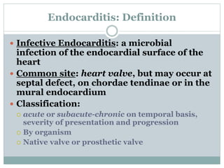
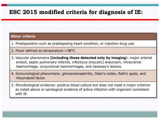
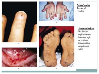
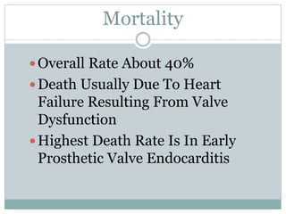
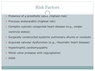
D R I H A B S U L I M A N
3 / 1 0 / 2 0 1 9
Endocarditis Presentation to
Internal Medicine
 https://www.aae.org/uploadedfiles/clinical_resourc
es/guidelines_and_position_statements/aae_antibi
otic-prophylaxis-2017update.pdf
Q1
 Which of the following is accurate about the etiology of
infective endocarditis?
 The most common underlying cause of native valve
endocarditis (NVE) is congenital heart disease
 Early prosthetic valve endocarditis (PVE) and late PVE
both have the same bacteriology and prognosis, with a
subacute presentation similar to that of NVE
 Overall, Staphylococcus aureus is the most common
cause of infective endocarditis, and more than one half of
cases are not associated with underlying valvular disease
 Pseudomonas aeruginosa is the most common etiologic
organism in patients with intravenous drug abuse
(IVDA) infective endocarditis
A1
 Overall, Staphylococcus aureus is the most common
cause of infective endocarditis, and more than one
half of cases are not associated with underlying
valvular disease
Explanation
 Overall, S aureus infection is the most common cause of
infective endocarditis, including PVE, acute infective
endocarditis, and IVDA infective endocarditis.
Approximately 35%-60.5% of staphylococcal bacteremias
are complicated by infective endocarditis. More than one
half of all cases are not associated with underlying
valvular disease.
 The mortality rate of S aureus infective endocarditis is
40%-50%. S aureus infection is the second most
common cause of nosocomial bloodstream infections,
second only to coagulase-negative staphylococci
infection.
Q2
 Which of the following is accurate about the presentation
of infective endocarditis?
 Dyspnea, cough, and chest pain are the most common
symptoms across all types of infective endocarditis
 Cerebrovascular accidents and congestive heart failure
are common developments associated with subacute
endocarditis
 Most cases of subacute infective endocarditis are caused
by dental procedures, with symptoms appearing within
3-4 days of such procedures in 85% of patients
 Nosocomial infective endocarditis often manifests with
elements of sepsis and mostly occurs in patients with
prosthetic valves
A2
 Nosocomial infective endocarditis often manifests with elements of sepsis and mostly occurs in patients with
prosthetic valves
 Nosocomial infective endocarditis commonly manifests with elements of a sepsis syndrome (ie, hypotension,
metabolic acidosis fever, leukocytosis, and multiple organ failure). The source of bacteremia may develop from
an infection in another organ (eg, pneumonia, pyelonephritis) or from a central venous catheter. Most often,
these patients are in the intensive care unit. The aging of the population is associated with an increased
incidence of staphylococcal healthcare-associated endocarditis, in addition to an increased mortality rate
associated with the disease. Approximately 45% of cases of nosocomial/healthcare-associated infective
endocarditis occur in patients with prosthetic valves.
 Fever and chills are the most common symptoms; anorexia, weight loss, malaise, headache, myalgias, night
sweats, shortness of breath, cough, or joint pains are common symptoms as well. Dyspnea, cough, and chest
pain are common symptoms in intravenous drug users with infective endocarditis. This is probably related to
the predominance of tricuspid valve endocarditis in this group and secondary embolic showering of the
pulmonary vasculature.
 A key concern is the distinction between subacute and acute infective endocarditis. The diagnosis of subacute
infective endocarditis is suggested by a history of an indolent process characterized by fever, fatigue, anorexia,
back pain, and weight loss. Less common developments include a cerebrovascular accident or congestive heart
failure.
 The patient should be questioned about invasive procedures and recreational drug use that may be causing the
bacteremia. Most subacute disease caused by Streptococcus viridans infection is related to dental disease.
Most cases are caused not by dental procedures, but by transient bacteremias secondary to gingivitis. In 85%
of patients, symptoms of endocarditis appear within 2 weeks of dental or other procedures.
Q3
 Which of the following is accurate about the physical
examination and associated findings in patients with infective
endocarditis?
 Heart murmurs are heard in the majority of patients with
infective endocarditis
 Multiple embolic pulmonary infections or infarctions are due
to left heart disease, whereas signs of systemic septic emboli
are due to right heart disease
 Purulent meningitis is observed in patients with subacute
disease compared with the aseptic type observed in patients
with acute infective endocarditis
 Splenomegaly is observed more commonly in patients with
recent subacute disease compared with long-standing
subacute disease
A3
 Heart murmurs are heard in the majority of patients with infective endocarditis.
 Heart murmurs are heard in approximately 85% of patients. A change in the
characteristics of a previously noted murmur occurs in 10% of these patients and
increases the likelihood of secondary congestive heart failure.
 Signs of systemic septic emboli are due to left heart disease and are more commonly
associated with mitral valve vegetations. Multiple embolic pulmonary infections or
infarctions are due to right heart disease.
 Purulent meningitis may be observed in patients with acute infective endocarditis,
compared with the aseptic type observed in patients with subacute disease. Other
neurologic findings are similar to those observed in patients with subacute disease.
 Splenomegaly is observed more commonly in patients with long-standing subacute
disease. It may persist long after successful therapy.
Q4
 Which of the following is accurate about the workup of
infective endocarditis?
 The presence of a continuous bacteremia is sufficient to
diagnose an infected valvular vegetation
 The criterion standard test for diagnosing infective
endocarditis is documentation of continuous bacteremia
(> 30 minutes) on the basis of blood culture results
 Leukocytosis is commonly observed in subacute
endocarditis, with anemia common in acute endocarditis
 A diagnosis of infective endocarditis can be excluded on
the basis of negative echocardiography findings, either
from transesophageal echocardiography or transthoracic
echocardiography
A4
 The criterion standard test for diagnosing infective endocarditis is
documentation of continuous bacteremia (>30 minutes) on the basis of
blood culture results. Although blood cultures remain key in making the
diagnosis of infective endocarditis, the need for indirect diagnostic
techniques that are both specific and sensitive is increasing. This is because
the nature of valvular infections has changed over the years. The number of
fastidious organisms has increased, and the rate of the classic peripheral
stigmata of infective endocarditis is much lower. Patients who are elderly,
chronically ill, or immunosuppressed are often afebrile and do not mount a
significant fever or exhibit the classic stigmata of valvular infection.
 Because S aureus can produce endotheliosis, the presence of a continuous
bacteremia does not necessarily imply an infected valvular vegetation.
 Anemia is common in subacute endocarditis. Leukocytosis is observed in
acute endocarditis. Erythrocyte sedimentation rate, although not specific, is
elevated in more than 90% of cases. Decreased C3, C4, and CH50 are
evident in subacute endocarditis.
Q5
 Which of the following is accurate about the treatment of
infective endocarditis?
 Patients with subacute infective endocarditis must be
treated with antibiotics before culture and sensitivity
results, owing to the increased risk for complication over
time
 Patients with PVE should receive monotherapy with
rifampin as the first-line treatment
 Congestive heart failure in a patient with NVE is the
primary indication for surgery
 Continuously infused vancomycin is the best therapy for
aminoglycoside-resistant enterococci
A5
 Approximately 15%-25% of patients with infective endocarditis
eventually require surgery. Indications for surgical intervention in
patients with NVE are as follows:
 Congestive heart failure that is refractory to standard medical
therapy
 Fungal infective endocarditis (except that caused by Histoplasma
capsulatum)
 Persistent sepsis after 72 hours of appropriate antibiotic treatment
 Recurrent septic emboli, especially after 2 weeks of antibiotic
treatment
 Rupture of an aneurysm of the sinus of Valsalva
 Conduction disturbances caused by a septal abscess
 Kissing infection of the anterior mitral leaflet in patients with
infective endocarditis of the aortic valve
•Conduction disturbances caused by a septal abscess
•Kissing infection of the anterior mitral leaflet in patients with infective endocarditis of the aortic valve
Endocarditis: Definition
 Infective Endocarditis: a microbial
infection of the endocardial surface of the
heart
 Common site: heart valve, but may occur at
septal defect, on chordae tendinae or in the
mural endocardium
 Classification:
 acute or subacute-chronic on temporal basis,
severity of presentation and progression
 By organism
 Native valve or prosthetic valve
Oslers’ nodes
Tender, s/c
nodules
Janeway lesions
Nontender
erythematous,
haemorrhagic,
or pustular
lesions often
on palms or
soles.
General Lesions
 Enlarged Spleen
 Arthritis
 Clubbing Of Fingers
 Cardiac Failure
 Conduction Abnormalities
 Stroke
 Psychiatric Disease
 Renal Failure
Mortality
Overall Rate About 40%
Death Usually Due To Heart
Failure Resulting From Valve
Dysfunction
Highest Death Rate Is In Early
Prosthetic Valve Endocarditis
Risk Factors
 Presence of a prosthetic valve (highest risk)
 Previous endocarditis (highest risk)
 Complex cyanotic congenital heart disease (e.g., single-
ventricle states)
 Surgically constructed systemic pulmonary shunts or conduits
 Acquired valvular dysfunction (e.g., rheumatic heart disease)
 Hypertrophic cardiomyopathy
 Mitral valve prolapse with regurgitation
 IVDA
Q1
A1
 C- VALVULAR Aortic Regurgitaion
Q2
A2
 B- Amyloidosis
Q3
A3
 B- Suppurative Pericarditis
Q4
A4
 D- Large ASD
Q5
 B- The presence of prosthetic valve
Q6
A6
 D-Candida
Q7
A7
 A-Viridans Streptococci
Q8
A8
 C-STAPH EPEDERMIDIS
Q9
A9
 D)Enterococcus fecalis.
Q10
A10
 C)Decreased erythrocyte sedimentation rate
Q11
A11
 B)positive blood cultures and echocardiographic
changes.
Q12
A12
 B)subacute, left-sided IE.
Q13
A13
 B)Clinical cure can only occur if synergistic
aminoglycosides are used in combination with a β-
lactam agent.
Q14
A14
B) Penicillin G 12-18 million units
every 24 hours for 4 weeks
Q15
A15
 B)yes, clindamycin 600 mg orally 1 hour before the
procedure.
Q16
 Which of these wouldn't be an appropriate
investigation of IE?
 A Blood Culture
 B Blood Tests
 C Cardiac Angiogram
 D ECG
 E Echocardiogram
A16
 C Cardiac Angiogram
 A blood culture is important to find the causative
organism. An ECG can be useful, as sometimes there
is a lengthened PR Interval with IE. An
echocardiogram can show the vegetations on the
valves. Blood tests will show a raised ESR/CRP with
infection, and there may also be an anaemia.
Q17
 Which of these isn't a common cause of infective
endocarditis?
 A Haemophilus parainfluenzae
 B Staphylococcus Aureus
 C Staphylococcus pidermidis
 D Streprococcus Viridans
A17
 A Haemophilus parainfluenzae
 Staphylococcus Aureus is a very common cause of
infective endocarditis (IE), as is streptococcus
viridans. Haemophilus parainfluenzae is one of the
HACEK organisms, a group of gram-negative
bacteria that can cause IE but are rarer. HACEK -
Haemophilus, Aggregatibacter, Cardiobacterium,
Eikenella, Kingella.
Q18
 Which of these is NOT a risk factor for IE?
 A IV Drug Use
 B Mitral Valve Disease
 C None - they are all risk factors
 D Prosthetic Valves
A18
 C None - they are all risk factors
 All of these options result in damage to valves, and
therefore increase the risk of IE. Although, 50% of IE
occurs on normal valves. Endocarditis normally
follows an acute course, but on abnormal valves it
tends to run a subacute course.
Q19
 Which of these is not a sign of infective endocarditis?
 A Aortic Regurgitation
 B Butterfly Rash
 C Fever
 D Janeway Lesions
 E Splinter Haemorrhages
A19
 B Butterfly Rash
 The signs of IE can be grouped into 4 main strands:
1. Septic Signs. This includes fever, rigors, night
sweats, malaise, weight loss, anaemia, splenomegaly
and clubbing 2. Cardiac lesions. The vegetations on
the valves may cause regurgitation or obstruction. 3.
Immune complex deposition: splinter haemorrhages,
Osler’s nodes (painful pulp infarcts in fingers or
toes). 4. Embolic phenomena: Janeway lesions
(painless palmar macules), which, together with
Osler’s nodes, are pathognomonic of IE.
Q20
 A 30 year old IV drug user presents with shortness of
breath, night sweats and a fever. On examination, you
find that he has splinter haemorrhages. Which of these
investigations is needed LEAST to confirm infective
endocarditis?
 A A single positive blood culture
 B An echocardiogram showing vegetations
 C An echocardiogram showing vegetations and two
positive blood cultures
 D ECG showing a long PR Interva
 B An echocardiogram showing vegetations
 The Duke criteria can be used to diagnose infective
endocarditis. The criteria consist of major and minor criteria,
and to diagnose IE, you need 2 major criteria, 1 major and 3
minor criteria or all 5 minor criteria. Major criteria: - Typical
organism in 2 separate cultures or persistently positive blood
cultures - Positive echocardiogram Minor Criteria: -
Predisposition (cardiac lesion; IV drug abuse) - Fever (>38) -
Vascular/immunological signs - Positive blood culture that
doesn't meet major criteria - Positive echocardiogram that
does not meet major criteria This patient has 3 minor criteria.
Therefore, they only need 1 major for a diagnosis - an
echocardiogram showing vegetations. They don't need both.

Endocarditis presentation to internal medicine2019

  • 1.
    D R IH A B S U L I M A N 3 / 1 0 / 2 0 1 9 Endocarditis Presentation to Internal Medicine
  • 2.
  • 3.
    Q1  Which ofthe following is accurate about the etiology of infective endocarditis?  The most common underlying cause of native valve endocarditis (NVE) is congenital heart disease  Early prosthetic valve endocarditis (PVE) and late PVE both have the same bacteriology and prognosis, with a subacute presentation similar to that of NVE  Overall, Staphylococcus aureus is the most common cause of infective endocarditis, and more than one half of cases are not associated with underlying valvular disease  Pseudomonas aeruginosa is the most common etiologic organism in patients with intravenous drug abuse (IVDA) infective endocarditis
  • 4.
    A1  Overall, Staphylococcusaureus is the most common cause of infective endocarditis, and more than one half of cases are not associated with underlying valvular disease
  • 5.
    Explanation  Overall, Saureus infection is the most common cause of infective endocarditis, including PVE, acute infective endocarditis, and IVDA infective endocarditis. Approximately 35%-60.5% of staphylococcal bacteremias are complicated by infective endocarditis. More than one half of all cases are not associated with underlying valvular disease.  The mortality rate of S aureus infective endocarditis is 40%-50%. S aureus infection is the second most common cause of nosocomial bloodstream infections, second only to coagulase-negative staphylococci infection.
  • 6.
    Q2  Which ofthe following is accurate about the presentation of infective endocarditis?  Dyspnea, cough, and chest pain are the most common symptoms across all types of infective endocarditis  Cerebrovascular accidents and congestive heart failure are common developments associated with subacute endocarditis  Most cases of subacute infective endocarditis are caused by dental procedures, with symptoms appearing within 3-4 days of such procedures in 85% of patients  Nosocomial infective endocarditis often manifests with elements of sepsis and mostly occurs in patients with prosthetic valves
  • 7.
    A2  Nosocomial infectiveendocarditis often manifests with elements of sepsis and mostly occurs in patients with prosthetic valves  Nosocomial infective endocarditis commonly manifests with elements of a sepsis syndrome (ie, hypotension, metabolic acidosis fever, leukocytosis, and multiple organ failure). The source of bacteremia may develop from an infection in another organ (eg, pneumonia, pyelonephritis) or from a central venous catheter. Most often, these patients are in the intensive care unit. The aging of the population is associated with an increased incidence of staphylococcal healthcare-associated endocarditis, in addition to an increased mortality rate associated with the disease. Approximately 45% of cases of nosocomial/healthcare-associated infective endocarditis occur in patients with prosthetic valves.  Fever and chills are the most common symptoms; anorexia, weight loss, malaise, headache, myalgias, night sweats, shortness of breath, cough, or joint pains are common symptoms as well. Dyspnea, cough, and chest pain are common symptoms in intravenous drug users with infective endocarditis. This is probably related to the predominance of tricuspid valve endocarditis in this group and secondary embolic showering of the pulmonary vasculature.  A key concern is the distinction between subacute and acute infective endocarditis. The diagnosis of subacute infective endocarditis is suggested by a history of an indolent process characterized by fever, fatigue, anorexia, back pain, and weight loss. Less common developments include a cerebrovascular accident or congestive heart failure.  The patient should be questioned about invasive procedures and recreational drug use that may be causing the bacteremia. Most subacute disease caused by Streptococcus viridans infection is related to dental disease. Most cases are caused not by dental procedures, but by transient bacteremias secondary to gingivitis. In 85% of patients, symptoms of endocarditis appear within 2 weeks of dental or other procedures.
  • 8.
    Q3  Which ofthe following is accurate about the physical examination and associated findings in patients with infective endocarditis?  Heart murmurs are heard in the majority of patients with infective endocarditis  Multiple embolic pulmonary infections or infarctions are due to left heart disease, whereas signs of systemic septic emboli are due to right heart disease  Purulent meningitis is observed in patients with subacute disease compared with the aseptic type observed in patients with acute infective endocarditis  Splenomegaly is observed more commonly in patients with recent subacute disease compared with long-standing subacute disease
  • 9.
    A3  Heart murmursare heard in the majority of patients with infective endocarditis.  Heart murmurs are heard in approximately 85% of patients. A change in the characteristics of a previously noted murmur occurs in 10% of these patients and increases the likelihood of secondary congestive heart failure.  Signs of systemic septic emboli are due to left heart disease and are more commonly associated with mitral valve vegetations. Multiple embolic pulmonary infections or infarctions are due to right heart disease.  Purulent meningitis may be observed in patients with acute infective endocarditis, compared with the aseptic type observed in patients with subacute disease. Other neurologic findings are similar to those observed in patients with subacute disease.  Splenomegaly is observed more commonly in patients with long-standing subacute disease. It may persist long after successful therapy.
  • 10.
    Q4  Which ofthe following is accurate about the workup of infective endocarditis?  The presence of a continuous bacteremia is sufficient to diagnose an infected valvular vegetation  The criterion standard test for diagnosing infective endocarditis is documentation of continuous bacteremia (> 30 minutes) on the basis of blood culture results  Leukocytosis is commonly observed in subacute endocarditis, with anemia common in acute endocarditis  A diagnosis of infective endocarditis can be excluded on the basis of negative echocardiography findings, either from transesophageal echocardiography or transthoracic echocardiography
  • 11.
    A4  The criterionstandard test for diagnosing infective endocarditis is documentation of continuous bacteremia (>30 minutes) on the basis of blood culture results. Although blood cultures remain key in making the diagnosis of infective endocarditis, the need for indirect diagnostic techniques that are both specific and sensitive is increasing. This is because the nature of valvular infections has changed over the years. The number of fastidious organisms has increased, and the rate of the classic peripheral stigmata of infective endocarditis is much lower. Patients who are elderly, chronically ill, or immunosuppressed are often afebrile and do not mount a significant fever or exhibit the classic stigmata of valvular infection.  Because S aureus can produce endotheliosis, the presence of a continuous bacteremia does not necessarily imply an infected valvular vegetation.  Anemia is common in subacute endocarditis. Leukocytosis is observed in acute endocarditis. Erythrocyte sedimentation rate, although not specific, is elevated in more than 90% of cases. Decreased C3, C4, and CH50 are evident in subacute endocarditis.
  • 12.
    Q5  Which ofthe following is accurate about the treatment of infective endocarditis?  Patients with subacute infective endocarditis must be treated with antibiotics before culture and sensitivity results, owing to the increased risk for complication over time  Patients with PVE should receive monotherapy with rifampin as the first-line treatment  Congestive heart failure in a patient with NVE is the primary indication for surgery  Continuously infused vancomycin is the best therapy for aminoglycoside-resistant enterococci
  • 13.
    A5  Approximately 15%-25%of patients with infective endocarditis eventually require surgery. Indications for surgical intervention in patients with NVE are as follows:  Congestive heart failure that is refractory to standard medical therapy  Fungal infective endocarditis (except that caused by Histoplasma capsulatum)  Persistent sepsis after 72 hours of appropriate antibiotic treatment  Recurrent septic emboli, especially after 2 weeks of antibiotic treatment  Rupture of an aneurysm of the sinus of Valsalva  Conduction disturbances caused by a septal abscess  Kissing infection of the anterior mitral leaflet in patients with infective endocarditis of the aortic valve •Conduction disturbances caused by a septal abscess •Kissing infection of the anterior mitral leaflet in patients with infective endocarditis of the aortic valve
  • 18.
    Endocarditis: Definition  InfectiveEndocarditis: a microbial infection of the endocardial surface of the heart  Common site: heart valve, but may occur at septal defect, on chordae tendinae or in the mural endocardium  Classification:  acute or subacute-chronic on temporal basis, severity of presentation and progression  By organism  Native valve or prosthetic valve
  • 22.
    Oslers’ nodes Tender, s/c nodules Janewaylesions Nontender erythematous, haemorrhagic, or pustular lesions often on palms or soles.
  • 23.
    General Lesions  EnlargedSpleen  Arthritis  Clubbing Of Fingers  Cardiac Failure  Conduction Abnormalities  Stroke  Psychiatric Disease  Renal Failure
  • 24.
    Mortality Overall Rate About40% Death Usually Due To Heart Failure Resulting From Valve Dysfunction Highest Death Rate Is In Early Prosthetic Valve Endocarditis
  • 25.
    Risk Factors  Presenceof a prosthetic valve (highest risk)  Previous endocarditis (highest risk)  Complex cyanotic congenital heart disease (e.g., single- ventricle states)  Surgically constructed systemic pulmonary shunts or conduits  Acquired valvular dysfunction (e.g., rheumatic heart disease)  Hypertrophic cardiomyopathy  Mitral valve prolapse with regurgitation  IVDA
  • 26.
  • 27.
    A1  C- VALVULARAortic Regurgitaion
  • 28.
  • 29.
  • 30.
  • 31.
  • 32.
  • 33.
  • 34.
  • 35.
     B- Thepresence of prosthetic valve
  • 36.
  • 37.
  • 38.
  • 39.
  • 40.
  • 41.
  • 42.
  • 43.
  • 44.
  • 45.
  • 46.
  • 47.
    A11  B)positive bloodcultures and echocardiographic changes.
  • 48.
  • 49.
  • 50.
  • 51.
    A13  B)Clinical curecan only occur if synergistic aminoglycosides are used in combination with a β- lactam agent.
  • 52.
  • 53.
    A14 B) Penicillin G12-18 million units every 24 hours for 4 weeks
  • 54.
  • 55.
    A15  B)yes, clindamycin600 mg orally 1 hour before the procedure.
  • 56.
    Q16  Which ofthese wouldn't be an appropriate investigation of IE?  A Blood Culture  B Blood Tests  C Cardiac Angiogram  D ECG  E Echocardiogram
  • 57.
    A16  C CardiacAngiogram  A blood culture is important to find the causative organism. An ECG can be useful, as sometimes there is a lengthened PR Interval with IE. An echocardiogram can show the vegetations on the valves. Blood tests will show a raised ESR/CRP with infection, and there may also be an anaemia.
  • 58.
    Q17  Which ofthese isn't a common cause of infective endocarditis?  A Haemophilus parainfluenzae  B Staphylococcus Aureus  C Staphylococcus pidermidis  D Streprococcus Viridans
  • 59.
    A17  A Haemophilusparainfluenzae  Staphylococcus Aureus is a very common cause of infective endocarditis (IE), as is streptococcus viridans. Haemophilus parainfluenzae is one of the HACEK organisms, a group of gram-negative bacteria that can cause IE but are rarer. HACEK - Haemophilus, Aggregatibacter, Cardiobacterium, Eikenella, Kingella.
  • 60.
    Q18  Which ofthese is NOT a risk factor for IE?  A IV Drug Use  B Mitral Valve Disease  C None - they are all risk factors  D Prosthetic Valves
  • 61.
    A18  C None- they are all risk factors  All of these options result in damage to valves, and therefore increase the risk of IE. Although, 50% of IE occurs on normal valves. Endocarditis normally follows an acute course, but on abnormal valves it tends to run a subacute course.
  • 62.
    Q19  Which ofthese is not a sign of infective endocarditis?  A Aortic Regurgitation  B Butterfly Rash  C Fever  D Janeway Lesions  E Splinter Haemorrhages
  • 63.
    A19  B ButterflyRash  The signs of IE can be grouped into 4 main strands: 1. Septic Signs. This includes fever, rigors, night sweats, malaise, weight loss, anaemia, splenomegaly and clubbing 2. Cardiac lesions. The vegetations on the valves may cause regurgitation or obstruction. 3. Immune complex deposition: splinter haemorrhages, Osler’s nodes (painful pulp infarcts in fingers or toes). 4. Embolic phenomena: Janeway lesions (painless palmar macules), which, together with Osler’s nodes, are pathognomonic of IE.
  • 64.
    Q20  A 30year old IV drug user presents with shortness of breath, night sweats and a fever. On examination, you find that he has splinter haemorrhages. Which of these investigations is needed LEAST to confirm infective endocarditis?  A A single positive blood culture  B An echocardiogram showing vegetations  C An echocardiogram showing vegetations and two positive blood cultures  D ECG showing a long PR Interva
  • 65.
     B Anechocardiogram showing vegetations  The Duke criteria can be used to diagnose infective endocarditis. The criteria consist of major and minor criteria, and to diagnose IE, you need 2 major criteria, 1 major and 3 minor criteria or all 5 minor criteria. Major criteria: - Typical organism in 2 separate cultures or persistently positive blood cultures - Positive echocardiogram Minor Criteria: - Predisposition (cardiac lesion; IV drug abuse) - Fever (>38) - Vascular/immunological signs - Positive blood culture that doesn't meet major criteria - Positive echocardiogram that does not meet major criteria This patient has 3 minor criteria. Therefore, they only need 1 major for a diagnosis - an echocardiogram showing vegetations. They don't need both.