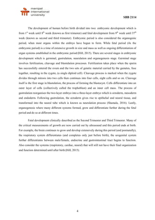
This document describes the key stages of human embryonic and fetal development. It begins with an introduction to embryology and the processes of gametogenesis that produce egg and sperm cells. There are then four main sections that outline the major developmental periods: 1) Germinal stage, which involves fertilization, cleavage of the zygote, and blastulation. 2) Gastrulation stage, where the embryo reorganizes into three germ layers. 3) Neurulation stage where the neural tube forms from ectoderm. 4) Organogenesis stage in which organs begin to form and differentiate. The document then describes fetal development in the second and third trimesters, focusing on continued growth and differentiation of organ systems.

![SBB 2114
5
2.0 EMBRYONIC DEVELOPMENT
The process of progressing from a single cell through the period of establishing organ
primordia (the first 8 weeks of human development) is called the period of embryogenesis. Human
embryonic development drived by cell division and cellular differentiation of the embryo that occurs
at first eight weeks after fertilization until it become a fetus. The staging of human embryos was
introduced in 1914 by Franklin P. Mall at the Department of Embryology of the Carnegie Institution
of Washington. Mall's sucessor, George L. Streeter, later refined the classification of human embryos
into 23 stages, or "developmental horizons" (Louisiana State University Health Sciences Center New
Orleans [LSUHSC], 2012). Embryonic development can be divided into different developmental
phases but there are four main stages should considered, which are germinal, gastrulation, neurulation
and organogenesis stages.
2.1 Germinal Stage:
The germinal stage is the prenatal, developmental stage that begins at conception and lasts
through the second week of pregnancy. During this time, the fertilized egg makes it way down the
fallopian tube, and begins to have cell reproduction. Eventually, the single celled zygote becomes a
multi celled ball that attaches itself to the wall of the uterus around the end of the second week, which
constitutes the beginning of the embryonic stage (Embryogenesis., n.d.). Germinal stage involves
fertilization, cleavage and blastulation processes.
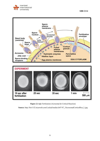
i. Fertilization (Figure 2.1 a):
Fertilization is the formation of a diploid zygote from a haploid egg and sperm (1st
day).
Sperm penetrate the protective layer around the egg by ezymatic reaction. Receptors on the
egg surface bind to molecules on the sperm surface. Changes at the egg surface prevent
polyspermy, the entry of multiple sperm nuclei into the egg. The acrosomal reaction is
triggered when the sperm meets the egg. The acrosome at the tip of the sperm releases
hydrolytic enzymes that digest material surrounding the egg. Gamete contact and/or fusion
depolarizes the egg cell membrane and sets up a fast block to polyspermy. Fusion of egg and
sperm also initiates the cortical reaction. Seconds after the sperm binds to the egg, vesicles
just beneath the egg plasma membrane release their contents and form a fertilization
envelope. The fertilization envelope acts as the slow block to polyspermy. The cortical
reaction requires a high concentration of Ca22+
ions in the egg. The reaction is triggered by a
change in Ca2+
concentration. Ca2+
spread across the egg correlates with the appearance of
the fertilization envelope (Pratheep, 2015).](https://image.slidesharecdn.com/dobassingment1-160822103546/85/Developmental-biology-6-320.jpg)


![SBB 2114
14
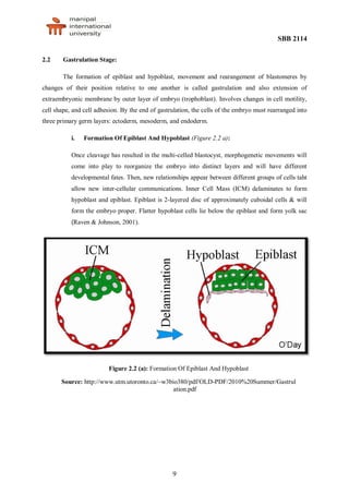
3.0 FETAL DEVELOPMENT
After the 8 week of embryonic development, the developing organism is called a fetus (fetal
development. All major structures are already formed in the fetus, but they continue to grow and
develop. Since the precursors of all the major organs are created by this time, the fetal period is
described both by organ and by a list of changes by weeks of gestational age. This development
divided into 2 main stages: second trimester (3 months) and third trimester (4 months).
3.1 Second Trimester:
During the second trimester (the next three months of pregnancy) the bones actively enlarge
and the brain develops a lot. During the second trimester until about 24 weeks, the fetus cannot live
outside of the body because its lungs, heart and blood systems have not developed enough. The face
looks more human, the baby has hair, the ears stand out, and the baby can hear the mother’s voice.
Between 16 and 20 weeks, the baby’s movements may be felt. The baby begins to store some
antibodies and this slowly increases until birth. Eyebrows and eyelashes appear. A fine downy hair
(lanugo) appears all over the baby’s body (BC Ministry of Health [BCMOH], 2011). The baby’s skin
is thin, shiny, and covered with a creamy protective coating called vernix. The baby’s legs lengthen,
and move well. Teeth develop-enamel and dentine are being formed. By the end of the fifth month the
baby is about half the length of a newborn. During the second trimester, meconium (the baby’s first
stool) begins to appear in the intestines. The fetus has grown to about 175 millimeters in length and
attained a weight of about 225 grams (Raven & Johnson, 2001).
3.2 Third Trimester:
During the third trimester (the last 3 months of pregnancy is predominantly a period of growth
rather than development. The weight of the fetus doubles several times, but this increase in bulk is not
the only kind of growth that occurs. Most of the major nerve tracts in the brain, as well as many new
neurons (nerve cells), are formed during this period. By week 28 the fetus can now store iron,
calcium, and other nutrients and the fetus can hear and respond to sounds. After week 32 the male
fetus’s testicles begin to drop into the scrotum and the pupils in the fetus’s eyes can react to light
(BCMOH, 2011). Neurological growth is far from complete at the end of the third trimester, when
birth takes place. The downy hair on the fetus’s body begins to disappear by week 36. The baby
continues to increase the store of antibodies and is able to resist some diseases. The testicles of male
fetus are now in the scrotum and the labia majora of female fetus are developed. The fetus is now full
term at week 40. The lungs of fetus completly grown and the fetus starts to breath and try to come out
from mother’s uterus (Meeks, 1982).](https://image.slidesharecdn.com/dobassingment1-160822103546/85/Developmental-biology-15-320.jpg)
![SBB 2114
16
REFERENCES
BC Ministry of Health, [BCMOH]. (2011). The best chance: Stages of pregnancy. Retrieved from:
http://www.bestchance.gov.bc.ca/pregnancy/2nd-trimester/stages-of-pregnancy/3rd-trimester-
baby.html.
Betsy, T.L.H. (2009). Longman Essential Revision Guide for Exam Success Biology. Malaysia:
Pearson Sdn. Bhd.
Brevini, T.A.L., & Pennarossa, G. (2013). Gametogenesis, Early Embryo Development, and Stem Cell
Derivation, SpringerBriefs in Stem Cells. DOI: 10.1007/978-1-4614-5532-5_2.
Danton, H.(2010). Gastrulation: formation of the primary germ layers [PDF document]. Retrieved
fromhttp://www.utm.utoronto.ca/~w3bio380/pdf/OLD-PDF/2010%20Summer/Gastrulation.pdf
Embryogenesis. (n.d.). In Wikipedia. Retrieved May 20, 2016, from
https://en.wikipedia.org/wiki/Embryogenesis
Gajaan, S., & Tossif, G. (2013). Organogenesis Critical period of Organogenesis [PowerPoint slides].
Retrieved from www.slideshare.net/applevsamsung/organogenesis.
Guus, V.D.B. (2011). Embryology: Early development from a phenomenological point of view.
Netherlands: Bolk’s Companions. Retrieved from
http://www.louisbolk.org/downloads/1281.pdf.
Hill, M.A. (2015, November 16). Embryology Embryonic Development. UNSW Embryology
[mediawiki]. Retrieved June 3, 2016, from
https://embryology.med.unsw.edu.au/embryology/index.php/Embryonic_Development
Louisiana State University Health Sciences Center New Orleans [LSUHSC]. (2012, November 30).
The Stages of Human Embryonic Development. Virtual Human Embryo Project
[www.ehd.org]. Retrieved from
http://virtualhumanembryo.lsuhsc.edu/DREM/Contact_info_EHD.html.
Marieb, E.N. (2006). Essential of Human Anatomy & Physiology. 8th ed. Addison Wesley Longman.
Meeks, J. (1982). Family Living and Human Reproduction. Columbus, OH: Charles E. Merrill Pub.
Co. Retrieved from http://www.edu.gov.mb.ca/k12/cur/physhlth/hs_k-8/blms/blm7-1-1.pdf.
Pratheep, S. (2015). Chapter 4: General embryology- Animal [PowerPoint slides]. Lecture Notes.
Raven, P.H., & Johnson, G.B. (2001). Biology: 6th edition. Retrieved from
www.mhhe.com/biosci/genbio/raven6b/graphics/raven06b/other/raven06_60.pdf
Sadler, T.W. (2012). Medical Embryology, 12th edition. Retrieved from
http://medfile.ir/iran%20anatomy%20files/text/Langman's%20Medical%20Embryology%2012t
h%20Edition%20(www.irananatomy.ir).pdf.
Shamala, M.(2016). Developmental Biology: Chapter 1 [PowerPoint slides]. Lecture Notes.
Taube, P.R.(n.d.). Ectoderm: Neurulation, Neural Tube, Neural Crest [PDF document]. Retrieved
from http://www.columbia.edu/itc/hs/medical/humandev/2004/Chapt4-Ectoderm.pdf.](https://image.slidesharecdn.com/dobassingment1-160822103546/85/Developmental-biology-17-320.jpg)