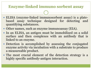
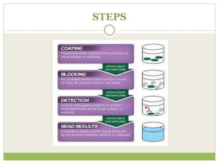
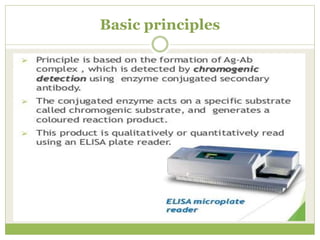
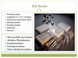
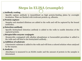
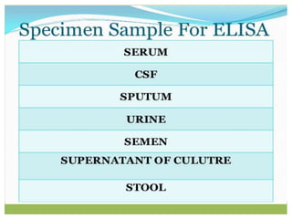
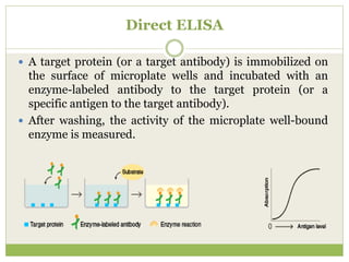
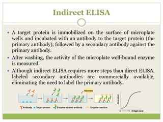
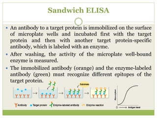
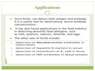
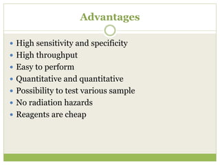
P R E S E N T E D B Y :
S Y E D A T A M A N N A Y A S M I N
M . S C . M I C R O B I O L O G Y ( 4 T H S E M E S T E R )
D E P T . O F L I F E S C I E N C E S
A S S A M D O N B O S C O U N I V E R S I T Y
ELISA
(Enzyme Linked Immuno Sorbent Assay)
Enzyme-linked immuno sorbent assay
 ELISA (enzyme-linked immunosorbent assay) is a plate-
based assay technique designed for detecting and
quantifying substances.
 Other names, such as enzyme immunoassay (EIA).
 In an ELISA, an antigen must be immobilized on a solid
surface and then complexes with an antibody that is
linked to an enzyme.
 Detection is accomplished by assessing the conjugated
enzyme activity via incubation with a substrate to produce
a measureable product.
 The most crucial element of the detection strategy is a
highly specific antibody-antigen interaction.
History
STEPS
Basic principles
Kit items
Steps in ELISA (example)
1.Antibody coating
Specific capture antibody is immobilized on high protein-binding plates by overnight
incubation. Plate are blocked with irrelevant protein e.g. albumin.
2.Protein capture
Samples and standard dilutions are added to the wells and will be captured by the bound
antibodies.
3.Detection antibody
Specific biotinylated detection antibody is added to the wells to enable detection of the
captured protein.
4.Streptavidin-enzyme conjugate
Streptavidin conjugated with alkaline phosphatase or horseradish peroxidase is added to
the wells and will bind to the biotinylated antibody. .
5.Addition of substrate
Colorimetric substrate is added to the wells and will form a colored solution when catalyzed
by the enzyme.
6.Analysis
Absorbance is measured in an ELISA reader and the amount of protein in the samples is
determined.
Direct ELISA
 A target protein (or a target antibody) is immobilized on
the surface of microplate wells and incubated with an
enzyme-labeled antibody to the target protein (or a
specific antigen to the target antibody).
 After washing, the activity of the microplate well-bound
enzyme is measured.
Indirect ELISA
 A target protein is immobilized on the surface of microplate
wells and incubated with an antibody to the target protein (the
primary antibody), followed by a secondary antibody against the
primary antibody.
 After washing, the activity of the microplate well-bound enzyme
is measured.
 Although indirect ELISA requires more steps than direct ELISA,
labeled secondary antibodies are commercially available,
eliminating the need to label the primary antibody.
Sandwich ELISA
 An antibody to a target protein is immobilized on the surface
of microplate wells and incubated first with the target
protein and then with another target protein-specific
antibody, which is labeled with an enzyme.
 After washing, the activity of the microplate well-bound
enzyme is measured.
 The immobilized antibody (orange) and the enzyme-labeled
antibody (green) must recognize different epitopes of the
target protein.
Competitive ELISA
 An antibody specific for a target protein is immobilized on
the surface of microplate wells and incubated with samples
containing the target protein and a known amount of
enzyme-labeled target protein.
 After the reaction, the activity of the microplate well-bound
enzyme is measured.
 When the antigen level in the sample is high, the level of
antibody-bound enzyme-labeled antigen is lower and the
color is lighter.
 Conversely, when it is low, the level of antibody-bound
enzyme-labeled antigen is higher and the color, darker. The
graph above and to the right illustrates the correlation
between absorption and antigen levels in samples.
Competitive ELISA
Protocol
1. Allow all reagents to reach room temperature before use. Gently mix all liquid reagents prior to use.
2. Add 50-100 µL of prepared standard and sample to wells. Cover plate and incubate at room
temperature for 2 hours.
3. Thoroughly aspirate or decant solution from wells and discard the liquid.
4. Wash wells 4 times using a squirt wash bottle or an automated 96-well plate washer.
5. Add 100 µL of diluted detection antibody to wells. Cover plate and incubate at room temperature for 1
hour.
6. Thoroughly aspirate or decant solution from wells and discard the liquid.
7. Wash wells 4 times.
8. Add 100 µL of diluted HRP conjugate to each well. Cover plate and incubate at room temperature for
30 minutes. Thoroughly aspirate or decant solution from wells and discard the liquid.
9. Wash wells 4 times.
10. Add 100 µL of chromogenic substrate to each well.
11. Develop plate at room temperature in the dark for 30 minutes.
12. Add 100 µL of stop solution to each well. The solution in the wells should change from blue to yellow.
13. The plate must be evaluated within 30 minutes of stopping the reaction. Read the absorbance of each
well at 450 nm and 550 nm. Subtract 550 nm values from 450 nm values to correct for optical
imperfections in the microplate.
14. Use curve-fitting statistical software to plot a four-parameter logistic curve fit to the standards and
then calculate results for the test samples.
Picture of plates
ELISA plate
before use
ELISA plate
after use
Applications
Advantages
 High sensitivity and specificity
 High throughput
 Easy to perform
 Quantitative and quantitative
 Possibility to test various sample
 No radiation hazards
 Reagents are cheap
Disadvantages
 Temporary readouts
 Limited antigen information
 Only monoclonal antibodies can be used
 Monoclonal antibodies cost more
 Negative control may indicate positive results if
blocking solution is ineffective
ELISA

ELISA

  • 1.
    P R ES E N T E D B Y : S Y E D A T A M A N N A Y A S M I N M . S C . M I C R O B I O L O G Y ( 4 T H S E M E S T E R ) D E P T . O F L I F E S C I E N C E S A S S A M D O N B O S C O U N I V E R S I T Y ELISA (Enzyme Linked Immuno Sorbent Assay)
  • 2.
    Enzyme-linked immuno sorbentassay  ELISA (enzyme-linked immunosorbent assay) is a plate- based assay technique designed for detecting and quantifying substances.  Other names, such as enzyme immunoassay (EIA).  In an ELISA, an antigen must be immobilized on a solid surface and then complexes with an antibody that is linked to an enzyme.  Detection is accomplished by assessing the conjugated enzyme activity via incubation with a substrate to produce a measureable product.  The most crucial element of the detection strategy is a highly specific antibody-antigen interaction.
  • 3.
  • 4.
  • 5.
  • 6.
  • 7.
    Steps in ELISA(example) 1.Antibody coating Specific capture antibody is immobilized on high protein-binding plates by overnight incubation. Plate are blocked with irrelevant protein e.g. albumin. 2.Protein capture Samples and standard dilutions are added to the wells and will be captured by the bound antibodies. 3.Detection antibody Specific biotinylated detection antibody is added to the wells to enable detection of the captured protein. 4.Streptavidin-enzyme conjugate Streptavidin conjugated with alkaline phosphatase or horseradish peroxidase is added to the wells and will bind to the biotinylated antibody. . 5.Addition of substrate Colorimetric substrate is added to the wells and will form a colored solution when catalyzed by the enzyme. 6.Analysis Absorbance is measured in an ELISA reader and the amount of protein in the samples is determined.
  • 10.
    Direct ELISA  Atarget protein (or a target antibody) is immobilized on the surface of microplate wells and incubated with an enzyme-labeled antibody to the target protein (or a specific antigen to the target antibody).  After washing, the activity of the microplate well-bound enzyme is measured.
  • 11.
    Indirect ELISA  Atarget protein is immobilized on the surface of microplate wells and incubated with an antibody to the target protein (the primary antibody), followed by a secondary antibody against the primary antibody.  After washing, the activity of the microplate well-bound enzyme is measured.  Although indirect ELISA requires more steps than direct ELISA, labeled secondary antibodies are commercially available, eliminating the need to label the primary antibody.
  • 12.
    Sandwich ELISA  Anantibody to a target protein is immobilized on the surface of microplate wells and incubated first with the target protein and then with another target protein-specific antibody, which is labeled with an enzyme.  After washing, the activity of the microplate well-bound enzyme is measured.  The immobilized antibody (orange) and the enzyme-labeled antibody (green) must recognize different epitopes of the target protein.
  • 13.
    Competitive ELISA  Anantibody specific for a target protein is immobilized on the surface of microplate wells and incubated with samples containing the target protein and a known amount of enzyme-labeled target protein.  After the reaction, the activity of the microplate well-bound enzyme is measured.  When the antigen level in the sample is high, the level of antibody-bound enzyme-labeled antigen is lower and the color is lighter.  Conversely, when it is low, the level of antibody-bound enzyme-labeled antigen is higher and the color, darker. The graph above and to the right illustrates the correlation between absorption and antigen levels in samples.
  • 14.
  • 15.
    Protocol 1. Allow allreagents to reach room temperature before use. Gently mix all liquid reagents prior to use. 2. Add 50-100 µL of prepared standard and sample to wells. Cover plate and incubate at room temperature for 2 hours. 3. Thoroughly aspirate or decant solution from wells and discard the liquid. 4. Wash wells 4 times using a squirt wash bottle or an automated 96-well plate washer. 5. Add 100 µL of diluted detection antibody to wells. Cover plate and incubate at room temperature for 1 hour. 6. Thoroughly aspirate or decant solution from wells and discard the liquid. 7. Wash wells 4 times. 8. Add 100 µL of diluted HRP conjugate to each well. Cover plate and incubate at room temperature for 30 minutes. Thoroughly aspirate or decant solution from wells and discard the liquid. 9. Wash wells 4 times. 10. Add 100 µL of chromogenic substrate to each well. 11. Develop plate at room temperature in the dark for 30 minutes. 12. Add 100 µL of stop solution to each well. The solution in the wells should change from blue to yellow. 13. The plate must be evaluated within 30 minutes of stopping the reaction. Read the absorbance of each well at 450 nm and 550 nm. Subtract 550 nm values from 450 nm values to correct for optical imperfections in the microplate. 14. Use curve-fitting statistical software to plot a four-parameter logistic curve fit to the standards and then calculate results for the test samples.
  • 16.
    Picture of plates ELISAplate before use ELISA plate after use
  • 17.
  • 19.
    Advantages  High sensitivityand specificity  High throughput  Easy to perform  Quantitative and quantitative  Possibility to test various sample  No radiation hazards  Reagents are cheap
  • 20.
    Disadvantages  Temporary readouts Limited antigen information  Only monoclonal antibodies can be used  Monoclonal antibodies cost more  Negative control may indicate positive results if blocking solution is ineffective