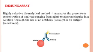
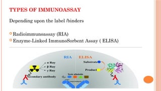
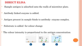
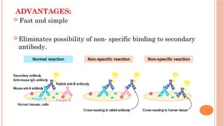
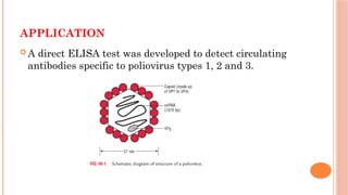
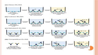
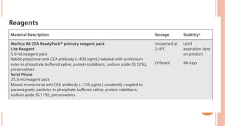
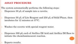
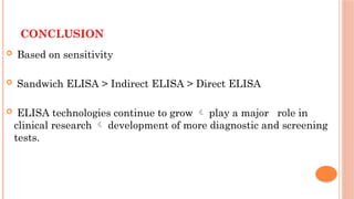
ENZYME-LINKED
IMMUNOSORBENT ASSAY
(ELISA)
Dr Dinakaran s
1st
year post graduate
Biochemistry
ESIC MC PGIMSR
OBJECTIVES
 Immunoassay definition and it’s types
 Principle/ definition
 Types of ELISA
 Applications
 Primary antibodies
 Detection strategies
IMMUNOASSAY
Highly selective bioanalytical method  measures the presence or
concentration of analytes ranging from micro to macromolecules in a
solution through the use of an antibody (usually) or an antigen
(sometimes).
TYPES OF IMMUNOASSAY
Depending upon the label /binders
 Radioimmunoassay (RIA)
 Enzyme-Linked ImmunoSorbent Assay ( ELISA)
ELISA
 It is an immunological assay.
 One of the reaction components is non specifically adsorbed
or covalently bound to the surface of a solid phase.
 ELISAs are typically performed in 96-well or 384-well
polystyrene plates.
PRINCIPLE:
 Detects interaction between specific antigen and antibody, by
enzyme labelled antibodies.
 Substrate of enzyme is added which gets coupled with coloured
reagent and change colourless reaction to coloured reaction.
 Intensity of colour is directly proportional to concentration of
Antigen/Antibody in given sample.
PRINCIPLE:
 Alkaline phosphatase
 Horse radish peroxidase
 Beta galactosidase
 Paranitrophenyl phosphate
 Ortho-
phenyldiaminedihydrochloride
 Tetramethylbenzidine
Dihydrochloride
Enzymes Substrates
SAMPLES USED IN ELISA
 Serum
 Plasma
 Saliva
 Urine
 Tissue lysates
1. Coating/capture - Immobilization of antigen / antibody
2. Plate blocking - Addition of molecule to cover all
unsaturated sites
3. Probing/detection- Incubation
4. Signal measurement-Detection of the signal generated.
General steps in ELISA
ELISA PLATES
 The first step is to optimize the plate-coating conditions for the
antigen or capture antibody.
 Clear polystyrene flat bottom plates - colorimetric signals
 Black or white opaque plates - fluorescent /chemiluminescent
signals.
ELISA PLATES
 Thermo Scientific ELISA Plates - Size of our choice.
 Plate coating is by passive adsorption  Hydrophobic interactions.
 Most common method for coating plates - Adding a 2–10 μg/ml solution
of protein dissolved in an phosphate-buffered saline (pH 7.4) or
carbonate-bicarbonate buffer (pH 9.4).
ELISA PLATES
 Incubation - several hours to overnight at 4–37° C.
 Coated plates can be used immediately or dried and stored at 4° C
for later use.
 With the exception of competitive ELISAs, the plates are coated
with less capture protein than can actually be bound during the
assay.
ELISA PLATES
 Proteins are best coated on plates at a concentration lower than
the maximum binding capacity - To prevent nonspecific binding
in later steps by a phenomenon called "hooking".
 When hooking nonspecifically traps detection of primary and
secondary antibodies  high background signal results 
lowering the signal to noise ratio and sensitivity.
ELISA
ELISA plate Coating Applications
Modified polymer surfaces Increase hydrophobicity or
hydrophilicity
Enhance passive binding of
biomolecules
Antibody-binding plates Protein A, G, L, or A/G properly orient antigen
binding capability
Biotin-binding plates Streptavidin or neutravidin Binds small biotinylated
peptides and other small
molecules  difficult to bind
by passive adsorption
Fusion-tag binding plates Glutathione (GST tag binding)
or
nickel or copper coated (His ta
g binding)
Study of genetically
engineered fusion proteins or
protein-protein interactions
TYPES OF ELISA
 Based on the basis of binding structure.
1. Direct ELISA (antigen coated plate, screening antigen)
2. Indirect ELISA (antigen coated plate, screening antibody)
3. Sandwich ELISA (antibody coated plate, screening antigen)
4. Competitive ELISA (screening antigen)
DIRECT ELISA
 Sample antigen is adsorbed onto the walls of microtiter plate.
 Antibody linked enzyme is added.
 Antigen present in sample binds to antibody- enzyme complex.
 Substrate is added for colour change
 The colour intensity is proportional to the antigen concentration
ADVANTAGES:
 Fast and simple
 Eliminates possibility of non- specific binding to secondary
antibody.
DISADVANTAGES:
 Immunoreactivity of the primary antibody might be
adversely affected
 Time-consuming
 Expensive – No flexibility
 Minimal signal amplification.
APPLICATION
 A direct ELISA test was developed to detect circulating
antibodies specific to poliovirus types 1, 2 and 3.
INDIRECT ELISA
Antibody can be detected or quantitatively determined.
Eg: Detection of HIV antibody
 A person’s serum - Applied to a multiwell (microtiter) plate.
 Antibodies in serum  Bind to antigen.
 Wash
 Secondary antibody conjugated with HRP is added
 Wash
 Plate will contain enzyme in proportion to the amount of
secondary antibody bound to the plate
ENZYME REACTION
A substrate containing H2O2 and diaminobenzidine for the
enzyme HRP is applied and catalysis by the enzyme leads to
change in the colour.
H2O2
HRP
H2O + (O)
Diaminobenzidine(DAB) + (O) Oxidised DAB
(colourless) (Brown colour)
The colour is directly proportional to the antibody concentrations
ADVANTAGES:
 Affordable.
 Versatile.
 Maximum immunoreactivity of the primary antibody is retained.
 Sensitivity is increased
 Signal will be amplified.
 Different detection methods can be used with the same primary
antibody (colorimetric, chemiluminescent, etc.).
DISADVANTAGES:
 Extra step.
 Time consuming.
 Cross- reactivity.
SANDWICH ELISA
 Detect the presence/measure the concentration of the target
antigen in samples.
 Detect the antigens between the two layers of antibodies
1) Capture antibody (polyclonal)
2) Detection antibody (monoclonal )
Antigens to be measured must also have at least two non-
overlapping epitopes capable of binding to two antibodies.
Eg: Hormonal assay (Thyroxine )
Tumour markers (CEA) *HRPO- Horseradish PerOxidase
*TMB- Tetramethylbenzidine
SANDWICH ELISA ADVANTAGES :
 High specificity because antigen/analyte is specially
captured and detected
 Flexible and sensitive.
 Suitable for complex samples as antigen does not require
any purification.
DISADVANTAGES:
 Possibility of error due to many steps
 Necessary use of “matched pair” (divalent /multivalent antigen)
and secondary antibody
 Antibody optimization is difficult if standard kit is not used.
 Time taking
 Expensive
COMPETITIVE ELISA
• Used to measure the concentration of an antigen in a sample.
• First incubate the antibody with a sample containing antigen.
• Antigen- antibody mixture is added to microtitre well which is coated with
antigen.
• The more the antigen is present in the sample, more the primary antibody
will bind to sample antigen.
• Therefore , there will be smaller amount of primary antibody available to bind
to antigen coated in well and wash have to give.
• Enzyme conjugated secondary antibody specific for isotype of the primary
antibody is added and Substrate is added
• The higher the concentration of antigen in the sample the lower is the
absorbance.
COMPETITIVE ELISA
ADVANTAGES:
 Less sample purification
 Commonly used for small molecules.
 Highly sensitive even the sample is in small amounts.
DISADVANTAGES:
 Less specific
 Cannot be used in diluent samples
APPLICATIONS OF ELISA
 Detect and Measure the Presence of Antibodies in
the Blood
 Detect and Estimate the Levels of Tumor Markers
 Detect and Estimate Hormone Levels
 Tracking Disease Outbreaks
 Detecting Past Exposures
 Screening Donated Blood for Possible Viral
Contaminants
 Detecting Drug Abuse
PRIMARY ANTIBODIES FOR ELISA
 Either monoclonal or polyclonal antibodies can be used as the
capture and detection antibodies.
 In addition to the use of traditional monoclonal antibodies,
recombinant monoclonal antibodies may also be utilized for ELISA.
 Compared to traditional monoclonal antibodies derived from
hybridomas, recombinant antibodies are not susceptible to cell-line
drift or lot-to-lot variation, thus allowing for peak antigen
specificity.
PRIMARY ANTIBODIES FOR ELISA
 For sandwich ELISA, the capture and detection antibodies must
recognize two different non-overlapping epitomes.
 When the antigen binds to the capture antibody, the epitome
recognized by the detection antibody must not be obscured or
altered.
 Capture and detection antibodies that do not interfere with one
another and can bind simultaneously are called "matched pairs" and
are suitable for developing a sandwich ELISA.
READY-TO-USE ELISA KITS AVAILABLE  DETECT
HUNDREDS OF SPECIFIC CYTOKINES, GROWTH FACTORS,
NEUROBIOLOGY ANALYTES PHOSPHORYLATED PROTEINS.
Antibody pair kits Uncoated ELISA kits
Coated ELISA
kits*
Instant ELISA kits
Need to coat plate Yes, an overnight coating
process is required
Yes, an overnight coating
process is required
No No
Incubation time** 24 h 24 h 2.5–4 h 3 h
Hands-on time 1 hr 30 mins 1 hr 30 mins 1 hr 40 mins
Readout HRP-TMB (colorimetric) HRP-TMB (colorimetric) HRP-TMB
(colorimetric)
HRP-TMB
(colorimetric)
Instrumentation
needed
Microplate reader,
absorbance
Microplate reader,
absorbance
Microplate reader,
absorbance
Microplate reader,
absorbance
Instrumentation read
time
2 min 2 min 2 min 2 min
ELISA KITS
DETECTION STRATEGIES FOR ELISA
Chromogenic
(colorimetric) Fluorescence Chemiluminescence
Sensitivity
Equipment required Standard absorbance
plate reader
Fluorometer Luminometer plate
reader
Enzyme HRP or AP Fluorescent tag or HRP
(with chemifluorescent
substrates)
HRP or AP
Advantages Direct visualization, high
reproducibility between
plates
High reproducibility
between plates, wide
dynamic range
Most sensitive detection
strategy, wide dynamic
range
Considerations Requires black
microplates
Requires opaque or
black microplates
DETECTION STRATEGIES FOR ELISA
 Chemiluminescence is a chemical reaction that generates energy released in
the form of light.
 Most chemiluminescent substrates are Horseradish PerOxidase (HRP)-
dependent, although some and Alkaline Phosphatase (AP) equivalents are
available.
 The most common approach is to use luminol in the presence of HRP and a
peroxide buffer.
 Light emission occurs only during the enzyme-substrate reaction, therefore
when the substrate becomes exhausted, the signal ceases.
DETECTION STRATEGIES FOR ELISA
 Chemiluminescent detection  more sensitive
 One drawback of using chemiluminescent substrates  the signal intensity
can vary with other substrates.
 For assays requiring many plates to be read, this can present a problem if the
signal begins to decay before plates are read.
 Make sure the assay has been optimized with the substrate in order to avoid
misinterpreting signal-fade in a sample as low antigen abundance.
 Chemiluminescent substrates for HRP include
Thermo Scientific Super Signal ELISA Pico and ELISA Femto substrates.
DETECTION STRATEGIES FOR ELISA
 Fluorescent ELISA substrates are not as common  require a fluorometer that
produces the correct excitation beam  signal emission to be generated from
the fluorescent tag.
 Chemifluorescent detection is also enzyme-based, but the generated product is
fluorescent rather than colorimetric.
 Examples of chemifluorescent substrates for HRP are Thermo Scientific
QuantaRed and QuantaBlu substrates.
CEA
 CarcinoEmbryonic Antigen(CEA) is normally found in embryonic
endodermal epithelium.
 Increased serum CEA levels have been detected in persons with
primary colorectal cancer and other malignancies.
 Elevated serum CEA levels have also been detected in patients with
nonmalignant disease, especially patients who are older or who are
smokers.
 So, CEA levels are not useful in screening the general population for
undetected cancers, But CEA is a useful tool for monitoring and
managing cancer therapy.
PRINCIPLES OF THE PROCEDURE
 The Atellica IM CEA assay is a 2-site sandwich immunoassay using
direct chemiluminometric technology.
 The first antibody, Lite Reagent  rabbit polyclonal anti-CEA
antibody labeled with acridinium ester.
 The second antibody, Solid Phasemouse monoclonal anti-CEA
antibody covalently coupled to paramagnetic particles.
 A direct relationship exists between the amount of CEA present
in sample and the amount of relative light units (RLUs)
detected by the system.
ASSAY PROCEDURE
The system automatically performs the following steps:
1. Dispenses 50 µL of sample into a cuvette.
2. Dispenses 50 µL of Lite Reagent and 250 µL of Solid Phase, then
incubates for 12 minutes at 37°C.
3. Washes the cuvette with special reagent water.
4. Dispenses 300 μL each of Atellica IM Acid and Atellica IM Base to
initiate the chemiluminescent reaction.
5. Reports results.
CONCLUSION
 Based on sensitivity
 Sandwich ELISA > Indirect ELISA > Direct ELISA
 ELISA technologies continue to grow  play a major role in
clinical research  development of more diagnostic and screening
tests.
REFERENCES
 Teitz fundamentals of clinical chemistry 6th
edition
 Textbook of biochemistry 4th
edition – Dr Rafi MD
 Textbook of biochemistry for medical students 10th
edition –DM
Vasudevan, Sreekumari S, Kannan Vaidyanathan.
 https://www.thermofisher.com/overview-elisa.html dated 27/02/24.
 Murthy N, Nair KM, Bhaskaram P. A direct ELISA technique to
detect antibodies against polioviruses. Indian J Biochem Biophys.
1995;32(5):249-253.
THANK YOU

ELISA presentation.pptx md biochemistry.

  • 1.
    ENZYME-LINKED IMMUNOSORBENT ASSAY (ELISA) Dr Dinakarans 1st year post graduate Biochemistry ESIC MC PGIMSR
  • 2.
    OBJECTIVES  Immunoassay definitionand it’s types  Principle/ definition  Types of ELISA  Applications  Primary antibodies  Detection strategies
  • 3.
    IMMUNOASSAY Highly selective bioanalyticalmethod  measures the presence or concentration of analytes ranging from micro to macromolecules in a solution through the use of an antibody (usually) or an antigen (sometimes).
  • 4.
    TYPES OF IMMUNOASSAY Dependingupon the label /binders  Radioimmunoassay (RIA)  Enzyme-Linked ImmunoSorbent Assay ( ELISA)
  • 5.
    ELISA  It isan immunological assay.  One of the reaction components is non specifically adsorbed or covalently bound to the surface of a solid phase.  ELISAs are typically performed in 96-well or 384-well polystyrene plates.
  • 6.
    PRINCIPLE:  Detects interactionbetween specific antigen and antibody, by enzyme labelled antibodies.  Substrate of enzyme is added which gets coupled with coloured reagent and change colourless reaction to coloured reaction.  Intensity of colour is directly proportional to concentration of Antigen/Antibody in given sample.
  • 7.
  • 8.
     Alkaline phosphatase Horse radish peroxidase  Beta galactosidase  Paranitrophenyl phosphate  Ortho- phenyldiaminedihydrochloride  Tetramethylbenzidine Dihydrochloride Enzymes Substrates
  • 9.
    SAMPLES USED INELISA  Serum  Plasma  Saliva  Urine  Tissue lysates
  • 10.
    1. Coating/capture -Immobilization of antigen / antibody 2. Plate blocking - Addition of molecule to cover all unsaturated sites 3. Probing/detection- Incubation 4. Signal measurement-Detection of the signal generated. General steps in ELISA
  • 11.
    ELISA PLATES  Thefirst step is to optimize the plate-coating conditions for the antigen or capture antibody.  Clear polystyrene flat bottom plates - colorimetric signals  Black or white opaque plates - fluorescent /chemiluminescent signals.
  • 12.
    ELISA PLATES  ThermoScientific ELISA Plates - Size of our choice.  Plate coating is by passive adsorption  Hydrophobic interactions.  Most common method for coating plates - Adding a 2–10 μg/ml solution of protein dissolved in an phosphate-buffered saline (pH 7.4) or carbonate-bicarbonate buffer (pH 9.4).
  • 13.
    ELISA PLATES  Incubation- several hours to overnight at 4–37° C.  Coated plates can be used immediately or dried and stored at 4° C for later use.  With the exception of competitive ELISAs, the plates are coated with less capture protein than can actually be bound during the assay.
  • 14.
    ELISA PLATES  Proteinsare best coated on plates at a concentration lower than the maximum binding capacity - To prevent nonspecific binding in later steps by a phenomenon called "hooking".  When hooking nonspecifically traps detection of primary and secondary antibodies  high background signal results  lowering the signal to noise ratio and sensitivity.
  • 15.
    ELISA ELISA plate CoatingApplications Modified polymer surfaces Increase hydrophobicity or hydrophilicity Enhance passive binding of biomolecules Antibody-binding plates Protein A, G, L, or A/G properly orient antigen binding capability Biotin-binding plates Streptavidin or neutravidin Binds small biotinylated peptides and other small molecules  difficult to bind by passive adsorption Fusion-tag binding plates Glutathione (GST tag binding) or nickel or copper coated (His ta g binding) Study of genetically engineered fusion proteins or protein-protein interactions
  • 16.
    TYPES OF ELISA Based on the basis of binding structure. 1. Direct ELISA (antigen coated plate, screening antigen) 2. Indirect ELISA (antigen coated plate, screening antibody) 3. Sandwich ELISA (antibody coated plate, screening antigen) 4. Competitive ELISA (screening antigen)
  • 17.
    DIRECT ELISA  Sampleantigen is adsorbed onto the walls of microtiter plate.  Antibody linked enzyme is added.  Antigen present in sample binds to antibody- enzyme complex.  Substrate is added for colour change  The colour intensity is proportional to the antigen concentration
  • 18.
    ADVANTAGES:  Fast andsimple  Eliminates possibility of non- specific binding to secondary antibody.
  • 19.
    DISADVANTAGES:  Immunoreactivity ofthe primary antibody might be adversely affected  Time-consuming  Expensive – No flexibility  Minimal signal amplification.
  • 20.
    APPLICATION  A directELISA test was developed to detect circulating antibodies specific to poliovirus types 1, 2 and 3.
  • 21.
    INDIRECT ELISA Antibody canbe detected or quantitatively determined. Eg: Detection of HIV antibody  A person’s serum - Applied to a multiwell (microtiter) plate.  Antibodies in serum  Bind to antigen.  Wash  Secondary antibody conjugated with HRP is added  Wash  Plate will contain enzyme in proportion to the amount of secondary antibody bound to the plate
  • 23.
    ENZYME REACTION A substratecontaining H2O2 and diaminobenzidine for the enzyme HRP is applied and catalysis by the enzyme leads to change in the colour. H2O2 HRP H2O + (O) Diaminobenzidine(DAB) + (O) Oxidised DAB (colourless) (Brown colour) The colour is directly proportional to the antibody concentrations
  • 24.
    ADVANTAGES:  Affordable.  Versatile. Maximum immunoreactivity of the primary antibody is retained.  Sensitivity is increased  Signal will be amplified.  Different detection methods can be used with the same primary antibody (colorimetric, chemiluminescent, etc.).
  • 25.
    DISADVANTAGES:  Extra step. Time consuming.  Cross- reactivity.
  • 26.
    SANDWICH ELISA  Detectthe presence/measure the concentration of the target antigen in samples.  Detect the antigens between the two layers of antibodies 1) Capture antibody (polyclonal) 2) Detection antibody (monoclonal ) Antigens to be measured must also have at least two non- overlapping epitopes capable of binding to two antibodies.
  • 27.
    Eg: Hormonal assay(Thyroxine ) Tumour markers (CEA) *HRPO- Horseradish PerOxidase *TMB- Tetramethylbenzidine
  • 28.
    SANDWICH ELISA ADVANTAGES:  High specificity because antigen/analyte is specially captured and detected  Flexible and sensitive.  Suitable for complex samples as antigen does not require any purification.
  • 29.
    DISADVANTAGES:  Possibility oferror due to many steps  Necessary use of “matched pair” (divalent /multivalent antigen) and secondary antibody  Antibody optimization is difficult if standard kit is not used.  Time taking  Expensive
  • 30.
    COMPETITIVE ELISA • Usedto measure the concentration of an antigen in a sample. • First incubate the antibody with a sample containing antigen. • Antigen- antibody mixture is added to microtitre well which is coated with antigen. • The more the antigen is present in the sample, more the primary antibody will bind to sample antigen. • Therefore , there will be smaller amount of primary antibody available to bind to antigen coated in well and wash have to give. • Enzyme conjugated secondary antibody specific for isotype of the primary antibody is added and Substrate is added • The higher the concentration of antigen in the sample the lower is the absorbance.
  • 31.
  • 32.
    ADVANTAGES:  Less samplepurification  Commonly used for small molecules.  Highly sensitive even the sample is in small amounts.
  • 33.
    DISADVANTAGES:  Less specific Cannot be used in diluent samples
  • 34.
    APPLICATIONS OF ELISA Detect and Measure the Presence of Antibodies in the Blood  Detect and Estimate the Levels of Tumor Markers  Detect and Estimate Hormone Levels  Tracking Disease Outbreaks  Detecting Past Exposures  Screening Donated Blood for Possible Viral Contaminants  Detecting Drug Abuse
  • 36.
    PRIMARY ANTIBODIES FORELISA  Either monoclonal or polyclonal antibodies can be used as the capture and detection antibodies.  In addition to the use of traditional monoclonal antibodies, recombinant monoclonal antibodies may also be utilized for ELISA.  Compared to traditional monoclonal antibodies derived from hybridomas, recombinant antibodies are not susceptible to cell-line drift or lot-to-lot variation, thus allowing for peak antigen specificity.
  • 37.
    PRIMARY ANTIBODIES FORELISA  For sandwich ELISA, the capture and detection antibodies must recognize two different non-overlapping epitomes.  When the antigen binds to the capture antibody, the epitome recognized by the detection antibody must not be obscured or altered.  Capture and detection antibodies that do not interfere with one another and can bind simultaneously are called "matched pairs" and are suitable for developing a sandwich ELISA.
  • 38.
    READY-TO-USE ELISA KITSAVAILABLE  DETECT HUNDREDS OF SPECIFIC CYTOKINES, GROWTH FACTORS, NEUROBIOLOGY ANALYTES PHOSPHORYLATED PROTEINS.
  • 39.
    Antibody pair kitsUncoated ELISA kits Coated ELISA kits* Instant ELISA kits Need to coat plate Yes, an overnight coating process is required Yes, an overnight coating process is required No No Incubation time** 24 h 24 h 2.5–4 h 3 h Hands-on time 1 hr 30 mins 1 hr 30 mins 1 hr 40 mins Readout HRP-TMB (colorimetric) HRP-TMB (colorimetric) HRP-TMB (colorimetric) HRP-TMB (colorimetric) Instrumentation needed Microplate reader, absorbance Microplate reader, absorbance Microplate reader, absorbance Microplate reader, absorbance Instrumentation read time 2 min 2 min 2 min 2 min ELISA KITS
  • 40.
    DETECTION STRATEGIES FORELISA Chromogenic (colorimetric) Fluorescence Chemiluminescence Sensitivity Equipment required Standard absorbance plate reader Fluorometer Luminometer plate reader Enzyme HRP or AP Fluorescent tag or HRP (with chemifluorescent substrates) HRP or AP Advantages Direct visualization, high reproducibility between plates High reproducibility between plates, wide dynamic range Most sensitive detection strategy, wide dynamic range Considerations Requires black microplates Requires opaque or black microplates
  • 41.
    DETECTION STRATEGIES FORELISA  Chemiluminescence is a chemical reaction that generates energy released in the form of light.  Most chemiluminescent substrates are Horseradish PerOxidase (HRP)- dependent, although some and Alkaline Phosphatase (AP) equivalents are available.  The most common approach is to use luminol in the presence of HRP and a peroxide buffer.  Light emission occurs only during the enzyme-substrate reaction, therefore when the substrate becomes exhausted, the signal ceases.
  • 42.
    DETECTION STRATEGIES FORELISA  Chemiluminescent detection  more sensitive  One drawback of using chemiluminescent substrates  the signal intensity can vary with other substrates.  For assays requiring many plates to be read, this can present a problem if the signal begins to decay before plates are read.  Make sure the assay has been optimized with the substrate in order to avoid misinterpreting signal-fade in a sample as low antigen abundance.  Chemiluminescent substrates for HRP include Thermo Scientific Super Signal ELISA Pico and ELISA Femto substrates.
  • 43.
    DETECTION STRATEGIES FORELISA  Fluorescent ELISA substrates are not as common  require a fluorometer that produces the correct excitation beam  signal emission to be generated from the fluorescent tag.  Chemifluorescent detection is also enzyme-based, but the generated product is fluorescent rather than colorimetric.  Examples of chemifluorescent substrates for HRP are Thermo Scientific QuantaRed and QuantaBlu substrates.
  • 44.
    CEA  CarcinoEmbryonic Antigen(CEA)is normally found in embryonic endodermal epithelium.  Increased serum CEA levels have been detected in persons with primary colorectal cancer and other malignancies.  Elevated serum CEA levels have also been detected in patients with nonmalignant disease, especially patients who are older or who are smokers.  So, CEA levels are not useful in screening the general population for undetected cancers, But CEA is a useful tool for monitoring and managing cancer therapy.
  • 45.
    PRINCIPLES OF THEPROCEDURE  The Atellica IM CEA assay is a 2-site sandwich immunoassay using direct chemiluminometric technology.  The first antibody, Lite Reagent  rabbit polyclonal anti-CEA antibody labeled with acridinium ester.  The second antibody, Solid Phasemouse monoclonal anti-CEA antibody covalently coupled to paramagnetic particles.  A direct relationship exists between the amount of CEA present in sample and the amount of relative light units (RLUs) detected by the system.
  • 47.
    ASSAY PROCEDURE The systemautomatically performs the following steps: 1. Dispenses 50 µL of sample into a cuvette. 2. Dispenses 50 µL of Lite Reagent and 250 µL of Solid Phase, then incubates for 12 minutes at 37°C. 3. Washes the cuvette with special reagent water. 4. Dispenses 300 μL each of Atellica IM Acid and Atellica IM Base to initiate the chemiluminescent reaction. 5. Reports results.
  • 48.
    CONCLUSION  Based onsensitivity  Sandwich ELISA > Indirect ELISA > Direct ELISA  ELISA technologies continue to grow  play a major role in clinical research  development of more diagnostic and screening tests.
  • 49.
    REFERENCES  Teitz fundamentalsof clinical chemistry 6th edition  Textbook of biochemistry 4th edition – Dr Rafi MD  Textbook of biochemistry for medical students 10th edition –DM Vasudevan, Sreekumari S, Kannan Vaidyanathan.  https://www.thermofisher.com/overview-elisa.html dated 27/02/24.  Murthy N, Nair KM, Bhaskaram P. A direct ELISA technique to detect antibodies against polioviruses. Indian J Biochem Biophys. 1995;32(5):249-253.
  • 50.