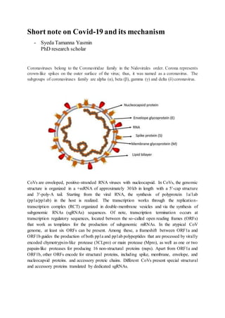
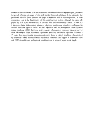
Short note on Covid-19 and its mechanism
- Syeda Tamanna Yasmin
PhD research scholar
Coronaviruses belong to the Coronaviridae family in the Nidovirales order. Corona represents
crown-like spikes on the outer surface of the virus; thus, it was named as a coronavirus. The
subgroups of coronaviruses family are alpha (α), beta (β), gamma (γ) and delta (δ) coronavirus.
CoVs are enveloped, positive-stranded RNA viruses with nucleocapsid. In CoVs, the genomic
structure is organized in a +ssRNA of approximately 30 kb in length with a 5′-cap structure
and 3′-poly-A tail. Starting from the viral RNA, the synthesis of polyprotein 1a/1ab
(pp1a/pp1ab) in the host is realized. The transcription works through the replication-
transcription complex (RCT) organized in double-membrane vesicles and via the synthesis of
subgenomic RNAs (sgRNAs) sequences. Of note, transcription termination occurs at
transcription regulatory sequences, located between the so-called open reading frames (ORFs)
that work as templates for the production of subgenomic mRNAs. In the atypical CoV
genome, at least six ORFs can be present. Among these, a frameshift between ORF1a and
ORF1b guides the production of both pp1a and pp1ab polypeptides that are processed by virally
encoded chymotrypsin-like protease (3CLpro) or main protease (Mpro), as well as one or two
papain-like proteases for producing 16 non-structural proteins (nsps). Apart from ORF1a and
ORF1b, other ORFs encode for structural proteins, including spike, membrane, envelope, and
nucleocapsid proteins. and accessory proteic chains. Different CoVs present special structural
and accessory proteins translated by dedicated sgRNAs.
Among the structural elements of CoVs, there are the spike glycoproteins composed of two
subunits (S1 and S2). Homotrimers of S proteins compose the spikes on the viral surface,
guiding the link to host receptors. Of note, in SARS-CoV-2, the S2 subunit containing a fusion
peptide, a transmembrane domain, and cytoplasmic domain is highly conserved. Thus, it could
be a target for antiviral (anti-S2) compounds. On the contrary, the spike receptor-binding
domain presents only a 40% amino acid identity with other SARS-CoVs.
Fig. : The life cycle of SARS-CoV-2 in host cells; begins its life cycle when S protein
binds to the cellular receptor ACE2. After receptor binding, the conformation change
in the S protein facilitates viral envelope fusion with the cell membrane through the
endosomal pathway. Then SARS-CoV-2 releases RNA into the host cell. Genome RNA
is translated into viral replicase polyproteins pp1aand 1ab, which are then cleaved into
small products by viral proteinases. The polymerase produces a series of subgenomic
mRNAs by discontinuous transcription and finally translated into relevant viral
proteins. Viral proteins and genome RNA are subsequently assembled into virions in
the ER and Golgi and then transported via vesicles and released out of the cell. ACE2,
angiotensin-converting enzyme 2; ER, endoplasmic reticulum; ERGIC, ER–Golgi
intermediate compartment.
The data so far available seem to indicate that the viral infection is capable of producing an
excessive immune reaction in the host. In some cases, a reaction takes place which as a whole
is labeled a 'cytokine storm'. The effect is extensive tissue damage. The protagonist of this
storm is interleukin 6 (IL-6). IL-6 is produced by activated leukocytes and acts on a large
number of cells and tissues. It is able to promote the differentiation of Blymphocytes, promotes
the growth of some categories of cells, and inhibits the growth of others. It also stimulates the
production of acute phase proteins and plays an important role in thermoregulation, in bone
maintenance and in the functionality of the central nervous system. Although the main role
played by IL-6 is pro-inflammatory, it can also have anti-inflammatory effects. In turn, IL-
6 increases during inflammatory diseases, infections, autoimmune disorders, cardiovascular
diseases and some types of cancer. It is also implicated into the pathogenesis of the cytokine
release syndrome (CRS) that is an acute systemic inflammatory syndrome characterized by
fever and multiple organ dysfunction syndromes (MODs). The clinical spectrum of COVID-
19 varies from asymptomatic or paucisymptomatic forms to clinical conditions characterized
by respiratory failure that necessitates mechanical ventilation and support in an intensive care
unit (ICU), to multiorgan and systemic manifestations in terms of sepsis, septic shock.
REFERENCES
1.Perlman S, Netland J. Coronaviruses post-SARS: update on replication and
pathogenesis. Nat. Rev. Microbiol. 2009 Jun;7(6):439-50.
2.Chan JF, To KK, Tse H, Jin DY, Yuen KY. Interspecies transmission and emergence of
novel viruses: lessons from bats and birds. Trends Microbiol. 2013 Oct;21(10):544-55.
3.Chen Y, Liu Q, Guo D. Emerging coronaviruses: Genome structure, replication, and
pathogenesis. J. Med. Virol. 2020 Apr;92(4):418-423.
4.Y. Wan, J. Shang, R. Graham, R.S. Baric, F. LiReceptor recognition by novel coronavirus
from Wuhan: an analysis based on decade-long structural studies of SARS, J Virol (2020)
5.A. Wu, Y. Peng, B. Huang, X. Ding, X. Wang, P. Niu, et al.Genome composition and
divergence of the novel coronavirus (2019-nCoV) originating in China,Cell Host
Microbe (2020)

short note on Covid-19

  • 1.
    Short note onCovid-19 and its mechanism - Syeda Tamanna Yasmin PhD research scholar Coronaviruses belong to the Coronaviridae family in the Nidovirales order. Corona represents crown-like spikes on the outer surface of the virus; thus, it was named as a coronavirus. The subgroups of coronaviruses family are alpha (α), beta (β), gamma (γ) and delta (δ) coronavirus. CoVs are enveloped, positive-stranded RNA viruses with nucleocapsid. In CoVs, the genomic structure is organized in a +ssRNA of approximately 30 kb in length with a 5′-cap structure and 3′-poly-A tail. Starting from the viral RNA, the synthesis of polyprotein 1a/1ab (pp1a/pp1ab) in the host is realized. The transcription works through the replication- transcription complex (RCT) organized in double-membrane vesicles and via the synthesis of subgenomic RNAs (sgRNAs) sequences. Of note, transcription termination occurs at transcription regulatory sequences, located between the so-called open reading frames (ORFs) that work as templates for the production of subgenomic mRNAs. In the atypical CoV genome, at least six ORFs can be present. Among these, a frameshift between ORF1a and ORF1b guides the production of both pp1a and pp1ab polypeptides that are processed by virally encoded chymotrypsin-like protease (3CLpro) or main protease (Mpro), as well as one or two papain-like proteases for producing 16 non-structural proteins (nsps). Apart from ORF1a and ORF1b, other ORFs encode for structural proteins, including spike, membrane, envelope, and nucleocapsid proteins. and accessory proteic chains. Different CoVs present special structural and accessory proteins translated by dedicated sgRNAs.
  • 2.
    Among the structuralelements of CoVs, there are the spike glycoproteins composed of two subunits (S1 and S2). Homotrimers of S proteins compose the spikes on the viral surface, guiding the link to host receptors. Of note, in SARS-CoV-2, the S2 subunit containing a fusion peptide, a transmembrane domain, and cytoplasmic domain is highly conserved. Thus, it could be a target for antiviral (anti-S2) compounds. On the contrary, the spike receptor-binding domain presents only a 40% amino acid identity with other SARS-CoVs. Fig. : The life cycle of SARS-CoV-2 in host cells; begins its life cycle when S protein binds to the cellular receptor ACE2. After receptor binding, the conformation change in the S protein facilitates viral envelope fusion with the cell membrane through the endosomal pathway. Then SARS-CoV-2 releases RNA into the host cell. Genome RNA is translated into viral replicase polyproteins pp1aand 1ab, which are then cleaved into small products by viral proteinases. The polymerase produces a series of subgenomic mRNAs by discontinuous transcription and finally translated into relevant viral proteins. Viral proteins and genome RNA are subsequently assembled into virions in the ER and Golgi and then transported via vesicles and released out of the cell. ACE2, angiotensin-converting enzyme 2; ER, endoplasmic reticulum; ERGIC, ER–Golgi intermediate compartment. The data so far available seem to indicate that the viral infection is capable of producing an excessive immune reaction in the host. In some cases, a reaction takes place which as a whole is labeled a 'cytokine storm'. The effect is extensive tissue damage. The protagonist of this storm is interleukin 6 (IL-6). IL-6 is produced by activated leukocytes and acts on a large
  • 3.
    number of cellsand tissues. It is able to promote the differentiation of Blymphocytes, promotes the growth of some categories of cells, and inhibits the growth of others. It also stimulates the production of acute phase proteins and plays an important role in thermoregulation, in bone maintenance and in the functionality of the central nervous system. Although the main role played by IL-6 is pro-inflammatory, it can also have anti-inflammatory effects. In turn, IL- 6 increases during inflammatory diseases, infections, autoimmune disorders, cardiovascular diseases and some types of cancer. It is also implicated into the pathogenesis of the cytokine release syndrome (CRS) that is an acute systemic inflammatory syndrome characterized by fever and multiple organ dysfunction syndromes (MODs). The clinical spectrum of COVID- 19 varies from asymptomatic or paucisymptomatic forms to clinical conditions characterized by respiratory failure that necessitates mechanical ventilation and support in an intensive care unit (ICU), to multiorgan and systemic manifestations in terms of sepsis, septic shock.
  • 4.
    REFERENCES 1.Perlman S, NetlandJ. Coronaviruses post-SARS: update on replication and pathogenesis. Nat. Rev. Microbiol. 2009 Jun;7(6):439-50. 2.Chan JF, To KK, Tse H, Jin DY, Yuen KY. Interspecies transmission and emergence of novel viruses: lessons from bats and birds. Trends Microbiol. 2013 Oct;21(10):544-55. 3.Chen Y, Liu Q, Guo D. Emerging coronaviruses: Genome structure, replication, and pathogenesis. J. Med. Virol. 2020 Apr;92(4):418-423. 4.Y. Wan, J. Shang, R. Graham, R.S. Baric, F. LiReceptor recognition by novel coronavirus from Wuhan: an analysis based on decade-long structural studies of SARS, J Virol (2020) 5.A. Wu, Y. Peng, B. Huang, X. Ding, X. Wang, P. Niu, et al.Genome composition and divergence of the novel coronavirus (2019-nCoV) originating in China,Cell Host Microbe (2020)