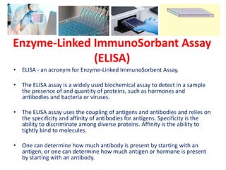
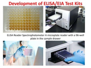
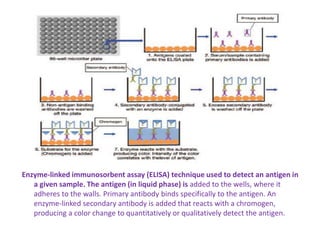
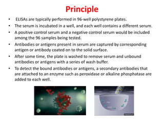
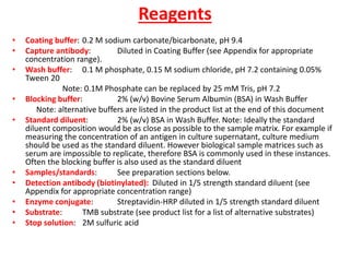
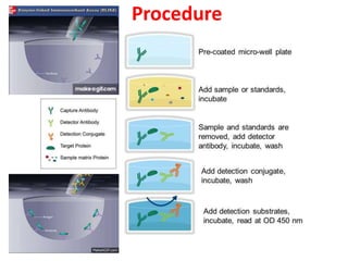
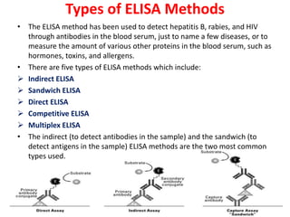
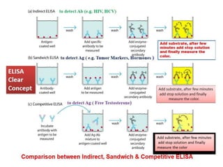
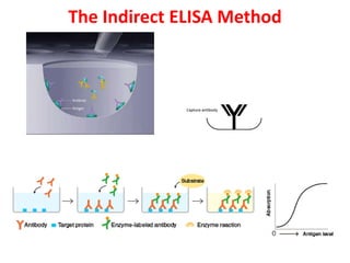
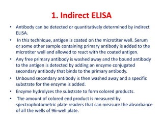
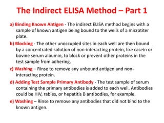
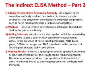
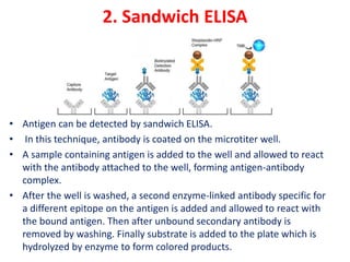
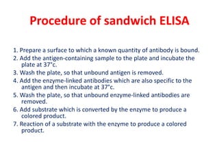
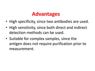
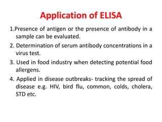
Enzyme-Linked ImmunoSorbant Assay
PRESENTED BY:
MADHU.J
PAMB 1321
Jr.Msc plant pathology
Enzyme-Linked ImmunoSorbant Assay
(ELISA)
• ELISA - an acronym for Enzyme-Linked ImmunoSorbent Assay.
• The ELISA assay is a widely used biochemical assay to detect in a sample
the presence of and quantity of proteins, such as hormones and
antibodies and bacteria or viruses.
• The ELISA assay uses the coupling of antigens and antibodies and relies on
the specificity and affinity of antibodies for antigens. Specificity is the
ability to discriminate among diverse proteins. Affinity is the ability to
tightly bind to molecules.
• One can determine how much antibody is present by starting with an
antigen, or one can determine how much antigen or hormone is present
by starting with an antibody.
What Are Antigens?
Antigens are any foreign substance in the body.
Antigens include “not-self” molecules and cells, such as:
a. foreign proteins
b. viruses
c. environmental pollutants and other foreign substances like asbestos, tattoo
ink, and cigarette smoke
d. bacteria and parasites (Protista, Fungi, Plantae, and Animalia cells)
e. foreign transplanted tissue
f. cancerous cells
What Are Antibodies and How Are
They Produced?
• Antibodies are large glycoprotein molecules produced by B-lymphocytes
during the humoral immune response to antigens introduced into the
body.
• Lymphocytes include B-lymphocytes (B-cells) and T-lymphocytes (T-cells)
which are white blood cells form from the hematopoietic (blood) stem
cells in the bone marrow.
• The immune system is made of two parts – humoral (antibody-mediated)
and cellular (cell-mediated).
Formation of B Lymphocytes and
T Lymphocytes
B Lymphocytes Mature in the Bone Marrow
and T Lymphocytes Mature in the Thymus
Gland
Humoral (Antibody-Mediated)
Immune System
• B-lymphocytes produce large glycoproteins called antibodies in response
to antigens (any foreign substance) and then mark those antigens-
antibody complex to be destroyed by the T-lymphocytes.
• Each B-cell makes its own distinct antibody in response to a specific
antigen which comes in contact with it. Each antibody is designed to bind
to a specific surface binding site or epitope on the antigen.
• There are millions of different types of antibodies circulating in an
individual’s bloodstream and they are based on exposure to antigens in
his/her environment.
• Over 80% of human glycoprotein
antibodies are in the immunoglobulin class
IgG. They are shaped like a Y and are
found in the blood, lymph, and intestine.
• IgG molecules have a molecular weight of
150,000 Daltons and are made of 2 long
(heavy) chains coded from chromosome
14, and 2 short (light) chains coded from
either chromosome 2 or 22, and then all
connected by disulphide bonds.
• Most of the molecule is composed of a
constant region that doesn’t change from
one IgG molecule to another. However, the
ends of the Y are variable, which accounts
for each IgG molecule binding only to a
specific antigen.
Structure of An Antibody
History of ELISA/EIA
• The development of the radioimmunoassay opened the door for
others to develop similar methods, like ELISA, to test for the
presence of proteins but without the use of radioactive substances.
• In 1971, Peter Permann and Eva Engvall in Stockholm published the
first paper on Enzyme-Linked ImmunoSorbent Assay (ELISA)
showing they could quantify the amount of IgG in rabbit serum
using alkaline phosphatase (an enzyme) as the reporter label.
• The same year, Anton Schuurs and Bauke Van Weemen in the
Netherlands published a paper showing that with an Enzyme
ImmunoAssay (EIA), they could quantify the amount of human
chorionic gonadotropin in urine with horseradish peroxidase (an
enzyme) coupled with glutaraldehyde as the reporter label.
• The assays were highly sensitive and compared favorably with
RadioimmunoAssays.
Development of ELISA/EIA Test Kits
ELISA Reader Spectrophotometer A microplate reader with a 96-well
plate in the sample drawer
Enzyme-linked immunosorbent assay (ELISA) technique used to detect an antigen in
a given sample. The antigen (in liquid phase) is added to the wells, where it
adheres to the walls. Primary antibody binds specifically to the antigen. An
enzyme-linked secondary antibody is added that reacts with a chromogen,
producing a color change to quantitatively or qualitatively detect the antigen.
Principle
• ELISAs are typically performed in 96-well polystyrene plates.
• The serum is incubated in a well, and each well contains a different serum.
• A positive control serum and a negative control serum would be included
among the 96 samples being tested.
• Antibodies or antigens present in serum are captured by corresponding
antigen or antibody coated on to the solid surface.
• After some time, the plate is washed to remove serum and unbound
antibodies or antigens with a series of wash buffer.
• To detect the bound antibodies or antigens, a secondary antibodies that
are attached to an enzyme such as peroxidase or alkaline phosphatase are
added to each well.
• After an incubation period, the unbound secondary antibodies are
washed off.
• When a suitable substrate is added, the enzyme reacts with it to
produce a color.
• This color produced is measurable as a function or quantity of
antigens or antibodies present in the given sample.
• The intensity of color/ optical density is measured at 450nm.
• The intensity of the color gives an indication of the amount of
antigen or antibody.
Reagents
• Coating buffer: 0.2 M sodium carbonate/bicarbonate, pH 9.4
• Capture antibody: Diluted in Coating Buffer (see Appendix for appropriate
concentration range).
• Wash buffer: 0.1 M phosphate, 0.15 M sodium chloride, pH 7.2 containing 0.05%
Tween 20
Note: 0.1M Phosphate can be replaced by 25 mM Tris, pH 7.2
• Blocking buffer: 2% (w/v) Bovine Serum Albumin (BSA) in Wash Buffer
Note: alternative buffers are listed in the product list at the end of this document
• Standard diluent: 2% (w/v) BSA in Wash Buffer. Note: Ideally the standard
diluent composition would be as close as possible to the sample matrix. For example if
measuring the concentration of an antigen in culture supernatant, culture medium
should be used as the standard diluent. However biological sample matrices such as
serum are impossible to replicate, therefore BSA is commonly used in these instances.
Often the blocking buffer is also used as the standard diluent
• Samples/standards: See preparation sections below.
• Detection antibody (biotinylated): Diluted in 1/5 strength standard diluent (see
Appendix for appropriate concentration range)
• Enzyme conjugate: Streptavidin-HRP diluted in 1/5 strength standard diluent
• Substrate: TMB substrate (see product list for a list of alternative substrates)
• Stop solution: 2M sulfuric acid
Procedure
Procedure
• Important: Do not allow the plate to dry at any point.
• Dilute the capture antibody to the appropriate concentration allowing
sufficient volume for 50-100 l per well.
• Add the diluted capture antibody to the plate, cover and incubate for
2 hours at room temperature (RT).
• Remove the solution and wash the plate with 200 l per well wash
buffer for 3 x 5 minutes on a shaking platform.
• Add 300 l blocking buffer per well, cover the plate and incubate for 1
hour at room temperature. Alternatively block overnight at 4 C.
• Prepare the samples and standards. The volume per well should be
the same as the capture antibody used in step 1.
• Remove the blocking buffer and add the samples and standards.
Cover the plate and incubate for 1 hour at RT.
• Remove the solution and wash the plate with 200 l per well wash
buffer for 3 x 5 minutes on a shaking platform.
• Dilute the biotinylated detection antibody to the appropriate
concentration. The volume per well should be the same as the
capture antibody used in step 1.
• Add the diluted detection antibody to the plate, cover and incubate
for 1 hour at RT.
• Remove the solution and wash the plate with 200 l per well wash
buffer for 3 x 5 minutes on a shaking platform.
• Dilute the enzyme conjugate to the appropriate concentration. The
volume per well should be the same as the capture antibody used
in step 1.
• Add the diluted enzyme conjugate to the plate, cover and incubate
for 1 hour at RT.
• Remove the solution and wash the plate with 200 l per well wash
buffer for 6 x 5 minutes on a shaking platform
• Add substrate solution to the plate. The volume per well should be
the same as the capture antibody used in step 1.
• Incubate the plate at RT until the desired color intensity is reached.
Ideally a clear gradient will result for the standards.
• Stop the reaction by adding an equal amount of stop solution.
• If using TMB, measure the absorbance at 450 nm. For other
substrates use the appropriate detection techniq
Types of ELISA Methods
• The ELISA method has been used to detect hepatitis B, rabies, and HIV
through antibodies in the blood serum, just to name a few diseases, or to
measure the amount of various other proteins in the blood serum, such as
hormones, toxins, and allergens.
• There are five types of ELISA methods which include:
 Indirect ELISA
 Sandwich ELISA
 Direct ELISA
 Competitive ELISA
 Multiplex ELISA
• The indirect (to detect antibodies in the sample) and the sandwich (to
detect antigens in the sample) ELISA methods are the two most common
types used.
The Indirect ELISA Method
1. Indirect ELISA
• Antibody can be detected or quantitatively determined by indirect
ELISA.
• In this technique, antigen is coated on the microtiter well. Serum
or some other sample containing primary antibody is added to the
microtiter well and allowed to react with the coated antigen.
• Any free primary antibody is washed away and the bound antibody
to the antigen is detected by adding an enzyme conjugated
secondary antibody that binds to the primary antibody.
• Unbound secondary antibody is then washed away and a specific
substrate for the enzyme is added.
• Enzyme hydrolyzes the substrate to form colored products.
• The amount of colored end product is measured by
spectrophotometric plate readers that can measure the absorbance
of all the wells of 96-well plate.
Procedure
1. Coat the micro titer plate wells with antigen.
2. Block all unbound sites to prevent false positive results.
3. Add sample containing antibody (e.g. rabbit monoclonal antibody)
to the wells and incubate the plate at 37°c.
4. Wash the plate, so that unbound antibody is removed.
5. Add secondary antibody conjugated to an enzyme (e.g. anti- mouse
IgG).
6. Wash the plate, so that unbound enzyme-linked antibodies are
removed.
7. Add substrate which is converted by the enzyme to produce a
colored product.
8. Reaction of a substrate with the enzyme to produce a colored
product.
Advantages
• Increased sensitivity, since more than one labeled antibody is bound per
primary antibody.
• A wide variety of labeled secondary antibodies are available commercially.
• Maximum immunoreactivity of the primary antibody is retained because it
is not labeled.
• Versatile because many primary antibodies can be made in one species
and the same labeled secondary antibody can be used for detection.
• Flexibility, since different primary detection antibodies can be used with a
single labeled secondary antibody.
• Cost savings, since fewer labeled antibodies are required.
• Different visualization markers can be used with the same primary
antibody.
• Cross-reactivity might occur with the secondary antibody, resulting in
nonspecific signal.
• An extra incubation step is required in the procedure.
Disadvantages
The Indirect ELISA Method – Part 1
a) Binding Known Antigen - The indirect ELISA method begins with a
sample of known antigen being bound to the wells of a microtiter
plate.
b) Blocking - The other unoccupied sites in each well are then bound
by a concentrated solution of non-interacting protein, like casein or
bovine serum albumin, to block or prevent other proteins in the
test sample from adhering.
c) Washing – Rinse to remove any unbound antigen and non-
interacting protein.
d) Adding Test Sample Primary Antibody - The test sample of serum
containing the primary antibodies is added to each well. Antibodies
could be HIV, rabies, or hepatitis B antibodies, for example.
e) Washing – Rinse to remove any antibodies that did not bind to the
known antigen.
The Indirect ELISA Method – Part 2
f) Adding Enzyme-linked Secondary Antibody - An enzyme-linked
secondary antibody is added next to bind to the test sample
antibodies. The enzyme on the secondary antibodies are proteins,
such as horse radish peroxidase or alkaline phosphatase.
g) Washing – Rinse to remove any secondary antibodies that did not
bind to the primary antibody.
h) Adding Substrate - A substrate is then applied which is converted by
the enzyme to give a color or fluorescence or electrochemical
signal. In the presence of horse radish peroxidase, ABTS turns
green, OPD turns orange, and TMB turns blue. In the presence of
alkaline phosphatase, pNPP turns yellow.
i) Reading Results - By using a spectrophotometer, spectrofluorometer,
or electrochemical device, the results can be read and recorded.
The amount of color produced is proportional to the amount of
primary antibody bound to the antigen proteins on the bottom of
the wells.
2. Sandwich ELISA
• Antigen can be detected by sandwich ELISA.
• In this technique, antibody is coated on the microtiter well.
• A sample containing antigen is added to the well and allowed to react
with the antibody attached to the well, forming antigen-antibody
complex.
• After the well is washed, a second enzyme-linked antibody specific for
a different epitope on the antigen is added and allowed to react with
the bound antigen. Then after unbound secondary antibody is
removed by washing. Finally substrate is added to the plate which is
hydrolyzed by enzyme to form colored products.
Procedure of sandwich ELISA
1. Prepare a surface to which a known quantity of antibody is bound.
2. Add the antigen-containing sample to the plate and incubate the
plate at 37°c.
3. Wash the plate, so that unbound antigen is removed.
4. Add the enzyme-linked antibodies which are also specific to the
antigen and then incubate at 37°c.
5. Wash the plate, so that unbound enzyme-linked antibodies are
removed.
6. Add substrate which is converted by the enzyme to produce a
colored product.
7. Reaction of a substrate with the enzyme to produce a colored
product.
Advantages
• High specificity, since two antibodies are used
the antigen is specifically captured and
detected.
• Suitable for complex samples, since the
antigen does not require purification prior to
measurement.
• Flexibility and sensitivity, since both direct and
indirect detection methods can be used.
Sandwich ELISA
• As the substrate is added, the enzyme converts it into
a colored product. The rate of color formation is
proportional to the amount of antigen present.
• For a diagram showing both a positive and negative
result if testing for an antigen using the sandwich
ELISA method, see
• https://cellularphysiology.wikispaces.com/Enzyme-
linked+immunosorbent+assay+(ELISA)
3. Competitive ELISA
• This test is used to measure the concentration of an
antigen in a sample.
• In this test, antibody is first incubated in solution with a
sample containing antigen. The antigen-antibody
mixture is then added to the microtitre well which is
coated with antigen. The more the antigen present in
the sample, the less free antibody will be available to
bind to the antigen-coated well. After the well is
washed, enzyme conjugated secondary antibody
specific for isotype of the primary antibody is added to
determine the amount of primary antibody bound to
the well. The higher the concentration of antigen in the
sample, the lower the absorbance.
Procedure
1. Antibody is incubated with sample containing antigen.
2. Antigen-antibody complex are added to the microtitre
well which are pre-coated with the antigen.
3. Wash the plate to remove unbound antibody.
4. Enzyme linked secondary antibody which is specific to
the primary antibody is added.
5. Wash the plate, so that unbound enzyme-linked
antibodies are removed.
6. Add substrate which is converted by the enzyme into a
fluorescent signal.
Advantages
• High specificity, since two antibodies are used.
• High sensitivity, since both direct and indirect
detection methods can be used.
• Suitable for complex samples, since the
antigen does not require purification prior to
measurement.
Application of ELISA
1.Presence of antigen or the presence of antibody in a
sample can be evaluated.
2. Determination of serum antibody concentrations in a
virus test.
3. Used in food industry when detecting potential food
allergens.
4. Applied in disease outbreaks- tracking the spread of
disease e.g. HIV, bird flu, common, colds, cholera,
STD etc.

ELISA.pptx

  • 2.
    Enzyme-Linked ImmunoSorbant Assay PRESENTEDBY: MADHU.J PAMB 1321 Jr.Msc plant pathology
  • 3.
    Enzyme-Linked ImmunoSorbant Assay (ELISA) •ELISA - an acronym for Enzyme-Linked ImmunoSorbent Assay. • The ELISA assay is a widely used biochemical assay to detect in a sample the presence of and quantity of proteins, such as hormones and antibodies and bacteria or viruses. • The ELISA assay uses the coupling of antigens and antibodies and relies on the specificity and affinity of antibodies for antigens. Specificity is the ability to discriminate among diverse proteins. Affinity is the ability to tightly bind to molecules. • One can determine how much antibody is present by starting with an antigen, or one can determine how much antigen or hormone is present by starting with an antibody.
  • 4.
    What Are Antigens? Antigensare any foreign substance in the body. Antigens include “not-self” molecules and cells, such as: a. foreign proteins b. viruses c. environmental pollutants and other foreign substances like asbestos, tattoo ink, and cigarette smoke d. bacteria and parasites (Protista, Fungi, Plantae, and Animalia cells) e. foreign transplanted tissue f. cancerous cells
  • 5.
    What Are Antibodiesand How Are They Produced? • Antibodies are large glycoprotein molecules produced by B-lymphocytes during the humoral immune response to antigens introduced into the body. • Lymphocytes include B-lymphocytes (B-cells) and T-lymphocytes (T-cells) which are white blood cells form from the hematopoietic (blood) stem cells in the bone marrow. • The immune system is made of two parts – humoral (antibody-mediated) and cellular (cell-mediated).
  • 6.
    Formation of BLymphocytes and T Lymphocytes
  • 7.
    B Lymphocytes Maturein the Bone Marrow and T Lymphocytes Mature in the Thymus Gland
  • 8.
    Humoral (Antibody-Mediated) Immune System •B-lymphocytes produce large glycoproteins called antibodies in response to antigens (any foreign substance) and then mark those antigens- antibody complex to be destroyed by the T-lymphocytes. • Each B-cell makes its own distinct antibody in response to a specific antigen which comes in contact with it. Each antibody is designed to bind to a specific surface binding site or epitope on the antigen. • There are millions of different types of antibodies circulating in an individual’s bloodstream and they are based on exposure to antigens in his/her environment.
  • 9.
    • Over 80%of human glycoprotein antibodies are in the immunoglobulin class IgG. They are shaped like a Y and are found in the blood, lymph, and intestine. • IgG molecules have a molecular weight of 150,000 Daltons and are made of 2 long (heavy) chains coded from chromosome 14, and 2 short (light) chains coded from either chromosome 2 or 22, and then all connected by disulphide bonds. • Most of the molecule is composed of a constant region that doesn’t change from one IgG molecule to another. However, the ends of the Y are variable, which accounts for each IgG molecule binding only to a specific antigen. Structure of An Antibody
  • 10.
    History of ELISA/EIA •The development of the radioimmunoassay opened the door for others to develop similar methods, like ELISA, to test for the presence of proteins but without the use of radioactive substances. • In 1971, Peter Permann and Eva Engvall in Stockholm published the first paper on Enzyme-Linked ImmunoSorbent Assay (ELISA) showing they could quantify the amount of IgG in rabbit serum using alkaline phosphatase (an enzyme) as the reporter label. • The same year, Anton Schuurs and Bauke Van Weemen in the Netherlands published a paper showing that with an Enzyme ImmunoAssay (EIA), they could quantify the amount of human chorionic gonadotropin in urine with horseradish peroxidase (an enzyme) coupled with glutaraldehyde as the reporter label. • The assays were highly sensitive and compared favorably with RadioimmunoAssays.
  • 11.
    Development of ELISA/EIATest Kits ELISA Reader Spectrophotometer A microplate reader with a 96-well plate in the sample drawer
  • 12.
    Enzyme-linked immunosorbent assay(ELISA) technique used to detect an antigen in a given sample. The antigen (in liquid phase) is added to the wells, where it adheres to the walls. Primary antibody binds specifically to the antigen. An enzyme-linked secondary antibody is added that reacts with a chromogen, producing a color change to quantitatively or qualitatively detect the antigen.
  • 13.
    Principle • ELISAs aretypically performed in 96-well polystyrene plates. • The serum is incubated in a well, and each well contains a different serum. • A positive control serum and a negative control serum would be included among the 96 samples being tested. • Antibodies or antigens present in serum are captured by corresponding antigen or antibody coated on to the solid surface. • After some time, the plate is washed to remove serum and unbound antibodies or antigens with a series of wash buffer. • To detect the bound antibodies or antigens, a secondary antibodies that are attached to an enzyme such as peroxidase or alkaline phosphatase are added to each well.
  • 14.
    • After anincubation period, the unbound secondary antibodies are washed off. • When a suitable substrate is added, the enzyme reacts with it to produce a color. • This color produced is measurable as a function or quantity of antigens or antibodies present in the given sample. • The intensity of color/ optical density is measured at 450nm. • The intensity of the color gives an indication of the amount of antigen or antibody.
  • 15.
    Reagents • Coating buffer:0.2 M sodium carbonate/bicarbonate, pH 9.4 • Capture antibody: Diluted in Coating Buffer (see Appendix for appropriate concentration range). • Wash buffer: 0.1 M phosphate, 0.15 M sodium chloride, pH 7.2 containing 0.05% Tween 20 Note: 0.1M Phosphate can be replaced by 25 mM Tris, pH 7.2 • Blocking buffer: 2% (w/v) Bovine Serum Albumin (BSA) in Wash Buffer Note: alternative buffers are listed in the product list at the end of this document • Standard diluent: 2% (w/v) BSA in Wash Buffer. Note: Ideally the standard diluent composition would be as close as possible to the sample matrix. For example if measuring the concentration of an antigen in culture supernatant, culture medium should be used as the standard diluent. However biological sample matrices such as serum are impossible to replicate, therefore BSA is commonly used in these instances. Often the blocking buffer is also used as the standard diluent • Samples/standards: See preparation sections below. • Detection antibody (biotinylated): Diluted in 1/5 strength standard diluent (see Appendix for appropriate concentration range) • Enzyme conjugate: Streptavidin-HRP diluted in 1/5 strength standard diluent • Substrate: TMB substrate (see product list for a list of alternative substrates) • Stop solution: 2M sulfuric acid
  • 16.
  • 17.
    Procedure • Important: Donot allow the plate to dry at any point. • Dilute the capture antibody to the appropriate concentration allowing sufficient volume for 50-100 l per well. • Add the diluted capture antibody to the plate, cover and incubate for 2 hours at room temperature (RT). • Remove the solution and wash the plate with 200 l per well wash buffer for 3 x 5 minutes on a shaking platform. • Add 300 l blocking buffer per well, cover the plate and incubate for 1 hour at room temperature. Alternatively block overnight at 4 C. • Prepare the samples and standards. The volume per well should be the same as the capture antibody used in step 1. • Remove the blocking buffer and add the samples and standards. Cover the plate and incubate for 1 hour at RT.
  • 18.
    • Remove thesolution and wash the plate with 200 l per well wash buffer for 3 x 5 minutes on a shaking platform. • Dilute the biotinylated detection antibody to the appropriate concentration. The volume per well should be the same as the capture antibody used in step 1. • Add the diluted detection antibody to the plate, cover and incubate for 1 hour at RT. • Remove the solution and wash the plate with 200 l per well wash buffer for 3 x 5 minutes on a shaking platform. • Dilute the enzyme conjugate to the appropriate concentration. The volume per well should be the same as the capture antibody used in step 1.
  • 19.
    • Add thediluted enzyme conjugate to the plate, cover and incubate for 1 hour at RT. • Remove the solution and wash the plate with 200 l per well wash buffer for 6 x 5 minutes on a shaking platform • Add substrate solution to the plate. The volume per well should be the same as the capture antibody used in step 1. • Incubate the plate at RT until the desired color intensity is reached. Ideally a clear gradient will result for the standards. • Stop the reaction by adding an equal amount of stop solution. • If using TMB, measure the absorbance at 450 nm. For other substrates use the appropriate detection techniq
  • 20.
    Types of ELISAMethods • The ELISA method has been used to detect hepatitis B, rabies, and HIV through antibodies in the blood serum, just to name a few diseases, or to measure the amount of various other proteins in the blood serum, such as hormones, toxins, and allergens. • There are five types of ELISA methods which include:  Indirect ELISA  Sandwich ELISA  Direct ELISA  Competitive ELISA  Multiplex ELISA • The indirect (to detect antibodies in the sample) and the sandwich (to detect antigens in the sample) ELISA methods are the two most common types used.
  • 22.
  • 23.
    1. Indirect ELISA •Antibody can be detected or quantitatively determined by indirect ELISA. • In this technique, antigen is coated on the microtiter well. Serum or some other sample containing primary antibody is added to the microtiter well and allowed to react with the coated antigen. • Any free primary antibody is washed away and the bound antibody to the antigen is detected by adding an enzyme conjugated secondary antibody that binds to the primary antibody. • Unbound secondary antibody is then washed away and a specific substrate for the enzyme is added. • Enzyme hydrolyzes the substrate to form colored products. • The amount of colored end product is measured by spectrophotometric plate readers that can measure the absorbance of all the wells of 96-well plate.
  • 24.
    Procedure 1. Coat themicro titer plate wells with antigen. 2. Block all unbound sites to prevent false positive results. 3. Add sample containing antibody (e.g. rabbit monoclonal antibody) to the wells and incubate the plate at 37°c. 4. Wash the plate, so that unbound antibody is removed. 5. Add secondary antibody conjugated to an enzyme (e.g. anti- mouse IgG). 6. Wash the plate, so that unbound enzyme-linked antibodies are removed. 7. Add substrate which is converted by the enzyme to produce a colored product. 8. Reaction of a substrate with the enzyme to produce a colored product.
  • 25.
    Advantages • Increased sensitivity,since more than one labeled antibody is bound per primary antibody. • A wide variety of labeled secondary antibodies are available commercially. • Maximum immunoreactivity of the primary antibody is retained because it is not labeled. • Versatile because many primary antibodies can be made in one species and the same labeled secondary antibody can be used for detection. • Flexibility, since different primary detection antibodies can be used with a single labeled secondary antibody. • Cost savings, since fewer labeled antibodies are required. • Different visualization markers can be used with the same primary antibody. • Cross-reactivity might occur with the secondary antibody, resulting in nonspecific signal. • An extra incubation step is required in the procedure. Disadvantages
  • 26.
    The Indirect ELISAMethod – Part 1 a) Binding Known Antigen - The indirect ELISA method begins with a sample of known antigen being bound to the wells of a microtiter plate. b) Blocking - The other unoccupied sites in each well are then bound by a concentrated solution of non-interacting protein, like casein or bovine serum albumin, to block or prevent other proteins in the test sample from adhering. c) Washing – Rinse to remove any unbound antigen and non- interacting protein. d) Adding Test Sample Primary Antibody - The test sample of serum containing the primary antibodies is added to each well. Antibodies could be HIV, rabies, or hepatitis B antibodies, for example. e) Washing – Rinse to remove any antibodies that did not bind to the known antigen.
  • 27.
    The Indirect ELISAMethod – Part 2 f) Adding Enzyme-linked Secondary Antibody - An enzyme-linked secondary antibody is added next to bind to the test sample antibodies. The enzyme on the secondary antibodies are proteins, such as horse radish peroxidase or alkaline phosphatase. g) Washing – Rinse to remove any secondary antibodies that did not bind to the primary antibody. h) Adding Substrate - A substrate is then applied which is converted by the enzyme to give a color or fluorescence or electrochemical signal. In the presence of horse radish peroxidase, ABTS turns green, OPD turns orange, and TMB turns blue. In the presence of alkaline phosphatase, pNPP turns yellow. i) Reading Results - By using a spectrophotometer, spectrofluorometer, or electrochemical device, the results can be read and recorded. The amount of color produced is proportional to the amount of primary antibody bound to the antigen proteins on the bottom of the wells.
  • 28.
    2. Sandwich ELISA •Antigen can be detected by sandwich ELISA. • In this technique, antibody is coated on the microtiter well. • A sample containing antigen is added to the well and allowed to react with the antibody attached to the well, forming antigen-antibody complex. • After the well is washed, a second enzyme-linked antibody specific for a different epitope on the antigen is added and allowed to react with the bound antigen. Then after unbound secondary antibody is removed by washing. Finally substrate is added to the plate which is hydrolyzed by enzyme to form colored products.
  • 29.
    Procedure of sandwichELISA 1. Prepare a surface to which a known quantity of antibody is bound. 2. Add the antigen-containing sample to the plate and incubate the plate at 37°c. 3. Wash the plate, so that unbound antigen is removed. 4. Add the enzyme-linked antibodies which are also specific to the antigen and then incubate at 37°c. 5. Wash the plate, so that unbound enzyme-linked antibodies are removed. 6. Add substrate which is converted by the enzyme to produce a colored product. 7. Reaction of a substrate with the enzyme to produce a colored product.
  • 30.
    Advantages • High specificity,since two antibodies are used the antigen is specifically captured and detected. • Suitable for complex samples, since the antigen does not require purification prior to measurement. • Flexibility and sensitivity, since both direct and indirect detection methods can be used.
  • 31.
    Sandwich ELISA • Asthe substrate is added, the enzyme converts it into a colored product. The rate of color formation is proportional to the amount of antigen present. • For a diagram showing both a positive and negative result if testing for an antigen using the sandwich ELISA method, see • https://cellularphysiology.wikispaces.com/Enzyme- linked+immunosorbent+assay+(ELISA)
  • 32.
    3. Competitive ELISA •This test is used to measure the concentration of an antigen in a sample. • In this test, antibody is first incubated in solution with a sample containing antigen. The antigen-antibody mixture is then added to the microtitre well which is coated with antigen. The more the antigen present in the sample, the less free antibody will be available to bind to the antigen-coated well. After the well is washed, enzyme conjugated secondary antibody specific for isotype of the primary antibody is added to determine the amount of primary antibody bound to the well. The higher the concentration of antigen in the sample, the lower the absorbance.
  • 33.
    Procedure 1. Antibody isincubated with sample containing antigen. 2. Antigen-antibody complex are added to the microtitre well which are pre-coated with the antigen. 3. Wash the plate to remove unbound antibody. 4. Enzyme linked secondary antibody which is specific to the primary antibody is added. 5. Wash the plate, so that unbound enzyme-linked antibodies are removed. 6. Add substrate which is converted by the enzyme into a fluorescent signal.
  • 34.
    Advantages • High specificity,since two antibodies are used. • High sensitivity, since both direct and indirect detection methods can be used. • Suitable for complex samples, since the antigen does not require purification prior to measurement.
  • 35.
    Application of ELISA 1.Presenceof antigen or the presence of antibody in a sample can be evaluated. 2. Determination of serum antibody concentrations in a virus test. 3. Used in food industry when detecting potential food allergens. 4. Applied in disease outbreaks- tracking the spread of disease e.g. HIV, bird flu, common, colds, cholera, STD etc.