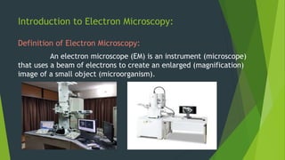
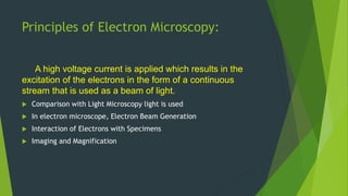
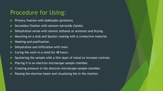
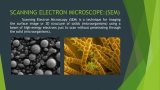
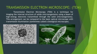
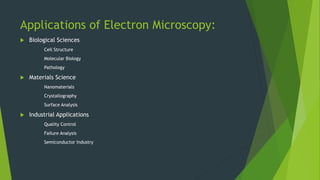
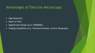
Electron Microscope
In the world of microscopes, the electron microscope is an advanced part of it.
NITHISH KRISHNA R
19.03.2024
Introduction to Electron Microscopy:
Definition of Electron Microscopy:
An electron microscope (EM) is an instrument (microscope)
that uses a beam of electrons to create an enlarged (magnification)
image of a small object (microorganism).
Principles of Electron Microscopy:
A high voltage current is applied which results in the
excitation of the electrons in the form of a continuous
stream that is used as a beam of light.
 Comparison with Light Microscopy light is used
 In electron microscope, Electron Beam Generation
 Interaction of Electrons with Specimens
 Imaging and Magnification
Procedure for Using:
 Primary fixation with aldehydes (proteins).
 Secondary fixation with osmium tetroxide (lipids).
 Dehydration series with solvent (ethanol or acetone) and Drying.
 Mounting on a stub and Sputter coating with a conductive material.
 Washing and postfixation.
 Dehydration and Infiltration with resin.
 Curing the resin in a mold for 48 hours.
 Sputtering the sample with a thin layer of metal to increase contrast.
 Placing it in an electron microscope sample chamber.
 Creating pressure in the electron microscope sample chamber.
 Passing the electron beam and visualizing the in the monitor.
Types of Electron Microscopes:
There are many types of electron microscopes the
main two types of electron microscopes are :
 Scanning Electron Microscope (SEM)
 Transmission Electron Microscope (TEM)
SCANNING ELECTRON MICROSCOPE:(SEM)
Scanning Electron Microscopy (SEM) is a technique for imaging
the surface image or 3D structure of solids (microorganisms) using a
beam of high-energy electrons just to scan without penetrating through
the solid (microorganisms).
TRANSMISSION ELECTRON MICROSCOPE: (TEM)
Transmission Electron Microscopy (TEM) is a technique for
imaging the internal structure of solids (microorganisms) using a beam of
high-energy electrons transmitted through the solid (microorganisms).
This arrangement can be compared to the basic optical microscope with
transmission illumination (sometimes called a biological microscope).
Applications of Electron Microscopy:
 Biological Sciences
Cell Structure
Molecular Biology
Pathology
 Materials Science
Nanomaterials
Crystallography
Surface Analysis
 Industrial Applications
Quality Control
Failure Analysis
Semiconductor Industry
Advantages of Electron Microscopy:
 High Resolution
 Depth of Field
 Magnification Range (up to 10000000x)
 Imaging Capabilities (e.g., Elemental Analysis, Surface Topography)
Limitations and Challenges:
 Sample Preparation Issues
 High Vacuum Requirements
 Complex Operation and Maintenance
 Cost
A detailed view on electron microscope..

A detailed view on electron microscope..

  • 1.
    Electron Microscope In theworld of microscopes, the electron microscope is an advanced part of it. NITHISH KRISHNA R 19.03.2024
  • 2.
    Introduction to ElectronMicroscopy: Definition of Electron Microscopy: An electron microscope (EM) is an instrument (microscope) that uses a beam of electrons to create an enlarged (magnification) image of a small object (microorganism).
  • 3.
    Principles of ElectronMicroscopy: A high voltage current is applied which results in the excitation of the electrons in the form of a continuous stream that is used as a beam of light.  Comparison with Light Microscopy light is used  In electron microscope, Electron Beam Generation  Interaction of Electrons with Specimens  Imaging and Magnification
  • 4.
    Procedure for Using: Primary fixation with aldehydes (proteins).  Secondary fixation with osmium tetroxide (lipids).  Dehydration series with solvent (ethanol or acetone) and Drying.  Mounting on a stub and Sputter coating with a conductive material.  Washing and postfixation.  Dehydration and Infiltration with resin.  Curing the resin in a mold for 48 hours.  Sputtering the sample with a thin layer of metal to increase contrast.  Placing it in an electron microscope sample chamber.  Creating pressure in the electron microscope sample chamber.  Passing the electron beam and visualizing the in the monitor.
  • 5.
    Types of ElectronMicroscopes: There are many types of electron microscopes the main two types of electron microscopes are :  Scanning Electron Microscope (SEM)  Transmission Electron Microscope (TEM)
  • 6.
    SCANNING ELECTRON MICROSCOPE:(SEM) ScanningElectron Microscopy (SEM) is a technique for imaging the surface image or 3D structure of solids (microorganisms) using a beam of high-energy electrons just to scan without penetrating through the solid (microorganisms).
  • 7.
    TRANSMISSION ELECTRON MICROSCOPE:(TEM) Transmission Electron Microscopy (TEM) is a technique for imaging the internal structure of solids (microorganisms) using a beam of high-energy electrons transmitted through the solid (microorganisms). This arrangement can be compared to the basic optical microscope with transmission illumination (sometimes called a biological microscope).
  • 8.
    Applications of ElectronMicroscopy:  Biological Sciences Cell Structure Molecular Biology Pathology  Materials Science Nanomaterials Crystallography Surface Analysis  Industrial Applications Quality Control Failure Analysis Semiconductor Industry
  • 9.
    Advantages of ElectronMicroscopy:  High Resolution  Depth of Field  Magnification Range (up to 10000000x)  Imaging Capabilities (e.g., Elemental Analysis, Surface Topography)
  • 10.
    Limitations and Challenges: Sample Preparation Issues  High Vacuum Requirements  Complex Operation and Maintenance  Cost