Electromyography is basically the study of motor unit activity.
In electromyography, the study of the electrical activity of contracting muscle provides information concerning the structure and function of the motor units.
INTRODUCTION
• Electromyography isbasically the study of
motor unit activity.
• In electromyography, the study of the
electrical activity of contracting muscle
provides information concerning the
structure and function of the motor units.
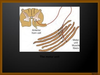
• Motor unitsare composed of one anterior
horn cell, one axon, its neuromuscular
junctions and all the muscle fibers
innervated by the axon
• The nerve cell and the muscle fiber it
supplies are defined as a motor unit.
DEFINITION
• Electromyography isthe extracellular
recording of bioelectrical activity generated
by muscle fibers.(JOSEPH MIZRAHI)
• Emg is the study of the electrical activity of
muscle.(MARC D.BINDER)
7.
HISTORY
• In 1771,galvanishowed that electrical
stimulation of animal muscle tissue
produced contraction
• In 1929,adrian devised a method to record
a single motor unitpotential by connecting
concentric needle electrodes to an
amplifier and a loud speaker
8.
• In 1938,denny-browndescribed the
fascicullation potentials and separate them
from fibrillations.
• In 1957,lambert and eaton described the
electrophysiological features of a new
myasthenic syndrome associated with
lung carcinoma.
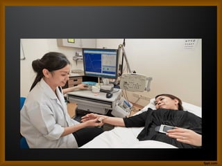
Diagnostic or clinical
electromyography
•It is used for the study of diseases of
muscles,neuromuscular junctions and
nerves. It is used for the purpose of
electrodiagnosis. The electric potentials
from the skeletal muscle fibers are
recorded and analysed for the study of
some disease processes.
15.
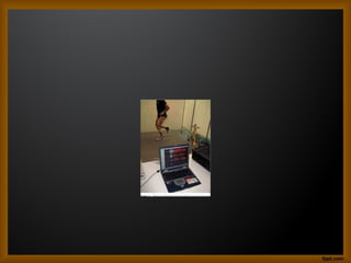
Kinesiological
electromyography
• It isused in the study of muscle activity
and to establish the role of various
muscles in specific activities.
Kinesiological EMG is beneficial for
producing the objective means for
documenting the effects of treatment on
muscle impairments.
17.
PHASES OF EMG
RECORDING
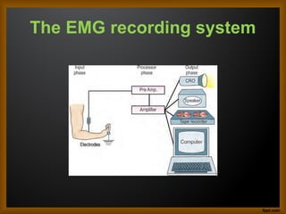
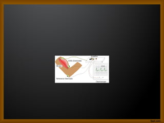
•Recording the EMG requires a three
phase system:
• 1. an input phase
• 2. a processor phase
• 3. an output phase.
18.
• An inputphase includes electrodes to pick
up electrical potential from contracting
muscle, a processor phase amplifies the
very small electrical potentials and an
output phase includes the display and
analysis of electrical potential by visual
and auditory means.
The Components ofElectromyography
• The components of electromyography
apparatus are:
• 1. Electrodes
• 2. Amplifier system
• 3. Display system.
21.
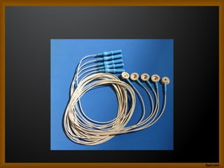
The Electrodes
• Theyare used in the input phase for
picking up of electrical potentials from the
contracting muscle fibres. The electrodes
are of following types:
22.
• a. Surfaceelectrodes
• b. Needle electrodes
• Fine wire indwelling electrodes
• Single fibre needle electrodes
• Macroelectrode
• Intra cellular electrode
• Multi lead electrode
23.
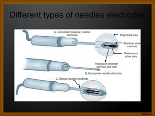
Needle electrodes
• Needleelectrodes are used for clinical
electromyography for recording single
motor unit potential from different parts of
a muscle. The different types of needle
electrodes used are:
• Concentric (coaxial) needle electrode
• Monopolar needle electrode
• Bipolar needle electrode
24.
• Concentric (coaxial)needle electrode:
This type of electrode consists of a
stainless steel cannula through which a
single wire of platinum or silver comes out.
The cannula shaft and wire are insulated
from each other and only their tips are
exposed.
25.
• Monopolar needleelectrode: These are
composed of single fine needle which is
insulated except at its tip. A second
surface electrode is placed on the skin
near the site of insertion which serves as a
reference electrode. These electrodes are
less painful than concentric electrodes
because they are much smaller in
diameter
26.
• Bipolar needleelectrode: These consist
of a cannula containing two insulated
wires with their bare tips. The bared tips of
both wires act as the two electrodes and
the needle serves as the ground
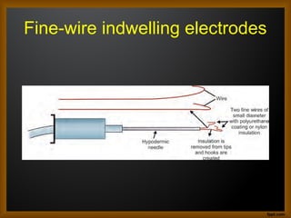
Fine wire indwelling
electrodes
•These are used for kinesiological study of
small and deep muscle. It is made by
using two fine wires of small diameter with
polyurethane coating or nylon insulation.
Insulation is removed from the tip of the
wires and hooks are created to keep the
wires imbedded while the needle is
removed from the muscle
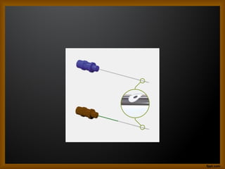
Single fibre needleelectrodes
• These are concentric wires of 25 μm
diameter and contain stainless steel
cannula of 0.5 mm diameter. Single fibre
EMG is employed to study neuromuscular
transmission abnormality and fibre density.
32.
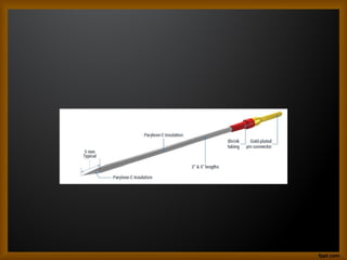
Macroelectrode
• Macroelectrode isa concentric needle
electrode of 15 mm shaft. It records from a
large number of motor units along the
shaft of the needle. The recording from
one motor unit is separated by using a
single fibre needle attached to
macroelectrode in the midshaft.
34.
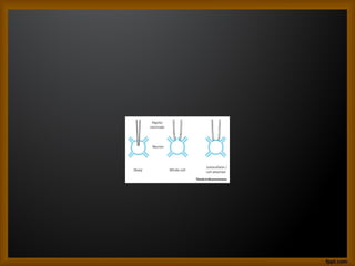
Intra cellular electrode
•This is an extremely fine electrode of
diameter 0.5 μm and is used to record the
potential changes inside the membrane
across a cell. It is made so fine so as to
penetrate deep inside a cell or intracellular
matrix.
36.
Multi lead electrode
•This electrode consists of a common steel
cannula which comprises of at least three
insulated electrodes at regular intervals
inside it.
38.
The Amplifier system
•Before the motor unit potential can be
visualized, it is necessary to amplify the
small myoelectric signals. An amplifier
converts the electric signal large enough
to be displayed.
41.
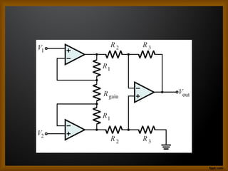
Differential amplifier
• Theelectric potential is composed of the EMG
signal from the muscle contraction and
unwanted noise from the static electricity in the
air and power lines. To control for the unwanted
part of the signal, the differential amplifier is
used, as noise is transmitted to the amplifier as
a common mode signal when the difference of
potential is reduced at both the ends, the noise
being cancelled out both the ends of amplifier.
42.
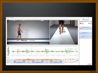
The display system
•The amplified or processed signal is
displayed in a useful manner.The form of
output used depends upon the desired
information and the instrumentation
available. The electrical signal can be
displayed visually on a cathode ray
oscilloscope or computer monitor for
analysis.
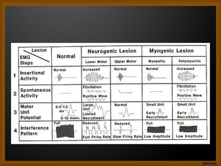
44.
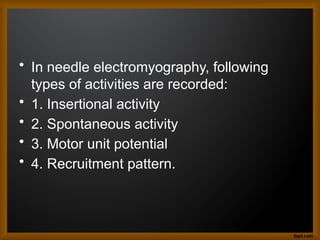
• In needleelectromyography, following
types of activities are recorded:
• 1. Insertional activity
• 2. Spontaneous activity
• 3. Motor unit potential
• 4. Recruitment pattern.
45.
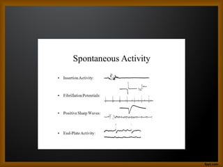
Abnormal spontaneous
potentials
• Asa normal muscle at rest exhibits
electrical silence,any activity seen during
the relaxed state is considered as
abnormal
• Fibrillation potential
• Positive sharp waves
• Fasciculation potential
• Repetitive discharges.
48.
COMPLICATIONS
• Complications rare
•Risk of bleeding
• Infexction
• Muscle soreness
• Nerve injury due to needle electrode
• Pain,tenderness,swelling at needle
insertion sites


![ELECTRO MYO GRAM [EMG]-1.pptx](https://cdn.slidesharecdn.com/ss_thumbnails/electromyogramemg-1-230303140416-5e58de8a-thumbnail.jpg?width=640&height=640&fit=bounds)


![CASE_PRESENTATION_ON_subdural_hematoma(SDH)[1 FINAL PPT]-1.pptx](https://cdn.slidesharecdn.com/ss_thumbnails/casepresentationonsubduralhematomasdh1finalppt-1-260129172522-d405d375-thumbnail.jpg?width=640&height=640&fit=bounds)


