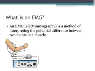
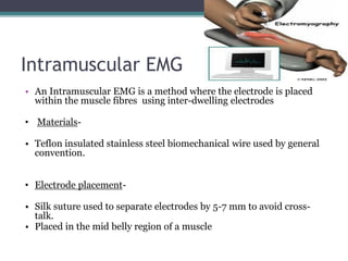
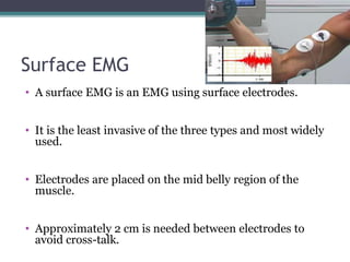
EMG
What is an EMG?
• An EMG (electromyography) is a method of
interpreting the potential difference between
two points in a muscle.
Types of EMG
• There are three types of EMG
• Intramuscular
• Subcutaneous
• Surface EMG
Intramuscular EMG
• An Intramuscular EMG is a method where the electrode is placed
within the muscle fibres using inter-dwelling electrodes
• Materials-
• Teflon insulated stainless steel biomechanical wire used by general
convention.
• Electrode placement-
• Silk suture used to separate electrodes by 5-7 mm to avoid cross-
talk.
• Placed in the mid belly region of a muscle
Subcutaneous
• A Subcutaneous EMG is the insertion of an
electrode into the subcutaneous layer between the
skin and muscle layer.
• The electrode by general convention is a stainless
steel needle.
• This method appears less effective as implied by the
reference source material when compared to the
other methods.
Surface EMG
• A surface EMG is an EMG using surface electrodes.
• It is the least invasive of the three types and most widely
used.
• Electrodes are placed on the mid belly region of the
muscle.
• Approximately 2 cm is needed between electrodes to
avoid cross-talk.
• Electrodes
•Differential Amplifier
•High and Low Pass Filters
•Precision Rectifiers
Differential Amplifier
High Pass Filter Low Pass Filter
Precision Rectifier
Our Model
• Version 1
Version 2
• The working model
EMG to Force
• There is a link between force and a spike in an EMG
signal.
• The precise quantitative nature is difficult to
calculate.
• Problems associated with this are
EMD(electromechanical delays) and the consistency
of the EMD throughout the EMG
-6
-5
-4
-3
-2
-1
0
1
2 s
.344
.692
1.04
1.388
1.736
2.084
2.432
2.78
3.128
3.476
3.824
4.172
4.52
4.868
5.216
5.564
5.912
6.26
6.608
6.956
7.304
7.652
8.00
8.348
8.696
9.044
9.392
9.74
Y-Values
Y-Values
EMG SIGNAL ACCUIRED
-6.00
-4.00
-2.00
.00
2.00
4.00
6.00
C
a
t
e
g
o
r
y
1
C
a
t
e
g
o
r
y
2
C
a
t
e
g
o
r
y
3
C
a
t
e
g
o
r
y
4
Series 2
Series 3
ISOMETRIC SIGNAL

Anatomy project emg