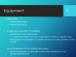
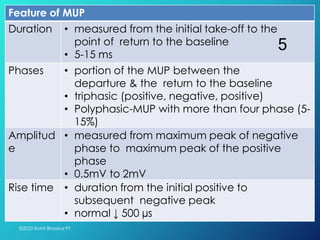
ELECTROMYOGRAPHY
©2020 Rohit Bhaskar PT
1
• Electromyography (EMG)
– is a technique for evaluating & recording the electrical
activity produced by skeletal muscles
– is performed using an instrument called an electromyograph,
to produce a record called an electromyogram
• a resting muscle does not show recordable electrical
potential but with increase force of contraction,
amplitude of potential increases
• an electromyograph detects electrical
potential generated by muscle cells when
these cells are electrically or neurologically
activated
©2020 Rohit Bhaskar PT
2
Equipment
• electrodes
– Surface electrodes
– Needle electrodes
• a high-gain amplifier (10-5000Hz)
– connected to an oscilloscope
• oscilloscopic traces may be photographed or stored on magnetic tape
– EMG signals may be fed to an audio unit for an on the spot feel of the
signals
• an arrangement for recording the output
– EMG is best done in a specially constructed shielded room to
prevent interference
3
• the sum of the action potentials produced in
the muscle
• characterized by its duration, number of
phases, amplitude, & rate of rise of first
component
©2020 Rohit Bhaskar PT
4
Motor unit potential (MUP)
Feature of MUP
Duration • measured from the initial take-off to the
point of return to the baseline
• 5-15 ms
Phases • portion of the MUP between the
departure & the return to the baseline
• triphasic (positive, negative, positive)
• Polyphasic-MUP with more than four phase (5-
15%)
Amplitud
e
• measured from maximum peak of negative
phase to maximum peak of the positive
phase
• 0.5mV to 2mV
Rise time • duration from the initial positive to
subsequent negative peak
• normal ↓ 500 µs
©2020 Rohit Bhaskar PT
5
Factors that effect MUP
• Technical factors
– Type of needle electrode
– Characteristics of recording surface
– Electrical characteristics of cable
– Preamplifier & amplifier
– Method of recording
• Physiological factors
– Age of the patient
– Muscle examined
– Temperature
6
Analysis of EMG
• Qualitative analysis
– visual inspection of the record
• Quantitative analysis
– amplitude, duration, & frequency
– power spectrum analysis
©2020 Rohit Bhaskar PT
7
Fibrillation Fasciculation
contraction of
individual muscle fiber
contraction of
individual motor units
cannot be seen through
the skin
visible through the skin
when muscle fibers lose
contact with their
innervating axon
occurs as a result of
additional nerve
impulses generated
10-100 µV in amplitude, 1-2
ms in duration, & 10 Hz in
frequency
50-500 µV in amplitude, 2-4
ms in duration, & 2-20 Hz in
frequency
Neuropathy, myopathy Neuropathy
©2020 Rohit Bhaskar PT
8
Thank You
©2020 Rohit Bhaskar PT
9

Electromyography (EMG) - Physiotherapy - Dr Rohit Bhaskar

  • 1.
  • 2.
    • Electromyography (EMG) –is a technique for evaluating & recording the electrical activity produced by skeletal muscles – is performed using an instrument called an electromyograph, to produce a record called an electromyogram • a resting muscle does not show recordable electrical potential but with increase force of contraction, amplitude of potential increases • an electromyograph detects electrical potential generated by muscle cells when these cells are electrically or neurologically activated ©2020 Rohit Bhaskar PT 2
  • 3.
    Equipment • electrodes – Surfaceelectrodes – Needle electrodes • a high-gain amplifier (10-5000Hz) – connected to an oscilloscope • oscilloscopic traces may be photographed or stored on magnetic tape – EMG signals may be fed to an audio unit for an on the spot feel of the signals • an arrangement for recording the output – EMG is best done in a specially constructed shielded room to prevent interference 3
  • 4.
    • the sumof the action potentials produced in the muscle • characterized by its duration, number of phases, amplitude, & rate of rise of first component ©2020 Rohit Bhaskar PT 4 Motor unit potential (MUP)
  • 5.
    Feature of MUP Duration• measured from the initial take-off to the point of return to the baseline • 5-15 ms Phases • portion of the MUP between the departure & the return to the baseline • triphasic (positive, negative, positive) • Polyphasic-MUP with more than four phase (5- 15%) Amplitud e • measured from maximum peak of negative phase to maximum peak of the positive phase • 0.5mV to 2mV Rise time • duration from the initial positive to subsequent negative peak • normal ↓ 500 µs ©2020 Rohit Bhaskar PT 5
  • 6.
    Factors that effectMUP • Technical factors – Type of needle electrode – Characteristics of recording surface – Electrical characteristics of cable – Preamplifier & amplifier – Method of recording • Physiological factors – Age of the patient – Muscle examined – Temperature 6
  • 7.
    Analysis of EMG •Qualitative analysis – visual inspection of the record • Quantitative analysis – amplitude, duration, & frequency – power spectrum analysis ©2020 Rohit Bhaskar PT 7
  • 8.
    Fibrillation Fasciculation contraction of individualmuscle fiber contraction of individual motor units cannot be seen through the skin visible through the skin when muscle fibers lose contact with their innervating axon occurs as a result of additional nerve impulses generated 10-100 µV in amplitude, 1-2 ms in duration, & 10 Hz in frequency 50-500 µV in amplitude, 2-4 ms in duration, & 2-20 Hz in frequency Neuropathy, myopathy Neuropathy ©2020 Rohit Bhaskar PT 8
  • 9.