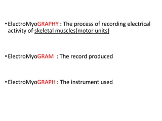
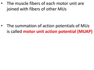
•ElectroMyoGRAPHY : The process of recording electrical
activity of skeletal muscles(motor units)
•ElectroMyoGRAM : The record produced
•ElectroMyoGRAPH : The instrument used
Motor unit : the
motor neuron and
the muscle fibers
it innervates.
Action Potential (AP)
• Cell membrane separates intracellular from extracellular
space, diffusion barrier which restricts ION flow. Concentration
of ions different inside vs. outside of cell membrane, results in
an electrical potential difference known as a MEMBRANE
POTENTIAL. Typical magnitude of membrane potential is
about −70 mV (interior of cell is negatively charged compared
to the outside) when the muscle cell is in resting state.
• When sufficient neurotransmitters are deposited at the motor
endplate, it opens up Na+ gates, causing an influx of Na+
ions, causing a rapid depolarization of the membrane near the
motor end plate. The membrane potential can change to +20
to +50 mV at the motor endplate within a fractions of a
second, which starts (all or none) a cascade of events.
AP Continued
• This changed potential, sets up local currents in adjacent areas of
membrane, which opens up more Na+ gates at the adjacent areas of
the membrane, depolarizing those areas. The newly depolarized
areas sets up electrical current with adjacent areas, and thus the
depolarization wave propagates along the entire length of the muscle
cell.
• Shortly after depolarization, the membrane is
again repolarized, by active transport
of ions across the membrane. As a result a
repolarization wave follows the
depolarization wave for the entire length of
the muscle.
• The muscle fibers of each motor unit are
joined with fibers of other MUs
• The summation of action potentials of MUs
is called motor unit action potential (MUAP)
Two types of electrodes have been used to acquire muscle signal:
1. Needle EMG (nEMG)
nEMG permits local recording from deep muscles by means of insertion
of a needle electrode into the muscle tissue. nEMG can be used to
assess individual MUs and has greater sensitivity and accuracy in the
recording of signals .
However, nEMG has several limitations. First, it reflects the activity of
only a small number of active MUs whose fibers are close to the
position of the detection site (not representative of all the fibers in the
MU, due to its small detection volume). An adequate sample is needed
to ensure adequate power (sensitivity and specificity) of the analysis of
MUAPs . Second, nEMG is painful especially during muscle activation,
and prolonged nEMG recording is not possible. In rare cases, local
trauma (e.g., pneumothorax) could occur during the examination of
some delicate regions.
Furthermore, nEMG is time and temperature sensitive. In this
regard, the detected signal in nEMG may vary as a function
elapsed time from the onset of the nerve injury . A low
temperature at the examination area modifies the parameters
and characteristics of the recorded signals .
2. Surface EMG (sEMG)
sEMG is a technique to measure muscle activity noninvasively
using surface electrodes placed on the skin overlying the muscle,
and has several advantages. First, sEMG recording is painless,
Quick and easy to apply . Furthermore, sEMG electrodes record
from a wide area of muscle territory providing a more global view
of MUs. Finally, it allows prolonged simultaneous recordings of
muscle activity from multiple sites.
However, sEMG has a relatively low-signal resolution, is highly
susceptible to movement artifacts. In addition, sEMG signals are
dominated by the contributions of superficial MUs, while deeper
MUs are not assessed; conditions that increase skin resistance
subsequently disturb the sEMG signal (e.g. obesity and edema).
EMG PROCEDURE
1. Clean the site of application of electrode
2. Insert needle/place surface electrodes at muscle belly
3. Record muscle activity at rest
4. Record muscle activity upon voluntary contraction of the muscle.
AT REST
EMG study
Muscle voluntary contraction recording In depolarization, the summation of action
potentials of the MUs (MUAPs) can be assessed by analysis of their parameters
Duration is measured from the initial deflection from baseline
to the terminal deflection back to baseline; it reflexes the
muscle fiber density in an MU. The average duration of
MUAPs increases from infancy to adult , and even more
during old age; the percentage depends on the specific
muscle.
Abnormalities of MUAP duration can be shown in
pathological conditions:
•Short-duration MUAPs are often detected in disorders with
loss of muscle fibers.
•Long-duration MUAPs are typically found in chronic
neuropathic disorders and polymyositis.
•A mixed pattern (coexisting MUAPs of long and short
duration) can be observed in rapidly progressing motor
neuron disease and chronic myositis.
Morphology(number of phases) is defined as
the number of baseline crossings of a MUAP
and reflects the firing synchrony of the muscle
fibers within a MU. Normally, a MUAP has two
to four phases. A MUAP of more than four
phases is named polyphasic potential.
MUAPs with abnormal morphology can be
recorded in neuromuscular disorders:
An abnormally increased polyphasia is a
non-specific signal of both myopathic and
neuropathic disorders
Amplitude is commonly measured from peak to peak.
provide a measure of the magnitude of muscle force
In other words, the stronger the muscle contraction and the
higher the number of activated muscles, the higher the
recorded voltage amplitude will be.(recruitment)
The progressive loss of MUs, results in MUAPs of smaller
amplitude .This phenomenon is especially noticeable in
extensor digitorum brevis muscle.
In chronic neuropathies, the MUAP amplitude can be
increased due to reinnervation process
Resting state recording
At resting state, muscle activity can be recorded using either
intramuscular (needle) or non-invasive (surface) detection
systems.
In a healthy muscle at rest, spontaneous physiological activity
can be recorded by means of nEMG:
Insertional activity: induced by mechanical depolarization of
muscle fiber due to needle electrode insertion
The most described abnormal spontaneous activities include:
•Fibrillation potentials, positive sharp waves, complex
repetitive with an unstable membrane potential that fire
individually without axonal stimulation.
•Fasciculation, spontaneous and continuous muscle fibre
activity generating from
disturbance of a group
of muscle fibers.
:
EMG analysis
The analysis of MUAPs can be performed on a qualitative or
quantitative basis. At least 20 samples from each studied muscle is
widely accepted as representative
1.Qualitative analysis
Visual recognition only provides limited information. The
effectiveness of this method depends on the experience of
the performer
2.Quantitative analysis
The parametric method establishes a comparison between
sample mean values and reference intervals (standard
deviation), while the nonparametric method considers both
extremes of the samples (“outliers”)
EMG.pdf

EMG.pdf

  • 1.
    •ElectroMyoGRAPHY : Theprocess of recording electrical activity of skeletal muscles(motor units) •ElectroMyoGRAM : The record produced •ElectroMyoGRAPH : The instrument used
  • 2.
    Motor unit :the motor neuron and the muscle fibers it innervates.
  • 4.
    Action Potential (AP) •Cell membrane separates intracellular from extracellular space, diffusion barrier which restricts ION flow. Concentration of ions different inside vs. outside of cell membrane, results in an electrical potential difference known as a MEMBRANE POTENTIAL. Typical magnitude of membrane potential is about −70 mV (interior of cell is negatively charged compared to the outside) when the muscle cell is in resting state. • When sufficient neurotransmitters are deposited at the motor endplate, it opens up Na+ gates, causing an influx of Na+ ions, causing a rapid depolarization of the membrane near the motor end plate. The membrane potential can change to +20 to +50 mV at the motor endplate within a fractions of a second, which starts (all or none) a cascade of events.
  • 5.
    AP Continued • Thischanged potential, sets up local currents in adjacent areas of membrane, which opens up more Na+ gates at the adjacent areas of the membrane, depolarizing those areas. The newly depolarized areas sets up electrical current with adjacent areas, and thus the depolarization wave propagates along the entire length of the muscle cell. • Shortly after depolarization, the membrane is again repolarized, by active transport of ions across the membrane. As a result a repolarization wave follows the depolarization wave for the entire length of the muscle.
  • 6.
    • The musclefibers of each motor unit are joined with fibers of other MUs • The summation of action potentials of MUs is called motor unit action potential (MUAP)
  • 7.
    Two types ofelectrodes have been used to acquire muscle signal: 1. Needle EMG (nEMG) nEMG permits local recording from deep muscles by means of insertion of a needle electrode into the muscle tissue. nEMG can be used to assess individual MUs and has greater sensitivity and accuracy in the recording of signals . However, nEMG has several limitations. First, it reflects the activity of only a small number of active MUs whose fibers are close to the position of the detection site (not representative of all the fibers in the MU, due to its small detection volume). An adequate sample is needed to ensure adequate power (sensitivity and specificity) of the analysis of MUAPs . Second, nEMG is painful especially during muscle activation, and prolonged nEMG recording is not possible. In rare cases, local trauma (e.g., pneumothorax) could occur during the examination of some delicate regions.
  • 8.
    Furthermore, nEMG istime and temperature sensitive. In this regard, the detected signal in nEMG may vary as a function elapsed time from the onset of the nerve injury . A low temperature at the examination area modifies the parameters and characteristics of the recorded signals .
  • 9.
    2. Surface EMG(sEMG) sEMG is a technique to measure muscle activity noninvasively using surface electrodes placed on the skin overlying the muscle, and has several advantages. First, sEMG recording is painless, Quick and easy to apply . Furthermore, sEMG electrodes record from a wide area of muscle territory providing a more global view of MUs. Finally, it allows prolonged simultaneous recordings of muscle activity from multiple sites. However, sEMG has a relatively low-signal resolution, is highly susceptible to movement artifacts. In addition, sEMG signals are dominated by the contributions of superficial MUs, while deeper MUs are not assessed; conditions that increase skin resistance subsequently disturb the sEMG signal (e.g. obesity and edema).
  • 10.
    EMG PROCEDURE 1. Cleanthe site of application of electrode 2. Insert needle/place surface electrodes at muscle belly 3. Record muscle activity at rest 4. Record muscle activity upon voluntary contraction of the muscle.
  • 11.
  • 12.
    EMG study Muscle voluntarycontraction recording In depolarization, the summation of action potentials of the MUs (MUAPs) can be assessed by analysis of their parameters
  • 14.
    Duration is measuredfrom the initial deflection from baseline to the terminal deflection back to baseline; it reflexes the muscle fiber density in an MU. The average duration of MUAPs increases from infancy to adult , and even more during old age; the percentage depends on the specific muscle. Abnormalities of MUAP duration can be shown in pathological conditions: •Short-duration MUAPs are often detected in disorders with loss of muscle fibers. •Long-duration MUAPs are typically found in chronic neuropathic disorders and polymyositis. •A mixed pattern (coexisting MUAPs of long and short duration) can be observed in rapidly progressing motor neuron disease and chronic myositis.
  • 15.
    Morphology(number of phases)is defined as the number of baseline crossings of a MUAP and reflects the firing synchrony of the muscle fibers within a MU. Normally, a MUAP has two to four phases. A MUAP of more than four phases is named polyphasic potential. MUAPs with abnormal morphology can be recorded in neuromuscular disorders: An abnormally increased polyphasia is a non-specific signal of both myopathic and neuropathic disorders
  • 16.
    Amplitude is commonlymeasured from peak to peak. provide a measure of the magnitude of muscle force In other words, the stronger the muscle contraction and the higher the number of activated muscles, the higher the recorded voltage amplitude will be.(recruitment) The progressive loss of MUs, results in MUAPs of smaller amplitude .This phenomenon is especially noticeable in extensor digitorum brevis muscle. In chronic neuropathies, the MUAP amplitude can be increased due to reinnervation process
  • 17.
    Resting state recording Atresting state, muscle activity can be recorded using either intramuscular (needle) or non-invasive (surface) detection systems. In a healthy muscle at rest, spontaneous physiological activity can be recorded by means of nEMG: Insertional activity: induced by mechanical depolarization of muscle fiber due to needle electrode insertion
  • 18.
    The most describedabnormal spontaneous activities include: •Fibrillation potentials, positive sharp waves, complex repetitive with an unstable membrane potential that fire individually without axonal stimulation. •Fasciculation, spontaneous and continuous muscle fibre activity generating from disturbance of a group of muscle fibers.
  • 19.
    : EMG analysis The analysisof MUAPs can be performed on a qualitative or quantitative basis. At least 20 samples from each studied muscle is widely accepted as representative 1.Qualitative analysis Visual recognition only provides limited information. The effectiveness of this method depends on the experience of the performer 2.Quantitative analysis The parametric method establishes a comparison between sample mean values and reference intervals (standard deviation), while the nonparametric method considers both extremes of the samples (“outliers”)