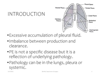
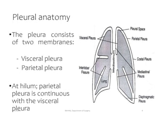
5/17/2023 MUHAS, Department of Surgery 1
(1.5 hours)
PLEURAL EFFUSION
& THORACIC EMPYEMA
Objectives
By the end of this session, students should be able
to,
1) Elaborate the pathophysiology and causes of
pleural effusion and empyema thoracis
2) Identify a patient with pleural effusion/empyema
thoracis based on clinical presentation
3) Investigate a patient and come to a final
diagnosis
4) Manage a patient with pleural effusion, thoracic
empyema and their complications.
5/17/2023 MUHAS, Department of Surgery 2
INTRODUCTION
•Excessive accumulation of pleural fluid.
•Imbalance between production and
clearance.
•PE is not a specific disease but it is a
reflection of underlying pathology.
•Pathology can be in the lungs, pleura or
systemic.
5/17/2023 MUHAS, Department of Surgery 3
Pleural anatomy
•The pleura consists
of two membranes:
- Visceral pleura
- Parietal pleura
•At hilum; parietal
pleura is continuous
with the visceral
pleura
5/17/2023 MUHAS, Department of Surgery 4
Anatomy cont.
Normally the pleural space contains:
 Clear pleural fluid ~0.3 ml/kg
 Low protein content
 Small number of mononuclear cells
 Pleural fluid glucose equal to serum
glucose level.
5/17/2023 MUHAS, Department of Surgery 5
Anatomy cont.
•Pleural lining is formed by mesothelial cells.
•Below the mesothelial lining is a thin
connective tissue lining which has Blood
vessels and lymphatics which are important in
dynamics of pleural fluid formation.
•Parietal pleura has stomata allowing
passageway for liquid from pleural space to
the lymphatic system.
5/17/2023 MUHAS, Department of Surgery 6
Anatomy cont.
•Sensory nerve endings are there in the parietal
and diaphragmatic pleura.
•Blood vessels supplying parietal pleural
surface originate from the systemic arterial
circulation-primarily Inter Costal Arteries.
•Venous blood from the parietal pleura drains
to systemic venous system.
5/17/2023 MUHAS, Department of Surgery 7
Anatomy cont.
•Visceral pleura is also supplied primarily by systemic
arteries, specifically branches of bronchial arterial
circulation, but the venous drainage is in to
pulmonary venous system.
•The Lymphatic vessels that drain the pleural surfaces
transport their fluid contents to different lymph
nodes.
•Ultimately the fluid is transported to the right
lymphatic or thoracic ducts, which empty in to the
systemic venous circulation
5/17/2023 MUHAS, Department of Surgery 8
Physiology
•Pleural space normally contains small amount
of fluid (10 ml).
•As per recent concept, formation of fluid is
ongoing primarily from the parietal pleural
surface, and fluid is reabsorbed through the
stomata into the lymphatic channels of the
parietal pleura.
•Normally rate of formation = absorption;
about 0.02 ml/kg/hour.
5/17/2023 MUHAS, Department of Surgery 9
Physiology cont.
•Pleural fluid is the ultra filtrate from the
pleural capillaries.
•There are counteracting forces which are
responsible for transport of fluid; Hydrostatic
pressure - P, and Colloid oncotic pressure -
COP.
•‘P’ promotes movement of fluid out of the
capillaries and COP prohibits the movement
from the capillaries.
5/17/2023 MUHAS, Department of Surgery 10
Physiology cont.
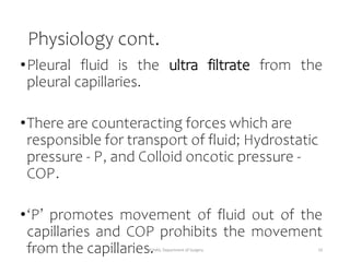
Fluid movement = K[(Pc –Pis ) – σ(COPc –COPis )]
Starlings equationcan be
applied to parietal pleura:
Pc = 30 cm H2 O,
Pis (mean intrapleural pressure)=
- 5 cm H2 O,
COPc = 32 cm H2 O,
COPis = 6 cm H2 O,
=1, K=1
Fluid movement at parietal
pleura = [30-(-5)]-1(32-6)
= 9 cm H2 O
5/17/2023 MUHAS, Department of Surgery 11
Pathogenesis
•A change in the magnitude of any of the
factors in the Starling equation can cause
sufficient imbalance in the pleural fluid
dynamics resulting in pleural fluid
accumulation.
•Interference with resorptive process like
blockage of lymphatic drainage as occurs
when tumor cells invade the lymphatic
channels or the draining LN.
5/17/2023 MUHAS, Department of Surgery 12
Pathogenesis cont.
•There are mainly two
categories of change
5/17/2023 MUHAS, Department of Surgery 13
Transudate vs. Exudate
5/17/2023 MUHAS, Department of Surgery 14
Aetiology
Causes of Transudative
Effusions
•Congestive heart failure
•Cirrhosis
•Nephrotic syndrome
•Hypoalbuminemia
•Fluid retention/overload
•Pulmonary embolism
•Lobar collapse
•Meigs’ syndrome
note
•Transudative effusions
occur because of
increased hydrostatic
pressure or decreased
oncotic pressures
•The etiology of pleural
effusion remains unclear in
nearly 20% of cases
5/17/2023 MUHAS, Department of Surgery 15
Aetiology cont.
Causes of Exudative Effusions
• Malignant
• Primary lung, pleural, or
metastatic carcinoma
• Lymphoma
• Mesothelioma
• Infectious
• Bacterial (parapneumonic)/
Empyema
• Tuberculosis
• Fungal
• Viral
• Parasitic
• Collagen-Vascular Disease related
• Rheumatoid arthritis
• Wegener’s granulomatosis
• Systemic lupus erythematosus
• Churg-Strauss syndrome
• Abdominal and Gastrointestinal
Disease–related
• Esophageal perforation
• Subphrenic abscess
• Pancreatitis, pancreatic
pseudocyst
• Meigs’ syndrome
5/17/2023 MUHAS, Department of Surgery 16
Aetiology cont.
Causes of Exudative Effusions
•Others
•Chylothorax
•Uremia
•Sarcoidosis
•After coronary artery bypass grafting
•Radiation / Trauma
•Dressler’s syndrome
•Pulmonary embolism with infarction
•Asbestosis related
5/17/2023 MUHAS, Department of Surgery 17
Pleural Empyema/ Pyothorax/ Purulent
Pleuritis/ Empyema Thoracis
•Accumulation of Pus in the Pleural cavity
5/17/2023 MUHAS, Department of Surgery 18
Other descriptions
Weese et al. defined an empyema as pleural fluid with
• a sp. Gravity >1.018,
• a WBC count >500 cells/mm3 or
• a protein level >2.5g/dl.
Vianna defined an empyema as pleural fluid on which
• the bacterial cultures are positive or
• the WBC count is > 15,000/mm3 and
• the protein level is >3.0g/dl.
5/17/2023 MUHAS, Department of Surgery 19
Thoracic Empyema
•Empyema is different from an abscess because the
later is the formation and collection of pus in a
newly formed cavity within the lung parenchyma.
•Any pleural effusion associated with bacterial
pneumonia, lung abscess or bronchiectasis is a
parapneumonic effusion.
•Many complicated parapneumonic effusions are
empyemas.
5/17/2023 MUHAS, Department of Surgery 20
Aetiology- Thoracic empyema
NON-TRAUMATIC
• Direct extension from an
adjacent site :lung
infection
• Aspiration pneumonia
• Post-obstructive
pneumonia
• Bronchiectasis, lung
abscess
TRAUMATIC
• Instrumentation and rupture of esophagus
• Leakage of an esophageal anastomosis after
resection
• Development of a bronchopleural fistula
following pneumonectomy
• Pleural aspiration/ tube drainage
Non- surgical trauma:
• Abdominal sepsis: subphrenic abscess , liver
abscess
• Sepsis in the pharynx, thoracic spine or chest
wall may extend into the pleura via tissue
planes or mediastinum
• Gun shot wounds, blast injuries and stab
wounds.
5/17/2023 MUHAS, Department of Surgery 21
5/17/2023 MUHAS, Department of Surgery 22
Aetiology - Bacteriology
•Streptococcus pneumonia: 15 – 20%
•Staphylococcus spp: 15 – 30%
•Streptococcus spp
•Gram Negatives: 20 – 50%
•Klebsiella, Enterobacter, pseudomonas,
Haemophilus, E. coli
•Anaerobes
•Fusobacterium, Bacteroides fragilis
5/17/2023 MUHAS, Department of Surgery 23
Pathology- Thoracic empyema
(1) Exudative STAGE (approx. 7days)
 Once infected by pathogenic organisms, the
connective tissue layers within the pleural
membranes become edematous and produce an
exudation of sterile proteinaceous fluid that starts
to fill the pleural cavity.
 At this stage, the pleural fluid is thin with a relatively
low white cell count and the visceral pleura and
underlying lung remain mobile.
 Fluid at this stage is having a low WBC count, low
LDH level and a normal glucose level and pH.
5/17/2023 MUHAS, Department of Surgery 24
Pathology cont.
(2) Fibrinopurulent STAGE
•Transitional stage, from 7 to 21 days
•Newly formed layers of fibrin become laid down on the
epithelial surface within the pleural cavity, particularly
on the pleural cavity.
• The empyema fluid now becomes more thicker and
more turbid, containing, a higher white cell count.
5/17/2023 MUHAS, Department of Surgery 25
Pathology cont.
Fibrinopurulent STAGE…
•With the deposition of fibrin on both pleural
surfaces, lung movements in this stage may
become increasingly restricted.
•The pleural fluid ph and glucose levels becomes
progressively lower and LDH level becomes
progressively higher.
5/17/2023 MUHAS, Department of Surgery 26
Pathology cont.
(3) Organizational STAGE
•Thickened fibrinous layers organize as collagen and
become vascularized by an ingrowth of capillaries.
•This stage may begin within two weeks but usually
takes 4-6 weeks to develop to a point at which the
empyema cavity becomes surrounded by a cortex,
peel or rind that may be more than 2 cm thick.
5/17/2023 MUHAS, Department of Surgery 27
Pathology cont.
Organizational STAGE…
• This inelastic pleural peel encases the lung and renders
it virtually functionless.
• By this time the empyema contains frank pus, which
may be viscid.
• Ultimately, an inadequately treated empyema cavity
may become obliterated and its rind may calcify,
producing a so-called fibrothorax, particularly in case
of old tuberculous pleural infection.
5/17/2023 MUHAS, Department of Surgery 28
Pathology cont.
Organizational STAGE…
•The inner layers of the thickened empyema cortex
continue to show a considerable inflammatory cell
infiltrate and
•The fibrous outermost layers exert an increasingly
restrictive effect, both compressing the underlying
lung (the so called “trapped lung” effect) and also
tending to draw the overlying ribs together,
ultimately producing a chest deformity with a dorsal
scoliosis that is concave towards the affected side.
5/17/2023 MUHAS, Department of Surgery 29
Pathology cont.
Organizational STAGE…
•Dry “sicca” pleuritis stage, the inflammatory
process of the pulmonary parenchyma extends to
the visceral pleura, causing a local pleuritic
reaction.
•This leads to a pleural rub and a characteristic
pleuritic chest pain which originates from the
sensitive innervations of the adjacent parietal
pleura.
5/17/2023 MUHAS, Department of Surgery 30
Clinical presentation
•Depends on the nature of infecting organism.
•Competence of patients immune system.
•Ranges from complete absence of symptoms
to a severe illness with all usual manifestations
of toxicity.
5/17/2023 MUHAS, Department of Surgery 31
Clinical presentation cont.
•Acute empyema:
•Fever, cough with pleuritic pain
•Features of fluid in pleural space
•Decreased chest expansion
•Stony dullness with decreased VF and VR
•Absent breath sounds
•Features of the underlying condition
5/17/2023 MUHAS, Department of Surgery .
Clinical presentation cont.
•Sub acute and chronic empyema:
•Less severe presentation as patient often has
been on antibiotics
•Slow convalescence after a pneumonic illness
•Clinical features of fluid as above
•Features of the underlying condition; eg
bronchiectasis, lung abscess, osteomyelitis of
ribs
•Features of bronchopleural fistula or empyema
necessitans
MUHAS, Department of Surgery
5/17/2023 33
Causes of Chronicity
•Inadequate Tube Drainage.
•Chronic pulmonary Disease (TB or Fungal
Infection)
•Immunosuppressed patients.
•Presence of Foreign body within the pleural space.
5/17/2023 MUHAS, Department of Surgery 34
Complications
•Spread to the subcutaneous tissue:
•Dissection through chest wall; Empyema Necessitans
•Dissection into abdominal cavity.
•Septicaemia & septic shock.
•Pleural thickening
•Chest deformity- scoliosis
•Rupture into the lung:
 Dissection into lung parenchyma
 BronchoPleural fistula & pyopneumothorax
5/17/2023 MUHAS, Department of Surgery 35
Treatment Benign pleural
effusion
•Transudative pleural effusions are managed by
treatment of the underlying disease and usually
resolve after it has been controlled.
•Occasionally, additive intervention is required,
either because the effusion is symptomatic or
because the underlying medical problem is
refractory to maximal medical treatment
Treatment- malignant PE
•The optimal treatment for malignant pleural
effusion is controversial
•malignant disease of long duration can
prevent full lung expansion because of;
 visceral pleural restriction
endobronchial obstruction
 parenchymal fibrosis
replacement by tumor.
•Treatment options
Drainage alone
Obliteration of pleural space
Drainage-Based Methods of Malignant
Effusion Control
• Serial thoracenteses appropriately control malignant
pleural effusions for some patients
temporarily relief, for poor prognosis pts.
• Occasionally, thoracentesis creates a hydropneumothorax
because of poor compliance or entrapment of the
ipsilateral lung.
• It can be achieved through;
1. serial percutaneous aspiration
2. Implantation of a long-term pleural catheter or
placement of a percutaneous medium durability pleural
catheter
THORACOCENTESIS
•Can be dose using large bore needle
•Applicable and curative in children
•May be sufficient in exudative phase
Limitation
•Thick pus
•Organized phase
•multiloculation
5/17/2023 MUHAS, Department of Surgery 40
Closed tube thoracostomy
•First step in adult
•Useful for fibrinopurulent and exudative stages
•Insert a thoracic catheter (relatively large 28 to
36F) tubes in 6th or 7th ICS between anterior and
posterior axillary lines and connect to UWSD
bottle
• Track above superior border of a rib to avoid damage to
neurovascular bundle
•Continuous controlled suction; reduce drainage
duration
5/17/2023 MUHAS, Department of Surgery 41
Closed tube thoracostomy…
•Limitation
•Tube blockage
•Multiloculation lead to inadeguate
drainage
•Presence of bronchopleural fistula
•Thick fibrous cortex limit lung expansion
5/17/2023 MUHAS, Department of Surgery 42
5/17/2023 MUHAS, Department of Surgery 43
Indications for removal of tube
•Volume of the pleural drainage is less than 50ml for
24 hrs and until the draining fluid becomes clear
yellow.
•The amount of sediment (representing WBCs and
debris) in the collection system should not be more
than 5ml.
•Tube ceases to work
Because it serves no useful purpose rather it as a
conduit for pleural super-infection.
NB; daily monitoring
Drainage fluid; colour, volume, patency and gas
bubbles
5/17/2023 MUHAS, Department of Surgery 44
Sclerosis-Based Pleural Effusion
Management
•accelerating the process of pleurodesis
•a substance that increases the inflammatory
response and thus causes intense adhesions
within the pleural envelope
•Examples talc, Cycline Drugs and Bleomycin
PROGNOSIS
•Favourable in patients started on
appropriate antibiotic.
•Early chest tube drainage is beneficial.
•Decortication or open drainage has
decreased mortality and morbidity.
•Mortality 6-12%
5/17/2023 MUHAS, Department of Surgery 46
References
•A Manual of Clinical Surgery by S. Das. 9th Edition
•Manipal Manual of Surgery by K. Rajgopal Shenoy
4th Edition
•Schwartz’s Principles of Surgery, 11th Edition
•www.uptodate.com
5/17/2023 MUHAS, Department of Surgery 47

effusion.pptx

  • 1.
    5/17/2023 MUHAS, Departmentof Surgery 1 (1.5 hours) PLEURAL EFFUSION & THORACIC EMPYEMA
  • 2.
    Objectives By the endof this session, students should be able to, 1) Elaborate the pathophysiology and causes of pleural effusion and empyema thoracis 2) Identify a patient with pleural effusion/empyema thoracis based on clinical presentation 3) Investigate a patient and come to a final diagnosis 4) Manage a patient with pleural effusion, thoracic empyema and their complications. 5/17/2023 MUHAS, Department of Surgery 2
  • 3.
    INTRODUCTION •Excessive accumulation ofpleural fluid. •Imbalance between production and clearance. •PE is not a specific disease but it is a reflection of underlying pathology. •Pathology can be in the lungs, pleura or systemic. 5/17/2023 MUHAS, Department of Surgery 3
  • 4.
    Pleural anatomy •The pleuraconsists of two membranes: - Visceral pleura - Parietal pleura •At hilum; parietal pleura is continuous with the visceral pleura 5/17/2023 MUHAS, Department of Surgery 4
  • 5.
    Anatomy cont. Normally thepleural space contains:  Clear pleural fluid ~0.3 ml/kg  Low protein content  Small number of mononuclear cells  Pleural fluid glucose equal to serum glucose level. 5/17/2023 MUHAS, Department of Surgery 5
  • 6.
    Anatomy cont. •Pleural liningis formed by mesothelial cells. •Below the mesothelial lining is a thin connective tissue lining which has Blood vessels and lymphatics which are important in dynamics of pleural fluid formation. •Parietal pleura has stomata allowing passageway for liquid from pleural space to the lymphatic system. 5/17/2023 MUHAS, Department of Surgery 6
  • 7.
    Anatomy cont. •Sensory nerveendings are there in the parietal and diaphragmatic pleura. •Blood vessels supplying parietal pleural surface originate from the systemic arterial circulation-primarily Inter Costal Arteries. •Venous blood from the parietal pleura drains to systemic venous system. 5/17/2023 MUHAS, Department of Surgery 7
  • 8.
    Anatomy cont. •Visceral pleurais also supplied primarily by systemic arteries, specifically branches of bronchial arterial circulation, but the venous drainage is in to pulmonary venous system. •The Lymphatic vessels that drain the pleural surfaces transport their fluid contents to different lymph nodes. •Ultimately the fluid is transported to the right lymphatic or thoracic ducts, which empty in to the systemic venous circulation 5/17/2023 MUHAS, Department of Surgery 8
  • 9.
    Physiology •Pleural space normallycontains small amount of fluid (10 ml). •As per recent concept, formation of fluid is ongoing primarily from the parietal pleural surface, and fluid is reabsorbed through the stomata into the lymphatic channels of the parietal pleura. •Normally rate of formation = absorption; about 0.02 ml/kg/hour. 5/17/2023 MUHAS, Department of Surgery 9
  • 10.
    Physiology cont. •Pleural fluidis the ultra filtrate from the pleural capillaries. •There are counteracting forces which are responsible for transport of fluid; Hydrostatic pressure - P, and Colloid oncotic pressure - COP. •‘P’ promotes movement of fluid out of the capillaries and COP prohibits the movement from the capillaries. 5/17/2023 MUHAS, Department of Surgery 10
  • 11.
    Physiology cont. Fluid movement= K[(Pc –Pis ) – σ(COPc –COPis )] Starlings equationcan be applied to parietal pleura: Pc = 30 cm H2 O, Pis (mean intrapleural pressure)= - 5 cm H2 O, COPc = 32 cm H2 O, COPis = 6 cm H2 O, =1, K=1 Fluid movement at parietal pleura = [30-(-5)]-1(32-6) = 9 cm H2 O 5/17/2023 MUHAS, Department of Surgery 11
  • 12.
    Pathogenesis •A change inthe magnitude of any of the factors in the Starling equation can cause sufficient imbalance in the pleural fluid dynamics resulting in pleural fluid accumulation. •Interference with resorptive process like blockage of lymphatic drainage as occurs when tumor cells invade the lymphatic channels or the draining LN. 5/17/2023 MUHAS, Department of Surgery 12
  • 13.
    Pathogenesis cont. •There aremainly two categories of change 5/17/2023 MUHAS, Department of Surgery 13
  • 14.
    Transudate vs. Exudate 5/17/2023MUHAS, Department of Surgery 14
  • 15.
    Aetiology Causes of Transudative Effusions •Congestiveheart failure •Cirrhosis •Nephrotic syndrome •Hypoalbuminemia •Fluid retention/overload •Pulmonary embolism •Lobar collapse •Meigs’ syndrome note •Transudative effusions occur because of increased hydrostatic pressure or decreased oncotic pressures •The etiology of pleural effusion remains unclear in nearly 20% of cases 5/17/2023 MUHAS, Department of Surgery 15
  • 16.
    Aetiology cont. Causes ofExudative Effusions • Malignant • Primary lung, pleural, or metastatic carcinoma • Lymphoma • Mesothelioma • Infectious • Bacterial (parapneumonic)/ Empyema • Tuberculosis • Fungal • Viral • Parasitic • Collagen-Vascular Disease related • Rheumatoid arthritis • Wegener’s granulomatosis • Systemic lupus erythematosus • Churg-Strauss syndrome • Abdominal and Gastrointestinal Disease–related • Esophageal perforation • Subphrenic abscess • Pancreatitis, pancreatic pseudocyst • Meigs’ syndrome 5/17/2023 MUHAS, Department of Surgery 16
  • 17.
    Aetiology cont. Causes ofExudative Effusions •Others •Chylothorax •Uremia •Sarcoidosis •After coronary artery bypass grafting •Radiation / Trauma •Dressler’s syndrome •Pulmonary embolism with infarction •Asbestosis related 5/17/2023 MUHAS, Department of Surgery 17
  • 18.
    Pleural Empyema/ Pyothorax/Purulent Pleuritis/ Empyema Thoracis •Accumulation of Pus in the Pleural cavity 5/17/2023 MUHAS, Department of Surgery 18
  • 19.
    Other descriptions Weese etal. defined an empyema as pleural fluid with • a sp. Gravity >1.018, • a WBC count >500 cells/mm3 or • a protein level >2.5g/dl. Vianna defined an empyema as pleural fluid on which • the bacterial cultures are positive or • the WBC count is > 15,000/mm3 and • the protein level is >3.0g/dl. 5/17/2023 MUHAS, Department of Surgery 19
  • 20.
    Thoracic Empyema •Empyema isdifferent from an abscess because the later is the formation and collection of pus in a newly formed cavity within the lung parenchyma. •Any pleural effusion associated with bacterial pneumonia, lung abscess or bronchiectasis is a parapneumonic effusion. •Many complicated parapneumonic effusions are empyemas. 5/17/2023 MUHAS, Department of Surgery 20
  • 21.
    Aetiology- Thoracic empyema NON-TRAUMATIC •Direct extension from an adjacent site :lung infection • Aspiration pneumonia • Post-obstructive pneumonia • Bronchiectasis, lung abscess TRAUMATIC • Instrumentation and rupture of esophagus • Leakage of an esophageal anastomosis after resection • Development of a bronchopleural fistula following pneumonectomy • Pleural aspiration/ tube drainage Non- surgical trauma: • Abdominal sepsis: subphrenic abscess , liver abscess • Sepsis in the pharynx, thoracic spine or chest wall may extend into the pleura via tissue planes or mediastinum • Gun shot wounds, blast injuries and stab wounds. 5/17/2023 MUHAS, Department of Surgery 21
  • 22.
  • 23.
    Aetiology - Bacteriology •Streptococcuspneumonia: 15 – 20% •Staphylococcus spp: 15 – 30% •Streptococcus spp •Gram Negatives: 20 – 50% •Klebsiella, Enterobacter, pseudomonas, Haemophilus, E. coli •Anaerobes •Fusobacterium, Bacteroides fragilis 5/17/2023 MUHAS, Department of Surgery 23
  • 24.
    Pathology- Thoracic empyema (1)Exudative STAGE (approx. 7days)  Once infected by pathogenic organisms, the connective tissue layers within the pleural membranes become edematous and produce an exudation of sterile proteinaceous fluid that starts to fill the pleural cavity.  At this stage, the pleural fluid is thin with a relatively low white cell count and the visceral pleura and underlying lung remain mobile.  Fluid at this stage is having a low WBC count, low LDH level and a normal glucose level and pH. 5/17/2023 MUHAS, Department of Surgery 24
  • 25.
    Pathology cont. (2) FibrinopurulentSTAGE •Transitional stage, from 7 to 21 days •Newly formed layers of fibrin become laid down on the epithelial surface within the pleural cavity, particularly on the pleural cavity. • The empyema fluid now becomes more thicker and more turbid, containing, a higher white cell count. 5/17/2023 MUHAS, Department of Surgery 25
  • 26.
    Pathology cont. Fibrinopurulent STAGE… •Withthe deposition of fibrin on both pleural surfaces, lung movements in this stage may become increasingly restricted. •The pleural fluid ph and glucose levels becomes progressively lower and LDH level becomes progressively higher. 5/17/2023 MUHAS, Department of Surgery 26
  • 27.
    Pathology cont. (3) OrganizationalSTAGE •Thickened fibrinous layers organize as collagen and become vascularized by an ingrowth of capillaries. •This stage may begin within two weeks but usually takes 4-6 weeks to develop to a point at which the empyema cavity becomes surrounded by a cortex, peel or rind that may be more than 2 cm thick. 5/17/2023 MUHAS, Department of Surgery 27
  • 28.
    Pathology cont. Organizational STAGE… •This inelastic pleural peel encases the lung and renders it virtually functionless. • By this time the empyema contains frank pus, which may be viscid. • Ultimately, an inadequately treated empyema cavity may become obliterated and its rind may calcify, producing a so-called fibrothorax, particularly in case of old tuberculous pleural infection. 5/17/2023 MUHAS, Department of Surgery 28
  • 29.
    Pathology cont. Organizational STAGE… •Theinner layers of the thickened empyema cortex continue to show a considerable inflammatory cell infiltrate and •The fibrous outermost layers exert an increasingly restrictive effect, both compressing the underlying lung (the so called “trapped lung” effect) and also tending to draw the overlying ribs together, ultimately producing a chest deformity with a dorsal scoliosis that is concave towards the affected side. 5/17/2023 MUHAS, Department of Surgery 29
  • 30.
    Pathology cont. Organizational STAGE… •Dry“sicca” pleuritis stage, the inflammatory process of the pulmonary parenchyma extends to the visceral pleura, causing a local pleuritic reaction. •This leads to a pleural rub and a characteristic pleuritic chest pain which originates from the sensitive innervations of the adjacent parietal pleura. 5/17/2023 MUHAS, Department of Surgery 30
  • 31.
    Clinical presentation •Depends onthe nature of infecting organism. •Competence of patients immune system. •Ranges from complete absence of symptoms to a severe illness with all usual manifestations of toxicity. 5/17/2023 MUHAS, Department of Surgery 31
  • 32.
    Clinical presentation cont. •Acuteempyema: •Fever, cough with pleuritic pain •Features of fluid in pleural space •Decreased chest expansion •Stony dullness with decreased VF and VR •Absent breath sounds •Features of the underlying condition 5/17/2023 MUHAS, Department of Surgery .
  • 33.
    Clinical presentation cont. •Subacute and chronic empyema: •Less severe presentation as patient often has been on antibiotics •Slow convalescence after a pneumonic illness •Clinical features of fluid as above •Features of the underlying condition; eg bronchiectasis, lung abscess, osteomyelitis of ribs •Features of bronchopleural fistula or empyema necessitans MUHAS, Department of Surgery 5/17/2023 33
  • 34.
    Causes of Chronicity •InadequateTube Drainage. •Chronic pulmonary Disease (TB or Fungal Infection) •Immunosuppressed patients. •Presence of Foreign body within the pleural space. 5/17/2023 MUHAS, Department of Surgery 34
  • 35.
    Complications •Spread to thesubcutaneous tissue: •Dissection through chest wall; Empyema Necessitans •Dissection into abdominal cavity. •Septicaemia & septic shock. •Pleural thickening •Chest deformity- scoliosis •Rupture into the lung:  Dissection into lung parenchyma  BronchoPleural fistula & pyopneumothorax 5/17/2023 MUHAS, Department of Surgery 35
  • 36.
    Treatment Benign pleural effusion •Transudativepleural effusions are managed by treatment of the underlying disease and usually resolve after it has been controlled. •Occasionally, additive intervention is required, either because the effusion is symptomatic or because the underlying medical problem is refractory to maximal medical treatment
  • 37.
    Treatment- malignant PE •Theoptimal treatment for malignant pleural effusion is controversial •malignant disease of long duration can prevent full lung expansion because of;  visceral pleural restriction endobronchial obstruction  parenchymal fibrosis replacement by tumor.
  • 38.
  • 39.
    Drainage-Based Methods ofMalignant Effusion Control • Serial thoracenteses appropriately control malignant pleural effusions for some patients temporarily relief, for poor prognosis pts. • Occasionally, thoracentesis creates a hydropneumothorax because of poor compliance or entrapment of the ipsilateral lung. • It can be achieved through; 1. serial percutaneous aspiration 2. Implantation of a long-term pleural catheter or placement of a percutaneous medium durability pleural catheter
  • 40.
    THORACOCENTESIS •Can be doseusing large bore needle •Applicable and curative in children •May be sufficient in exudative phase Limitation •Thick pus •Organized phase •multiloculation 5/17/2023 MUHAS, Department of Surgery 40
  • 41.
    Closed tube thoracostomy •Firststep in adult •Useful for fibrinopurulent and exudative stages •Insert a thoracic catheter (relatively large 28 to 36F) tubes in 6th or 7th ICS between anterior and posterior axillary lines and connect to UWSD bottle • Track above superior border of a rib to avoid damage to neurovascular bundle •Continuous controlled suction; reduce drainage duration 5/17/2023 MUHAS, Department of Surgery 41
  • 42.
    Closed tube thoracostomy… •Limitation •Tubeblockage •Multiloculation lead to inadeguate drainage •Presence of bronchopleural fistula •Thick fibrous cortex limit lung expansion 5/17/2023 MUHAS, Department of Surgery 42
  • 43.
  • 44.
    Indications for removalof tube •Volume of the pleural drainage is less than 50ml for 24 hrs and until the draining fluid becomes clear yellow. •The amount of sediment (representing WBCs and debris) in the collection system should not be more than 5ml. •Tube ceases to work Because it serves no useful purpose rather it as a conduit for pleural super-infection. NB; daily monitoring Drainage fluid; colour, volume, patency and gas bubbles 5/17/2023 MUHAS, Department of Surgery 44
  • 45.
    Sclerosis-Based Pleural Effusion Management •acceleratingthe process of pleurodesis •a substance that increases the inflammatory response and thus causes intense adhesions within the pleural envelope •Examples talc, Cycline Drugs and Bleomycin
  • 46.
    PROGNOSIS •Favourable in patientsstarted on appropriate antibiotic. •Early chest tube drainage is beneficial. •Decortication or open drainage has decreased mortality and morbidity. •Mortality 6-12% 5/17/2023 MUHAS, Department of Surgery 46
  • 47.
    References •A Manual ofClinical Surgery by S. Das. 9th Edition •Manipal Manual of Surgery by K. Rajgopal Shenoy 4th Edition •Schwartz’s Principles of Surgery, 11th Edition •www.uptodate.com 5/17/2023 MUHAS, Department of Surgery 47

Editor's Notes

  • #5 The parietal pleura, is separated from the visceral pleura, by a small amount of pleural fluid. The parietal pleura covers the chest wall, mediastinum, diaphragm, and pericardium. The visceral pleura covers the lung and separates the lobes from one another. The pleural space is a potential space that may compress the lungs or heart with fluid, tumor, or infection. The right and left pleural spaces are separated from one another by the mediastinum.
  • #12 K=filtration coefficient (a function of the permeability of the pleural surface), P= hydrostatic press, COP= colloid oncotic press, = measure of capillary permeability to protein (called the reflection coefficient) and subscripts “c” and “is” refers to capillary and pericapillary interstitial space.
  • #25 The deepest layers of the pleural membranes are relatively impervious so that infection tends to be contained within the pleural cavity itself and spread beyond it is unusual