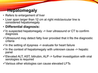
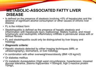
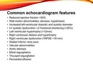
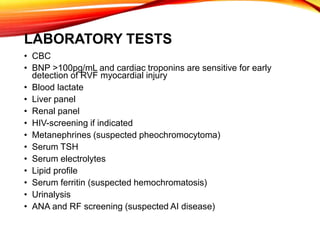
โ€
Differential diagnosis of
hepatomegaly in heart failure, viral
hepatitis and metabolic-associated
fatty liver disease
Hepatomegaly
โ€ข Refers to enlargement of liver
โ€ข Liver span larger than 12 cm at right midclavicular line is
considered hepatomegaly
โ€ข Differential diagnosis:
๏ƒผIn suspected hepatomegaly -> liver ultrasound or CT to confirm
diagnosis
๏ƒผUltrasound may detect fatty liver provided that it fits the diagnostic
criteria
๏ƒผIn the setting of dyspnea -> evaluate for heart failure
๏ƒผIn the context of hepatomegaly with unknown cause -> hepatic
panel
๏ƒผElevated ALT, AST, bilirubin, ALP -> further investigation with viral
serologies is required
๏ƒผVarious other etiologies can cause elevated LFTs
METABOLIC-ASSOCIATED FATTY LIVER
DISEASE
โ€ข Is defined on the presence of steatosis involving >5% of hepatocytes and the
absence of significant alcohol consumption or other causes of chronic liver
disease
โ€ข FL is the mildest form
โ€ข Steatohepatitis is defined as the presence of hepatic steatosis and
inflammation with hepatocyte injury (ballooning), Mallory hyaline, and mixed
lymphocytic and neutrophilic inflammatory infiltrate in perivenular areas with or
without fibrosis
โ€ข FL and steatohepatitis could only be distinguished by liver biopsy and
histology
โ€ข Diagnostic criteria:
๏ƒผ Hepatic steatosis detected by either imaging techniques (MRI, or
ultrasonography), biomarkers, or liver histology
๏ƒผ In combination with either overweightness/obesity (BMI >25 kg/m2)
๏ƒผ Or diabetes mellitus
๏ƒผ Or metabolic dysregulation (High waist circumference, hypertension, impaired
glucose tolerance, plasma triglycerides >150mg/dl, high c-reactive protein
>2mg/L
FL SH
Etiology
โ€ข Insulin resistance is the primary metabolic defect leading to NAFLD
โ€ข Insulin resistance increases influx of FFA into the liver
โ€ข Fat accumulates in the liver due to increased delivery of FFA
โ€ข Increased synthesis of fatty acids
โ€ข Decreased oxidation of FFA
โ€ข Or decreased synthesis of VLDL
โ€ข Other factors:
๏ƒผMedications (tamoxifen, amiodarone, methotrexate)
๏ƒผMetabolic abnormalities
๏ƒผAlcohol
๏ƒผWilsons disease or celiac sprue
History and evaluation
โ€ข Usually asymptomatic
โ€ข Nonspecific symptoms (fatigue, weightloss, etc), or right upper
quadrant discomfort.
โ€ข History must include alcohol use, family history, medications or
supplements, diet, physical activity, changes in weight
โ€ข Evaluation for hypertension, obesity, diabetes, hyperlipidemia
โ€ข LFTโ€™s: may be elevated ALT, AST, GGT, and occasionally, ALP
โ€ข GGT when elevated is a marker of increased mortality
โ€ข Serological tests to rule out viral hepatitis should be ordered
โ€ข Ultrasonography is the preferred method of diagnosis
โ€ข Ultrasonography features: increased echogenicity,
hepatomegaly, coarsened echotexture
VIRAL HEPATITIS
โ€ข Virally mediated liver inflammation
โ€ข There are numerous viruses may affect the liver
โ€ข Including Epstein-barr virus, herpes simplex,
cytomegalovirus
โ€ข More common hepatotropic viruses (A, B, C, D, E), with A, B,
and C being the most common
โ€ข Complications (cirrhosis, and hepatocellular carcinoma)
result in 1-4 million deaths / year / worldwide
Etiology
โ€ข Hepatitis A and E are transmitted via oral-fecal route
โ€ข Hepatitis B and C are primarily blood-borne
โ€ข Hepatitis B is a DNA virus, all others are RNA viruses
โ€ข HDV requires HBsAg to replicate
โ€ข History should include:
๏ƒผRecent travel to endemic areas
๏ƒผParenteral exposure (intravenous drug use, or blood transfusion
๏ƒผSexual contact with infected individuals, or are suffering from
jaundice
โ€ข Clinical picture: fever, anorexia, fatigue, vomiting, right upper
quadrant pain, jaundice, dark urine, pale stools
โ€ข Hepatomegaly, scleral icterus or jaundice, right upper quadrant
tenderness
EVALUATIONS
โ€ข Elevated aminotransferases and bilirubin
โ€ข Acute hepatitis presents with aminotransferases levels in
1000s
โ€ข ELISA serology and PCR are used for diagnosis
โ€ข Positive anti-HAV IgM indicates acute HAV infection
โ€ข HBV: acute infection is indicated by presence of HBsAg,
HBeAg, anti-HBc IgM, and elevated viral load
โ€ข Chronic HBV is denoted by presence of HBsAg >6 months,
anti-HBc IgG, and absence of anti-HBs antibodies
โ€ข HCV: indicated by HCV RNA, with or without anti-HCV IgM
โ€ข HDV: Viral load indicates current infection
โ€ข HEV: anti-HEV IgM, HEV antigens, and RNA viral load are
indicative of acute infection
HEART FAILURE
โ€ข Hepatomegaly secondary to right-sided heart failure due to hepatic
congestion
โ€ข Usually left-sided heart failure causes the right heart failure (i.e.
congestive heart failure)
โ€ข Right heart failure causes an increase in venous congestion and
increase of pressure in the hepatic sinusoids
โ€ข Heart failure is unlikely in the absence of dyspnea
โ€ข Common causes:
๏ƒผValvular disease (tricuspid regurgitation, mitral stenosis)
๏ƒผAdvanced left-sided heart failure
๏ƒผSevere pulmonary hypertension
๏ƒผCor pulmonale
๏ƒผCardiomyopathy
๏ƒผPericardial disease
๏ƒผCardiac tamponade
๏ƒผConstrictive pericarditis
Etiopathogenesis
โ€ข Heart failure can result from increased demand, systolic
dysfunction, or diastolic dysfunction.
โ€ข RVF is most often a result of LVF
โ€ข An increase in cardiac diastolic pressure or low cardiac
output and impaired perfusion
โ€ข Increase in preload or central venous pressure due to right
ventricular failure may cause direct liver damage which
generates an elevation in liver enzymes
โ€ข The elevated pressure in right heart chambers leads to
intrahepatic congestion, decreased perfusion and changes
in liver tissue like fibrosis, and atrophy of hepatocytes and
hemorrhage.
Clinical Presentation
โ€ข Hypoxemia and systemic venous congestion
โ€ข Dyspnea, orthopnea, peripheral edema
โ€ข Jugular venous distension
โ€ข Hepatojugular reflux
โ€ข Ascites
โ€ข Right-sided abdominal pain due to hepatic congestion
โ€ข Nausea, vomiting
โ€ข Constipation
โ€ข Jaundice in advanced disease
โ€ข Weight gain
EVALUATIONS
โ€ข Initial assessment: thorough history and physical examination
๏ƒผMedical history: anemia, CAD, valvular disease, diabetes mellitus,
hypertension, AI disease, hemochromatosis, hypercholesterolemia,
rheumatic fever, STD, hyperthyroidism, rheumatic fever,
๏ƒผSocial history: Travel, alcohol or drug abuse
๏ƒผFamily history: CAD, cardiomyopathies, arrhythmias
๏ƒผPhysical exam: bradycardia or tachycardia, bronze skin,
arrhythmia, pedal edema, diminished peripheral pulses, heart
murmurs, rales, weight loss or gain, jugular venous distension,
hepatomegaly, splenomegaly
โ€ข ECG
โ€ข Chest radiograph
โ€ข Echocardiogram
โ€ข Coronary angiography in CAD, and angina.
Common echocardiogram features
โ€ข Reduced ejection fraction <50%
โ€ข Wall motion abnormalities: akinesis, hypokinesis
โ€ข Increased left-ventricular diastolic and systolic diameter
โ€ข LV systolic dysfunction: LV fractional shortening (<25%)
โ€ข Left ventricular hypertrophy (>12mm)
โ€ข Right ventricular dilation and hypertrophy
โ€ข Right ventricular dysfunction (TAPSE <16 mm)
โ€ข Dilated inferior vena cava
โ€ข Valvular abnormalities
โ€ข Aortic stenosis
โ€ข Mitral regurgitation
โ€ข Tricuspid regurgitation
โ€ข Pericardial effusion
LABORATORY TESTS
โ€ข CBC
โ€ข BNP >100pg/mL and cardiac troponins are sensitive for early
detection of RVF myocardial injury
โ€ข Blood lactate
โ€ข Liver panel
โ€ข Renal panel
โ€ข HIV-screening if indicated
โ€ข Metanephrines (suspected pheochromocytoma)
โ€ข Serum TSH
โ€ข Serum electrolytes
โ€ข Lipid profile
โ€ข Serum ferritin (suspected hemochromatosis)
โ€ข Urinalysis
โ€ข ANA and RF screening (suspected AI disease)
Sources
โ€ข Ncbi.nlm.nih.gov
โ€ข Medscape
โ€ข Gastroenterologyadvisor.com
โ€ข Aafp.org
โ€ข Textbookofcardiology

Hepatomegaly differential diagnosis

  • 1.
    โ€ Differential diagnosis of hepatomegalyin heart failure, viral hepatitis and metabolic-associated fatty liver disease
  • 2.
    Hepatomegaly โ€ข Refers toenlargement of liver โ€ข Liver span larger than 12 cm at right midclavicular line is considered hepatomegaly โ€ข Differential diagnosis: ๏ƒผIn suspected hepatomegaly -> liver ultrasound or CT to confirm diagnosis ๏ƒผUltrasound may detect fatty liver provided that it fits the diagnostic criteria ๏ƒผIn the setting of dyspnea -> evaluate for heart failure ๏ƒผIn the context of hepatomegaly with unknown cause -> hepatic panel ๏ƒผElevated ALT, AST, bilirubin, ALP -> further investigation with viral serologies is required ๏ƒผVarious other etiologies can cause elevated LFTs
  • 3.
    METABOLIC-ASSOCIATED FATTY LIVER DISEASE โ€ขIs defined on the presence of steatosis involving >5% of hepatocytes and the absence of significant alcohol consumption or other causes of chronic liver disease โ€ข FL is the mildest form โ€ข Steatohepatitis is defined as the presence of hepatic steatosis and inflammation with hepatocyte injury (ballooning), Mallory hyaline, and mixed lymphocytic and neutrophilic inflammatory infiltrate in perivenular areas with or without fibrosis โ€ข FL and steatohepatitis could only be distinguished by liver biopsy and histology โ€ข Diagnostic criteria: ๏ƒผ Hepatic steatosis detected by either imaging techniques (MRI, or ultrasonography), biomarkers, or liver histology ๏ƒผ In combination with either overweightness/obesity (BMI >25 kg/m2) ๏ƒผ Or diabetes mellitus ๏ƒผ Or metabolic dysregulation (High waist circumference, hypertension, impaired glucose tolerance, plasma triglycerides >150mg/dl, high c-reactive protein >2mg/L
  • 4.
  • 5.
    Etiology โ€ข Insulin resistanceis the primary metabolic defect leading to NAFLD โ€ข Insulin resistance increases influx of FFA into the liver โ€ข Fat accumulates in the liver due to increased delivery of FFA โ€ข Increased synthesis of fatty acids โ€ข Decreased oxidation of FFA โ€ข Or decreased synthesis of VLDL โ€ข Other factors: ๏ƒผMedications (tamoxifen, amiodarone, methotrexate) ๏ƒผMetabolic abnormalities ๏ƒผAlcohol ๏ƒผWilsons disease or celiac sprue
  • 6.
    History and evaluation โ€ขUsually asymptomatic โ€ข Nonspecific symptoms (fatigue, weightloss, etc), or right upper quadrant discomfort. โ€ข History must include alcohol use, family history, medications or supplements, diet, physical activity, changes in weight โ€ข Evaluation for hypertension, obesity, diabetes, hyperlipidemia โ€ข LFTโ€™s: may be elevated ALT, AST, GGT, and occasionally, ALP โ€ข GGT when elevated is a marker of increased mortality โ€ข Serological tests to rule out viral hepatitis should be ordered โ€ข Ultrasonography is the preferred method of diagnosis โ€ข Ultrasonography features: increased echogenicity, hepatomegaly, coarsened echotexture
  • 7.
    VIRAL HEPATITIS โ€ข Virallymediated liver inflammation โ€ข There are numerous viruses may affect the liver โ€ข Including Epstein-barr virus, herpes simplex, cytomegalovirus โ€ข More common hepatotropic viruses (A, B, C, D, E), with A, B, and C being the most common โ€ข Complications (cirrhosis, and hepatocellular carcinoma) result in 1-4 million deaths / year / worldwide
  • 8.
    Etiology โ€ข Hepatitis Aand E are transmitted via oral-fecal route โ€ข Hepatitis B and C are primarily blood-borne โ€ข Hepatitis B is a DNA virus, all others are RNA viruses โ€ข HDV requires HBsAg to replicate โ€ข History should include: ๏ƒผRecent travel to endemic areas ๏ƒผParenteral exposure (intravenous drug use, or blood transfusion ๏ƒผSexual contact with infected individuals, or are suffering from jaundice โ€ข Clinical picture: fever, anorexia, fatigue, vomiting, right upper quadrant pain, jaundice, dark urine, pale stools โ€ข Hepatomegaly, scleral icterus or jaundice, right upper quadrant tenderness
  • 9.
    EVALUATIONS โ€ข Elevated aminotransferasesand bilirubin โ€ข Acute hepatitis presents with aminotransferases levels in 1000s โ€ข ELISA serology and PCR are used for diagnosis โ€ข Positive anti-HAV IgM indicates acute HAV infection โ€ข HBV: acute infection is indicated by presence of HBsAg, HBeAg, anti-HBc IgM, and elevated viral load โ€ข Chronic HBV is denoted by presence of HBsAg >6 months, anti-HBc IgG, and absence of anti-HBs antibodies โ€ข HCV: indicated by HCV RNA, with or without anti-HCV IgM โ€ข HDV: Viral load indicates current infection โ€ข HEV: anti-HEV IgM, HEV antigens, and RNA viral load are indicative of acute infection
  • 10.
    HEART FAILURE โ€ข Hepatomegalysecondary to right-sided heart failure due to hepatic congestion โ€ข Usually left-sided heart failure causes the right heart failure (i.e. congestive heart failure) โ€ข Right heart failure causes an increase in venous congestion and increase of pressure in the hepatic sinusoids โ€ข Heart failure is unlikely in the absence of dyspnea โ€ข Common causes: ๏ƒผValvular disease (tricuspid regurgitation, mitral stenosis) ๏ƒผAdvanced left-sided heart failure ๏ƒผSevere pulmonary hypertension ๏ƒผCor pulmonale ๏ƒผCardiomyopathy ๏ƒผPericardial disease ๏ƒผCardiac tamponade ๏ƒผConstrictive pericarditis
  • 11.
    Etiopathogenesis โ€ข Heart failurecan result from increased demand, systolic dysfunction, or diastolic dysfunction. โ€ข RVF is most often a result of LVF โ€ข An increase in cardiac diastolic pressure or low cardiac output and impaired perfusion โ€ข Increase in preload or central venous pressure due to right ventricular failure may cause direct liver damage which generates an elevation in liver enzymes โ€ข The elevated pressure in right heart chambers leads to intrahepatic congestion, decreased perfusion and changes in liver tissue like fibrosis, and atrophy of hepatocytes and hemorrhage.
  • 12.
    Clinical Presentation โ€ข Hypoxemiaand systemic venous congestion โ€ข Dyspnea, orthopnea, peripheral edema โ€ข Jugular venous distension โ€ข Hepatojugular reflux โ€ข Ascites โ€ข Right-sided abdominal pain due to hepatic congestion โ€ข Nausea, vomiting โ€ข Constipation โ€ข Jaundice in advanced disease โ€ข Weight gain
  • 13.
    EVALUATIONS โ€ข Initial assessment:thorough history and physical examination ๏ƒผMedical history: anemia, CAD, valvular disease, diabetes mellitus, hypertension, AI disease, hemochromatosis, hypercholesterolemia, rheumatic fever, STD, hyperthyroidism, rheumatic fever, ๏ƒผSocial history: Travel, alcohol or drug abuse ๏ƒผFamily history: CAD, cardiomyopathies, arrhythmias ๏ƒผPhysical exam: bradycardia or tachycardia, bronze skin, arrhythmia, pedal edema, diminished peripheral pulses, heart murmurs, rales, weight loss or gain, jugular venous distension, hepatomegaly, splenomegaly โ€ข ECG โ€ข Chest radiograph โ€ข Echocardiogram โ€ข Coronary angiography in CAD, and angina.
  • 14.
    Common echocardiogram features โ€ขReduced ejection fraction <50% โ€ข Wall motion abnormalities: akinesis, hypokinesis โ€ข Increased left-ventricular diastolic and systolic diameter โ€ข LV systolic dysfunction: LV fractional shortening (<25%) โ€ข Left ventricular hypertrophy (>12mm) โ€ข Right ventricular dilation and hypertrophy โ€ข Right ventricular dysfunction (TAPSE <16 mm) โ€ข Dilated inferior vena cava โ€ข Valvular abnormalities โ€ข Aortic stenosis โ€ข Mitral regurgitation โ€ข Tricuspid regurgitation โ€ข Pericardial effusion
  • 15.
    LABORATORY TESTS โ€ข CBC โ€ขBNP >100pg/mL and cardiac troponins are sensitive for early detection of RVF myocardial injury โ€ข Blood lactate โ€ข Liver panel โ€ข Renal panel โ€ข HIV-screening if indicated โ€ข Metanephrines (suspected pheochromocytoma) โ€ข Serum TSH โ€ข Serum electrolytes โ€ข Lipid profile โ€ข Serum ferritin (suspected hemochromatosis) โ€ข Urinalysis โ€ข ANA and RF screening (suspected AI disease)
  • 16.
    Sources โ€ข Ncbi.nlm.nih.gov โ€ข Medscape โ€ขGastroenterologyadvisor.com โ€ข Aafp.org โ€ข Textbookofcardiology