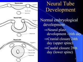
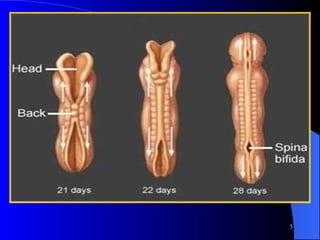
Hatem I. Badr   MD. ASS. Professor Neurosurgery Department of Neurosurgery Mansoura University, Egypt CNS congenital anomalies
Neural Tube Development Normal embryological development  Neural plate development -16th day Cranial closure 24th day (upper spine) Caudal closure 28th day (lower spine)
 
Spina Bifida Occulta Very mild & common form.  Very rarely causes disability.  Can only detected by x-ray or investigating a back injury.  May be associated with tethered cord
Usually associated with skin visible signs on the back.  Dimple  small hair growth  Nevus flaminous (red spot) or port wine Pad of subcutaneous fat
Dimple
Tuft of hair
Dimple with nauves port wine
Tethered cord  The spinal cord could be caught against the vertebrae  Normal cord ends at lower end of L 1 Motor weakness of lower limbs Sphincteric problems such as inefficient bladder control.
Autopsy of Infant with tethered cord
Meningocele Least common form  Sac contains meninges and cerebro-spinal fluid.  Cerebro-spinal fluid protects the brain and spinal cord.  The nerves are not badly damaged and able to function normally.  Limited disability is present .
Myelomeningocele Most serious and common  The cyst not only contains meninges and c.s.f but also the nerves and spinal cord.   The spinal cord is damaged or not properly developed resulting in motor and sensory deficit.  Majority have bowel and bladder problems.
Myelomeningocele
Intact Mylomeningocele Thin transparent membrane
Intact Mylomeningocele covered by thin membrane surrounded by hyper pigmentation
Financial Costs :   Average estimated lifetime cost of $532,000 for each infant born with spina bifida (CDC 1999) adds an estimated 19 million dollars every year to Missouri resident lifetime costs associated with spina bifida. Physical Costs :   Possible paralysis  (the leading cause of childhood paralysis),  bowel and bladder control problems, learning disabilities, hydrocephalus, surgical procedures, latex allergies, increased health problems with age Emotional Costs :   Miscarriage,  stillbirth, infant mortality (death before 1st birthday),  disability, feeling “different” The High Cost of NTDs
How do you get diagnosis?
preNatal detection of NTD Serum alpha-fetoprotein (AFP) Normal fetal glycoprotein (MW= 70,000) Present normally in amniotic fluid and mother serum  start 12 week increase steadily till 32 week High maternal serum AFP > 2 multiples of median for appropriate week of gestation is diagnostic 91%  sensitivity in spina bifida
Ultrasound Detect  90-95 % of cases of spina bifida  100% cases of anencephaly  In cases of elevated AFP diffrentiate NTD fron non-neurological causes of elevated AFP  e.g. omphalocele
Amniocentesis Indication: Pregnancies subsequent to NTD Elevated AFP with normal US Show elevated AFP between 12-15 week earlier than serum AFP Carries 6% risk of abortion and fetal loss
Is prophylaxis feasible?
Factors Associated With Increased Risk of NTDs. . . Family history of NTD A previous pregnancy affected with NTD Maternal insulin-dependent diabetes Maternal obesity Anti-epileptic drugs (Valporic Acid, Carbamazapine) Lower socioeconomic/educational level, dietry deficiency specially  folic acid
The only most significant risk factor associated with NTDs is folic acid deficiency
Folic Acid For Women As NTD occur before diagnosis of pregnancy. All women of childbearing age should receive 400 micrograms (0.4 mg) of folic acid daily.  Women who have had a previous child with NTD should receive 4000 micrograms (4 mg) of folic acid daily.
What is the proper management?
Nursing Care Like any other neonate with congenital anomalies efforts should be towards careful examination and investigations to rule out other anomalies. Nursed in Trendlenburg position aiming to reduce pressure and keep it away from cystic lesion. Much care not to disturb intact membrane (high incidence of infection and urgent surgery).
Cover lesion with Gauze ring soaked with normal saline or Ringer solution to prevent dryness Avoid antiseptics e.g betadine as it is Neurotoxic affecting functioning roots in placode Avoid mechanical trauma to placode  no need for ultra frequent dressing
General assessment Assess whether lesion is ruptured or unruptured Ruptured lesions start prophylactic antibiotic Urgent surgery Measure size and site of defect for proper planing for closure Evaluation by neonatologist  Other anomalies (average 2-2.5% additional anomalies) Condition oppose with surgery e.g lung immaturity Bladder  Start with on regular urinery catheterization Urological consultation Orthopedic consultation for sever kyphotic or scoliotic deformities and hip, knee and foot deformities
Neurological preop. Assessment Watch for spontaneous movement of lower limbs which associated with better outcome. Assess lowest level of neurological function Response to painful stimuli Differentiate between voluntary movement from reflex movement which is stereotyped and not persist after stimulus Evaluate other neurological associations Hydrocephalus Anterior fontanel Head circumference Chiari II Check for inspiratory stridor and apneic episodes
Intra-operative Cauda equina Dural edge placode
Post-operative
FOR THE FUTURE
Baby Samuel Armas's tiny fingers grasped the doctor's huge hand - - not at birth but, at 21 weeks during surgery for repair of MM Samuel Armas Photo 8/19/99; Born 12/2/99
Sara Switzer Photo: July 1 st  /99; Born August 22 nd  /99  BORN TWICE
Hydrocephalus
Thank You

Ee

  • 1.
    Hatem I. Badr MD. ASS. Professor Neurosurgery Department of Neurosurgery Mansoura University, Egypt CNS congenital anomalies
  • 2.
    Neural Tube DevelopmentNormal embryological development Neural plate development -16th day Cranial closure 24th day (upper spine) Caudal closure 28th day (lower spine)
  • 3.
  • 4.
    Spina Bifida OccultaVery mild & common form. Very rarely causes disability. Can only detected by x-ray or investigating a back injury. May be associated with tethered cord
  • 5.
    Usually associated withskin visible signs on the back. Dimple small hair growth Nevus flaminous (red spot) or port wine Pad of subcutaneous fat
  • 6.
  • 7.
  • 8.
  • 9.
    Tethered cord The spinal cord could be caught against the vertebrae Normal cord ends at lower end of L 1 Motor weakness of lower limbs Sphincteric problems such as inefficient bladder control.
  • 10.
    Autopsy of Infantwith tethered cord
  • 11.
    Meningocele Least commonform Sac contains meninges and cerebro-spinal fluid. Cerebro-spinal fluid protects the brain and spinal cord. The nerves are not badly damaged and able to function normally. Limited disability is present .
  • 12.
    Myelomeningocele Most seriousand common The cyst not only contains meninges and c.s.f but also the nerves and spinal cord. The spinal cord is damaged or not properly developed resulting in motor and sensory deficit. Majority have bowel and bladder problems.
  • 13.
  • 14.
    Intact Mylomeningocele Thintransparent membrane
  • 15.
    Intact Mylomeningocele coveredby thin membrane surrounded by hyper pigmentation
  • 16.
    Financial Costs : Average estimated lifetime cost of $532,000 for each infant born with spina bifida (CDC 1999) adds an estimated 19 million dollars every year to Missouri resident lifetime costs associated with spina bifida. Physical Costs : Possible paralysis (the leading cause of childhood paralysis), bowel and bladder control problems, learning disabilities, hydrocephalus, surgical procedures, latex allergies, increased health problems with age Emotional Costs : Miscarriage, stillbirth, infant mortality (death before 1st birthday), disability, feeling “different” The High Cost of NTDs
  • 17.
    How do youget diagnosis?
  • 18.
    preNatal detection ofNTD Serum alpha-fetoprotein (AFP) Normal fetal glycoprotein (MW= 70,000) Present normally in amniotic fluid and mother serum start 12 week increase steadily till 32 week High maternal serum AFP > 2 multiples of median for appropriate week of gestation is diagnostic 91% sensitivity in spina bifida
  • 19.
    Ultrasound Detect 90-95 % of cases of spina bifida 100% cases of anencephaly In cases of elevated AFP diffrentiate NTD fron non-neurological causes of elevated AFP e.g. omphalocele
  • 20.
    Amniocentesis Indication: Pregnanciessubsequent to NTD Elevated AFP with normal US Show elevated AFP between 12-15 week earlier than serum AFP Carries 6% risk of abortion and fetal loss
  • 21.
  • 22.
    Factors Associated WithIncreased Risk of NTDs. . . Family history of NTD A previous pregnancy affected with NTD Maternal insulin-dependent diabetes Maternal obesity Anti-epileptic drugs (Valporic Acid, Carbamazapine) Lower socioeconomic/educational level, dietry deficiency specially folic acid
  • 23.
    The only mostsignificant risk factor associated with NTDs is folic acid deficiency
  • 24.
    Folic Acid ForWomen As NTD occur before diagnosis of pregnancy. All women of childbearing age should receive 400 micrograms (0.4 mg) of folic acid daily. Women who have had a previous child with NTD should receive 4000 micrograms (4 mg) of folic acid daily.
  • 25.
    What is theproper management?
  • 26.
    Nursing Care Likeany other neonate with congenital anomalies efforts should be towards careful examination and investigations to rule out other anomalies. Nursed in Trendlenburg position aiming to reduce pressure and keep it away from cystic lesion. Much care not to disturb intact membrane (high incidence of infection and urgent surgery).
  • 27.
    Cover lesion withGauze ring soaked with normal saline or Ringer solution to prevent dryness Avoid antiseptics e.g betadine as it is Neurotoxic affecting functioning roots in placode Avoid mechanical trauma to placode no need for ultra frequent dressing
  • 28.
    General assessment Assesswhether lesion is ruptured or unruptured Ruptured lesions start prophylactic antibiotic Urgent surgery Measure size and site of defect for proper planing for closure Evaluation by neonatologist Other anomalies (average 2-2.5% additional anomalies) Condition oppose with surgery e.g lung immaturity Bladder Start with on regular urinery catheterization Urological consultation Orthopedic consultation for sever kyphotic or scoliotic deformities and hip, knee and foot deformities
  • 29.
    Neurological preop. AssessmentWatch for spontaneous movement of lower limbs which associated with better outcome. Assess lowest level of neurological function Response to painful stimuli Differentiate between voluntary movement from reflex movement which is stereotyped and not persist after stimulus Evaluate other neurological associations Hydrocephalus Anterior fontanel Head circumference Chiari II Check for inspiratory stridor and apneic episodes
  • 30.
    Intra-operative Cauda equinaDural edge placode
  • 31.
  • 32.
  • 33.
    Baby Samuel Armas'stiny fingers grasped the doctor's huge hand - - not at birth but, at 21 weeks during surgery for repair of MM Samuel Armas Photo 8/19/99; Born 12/2/99
  • 34.
    Sara Switzer Photo:July 1 st /99; Born August 22 nd /99 BORN TWICE
  • 35.
  • 36.