3. 1000 patients supported on ECMO at the University
of Michigan were reviewed (retrospectively)
VV-ECMO for respiratory failure provided survival
to discharge:
88% of 586 cases of respiratory failure in
neonates
70% for 132 cases of respiratory failure in
children
56% for 146 cases of respiratory failure in adults
4. INIZIALE ESPERIENZA ECMO in UTIC Careggi
07/11/2011 dr Valente
CASISTICA ECMO: 11
PAZIENTI
-Rottura di cuore in corso di
STEMI anteriore in 1 pz
-STEMI complicato da shock
cardiogeno: 3 pz
-Occlusione TC in corso di VCG: 1
pz
-NSTE-SCA (occlusione del TC): 1
pz
-Ipertrofica: in 1 pz
- Dilatativa: 2 pz
- Embolia polmonare: 1 pz
- Sindrome di Tako-Tsubo: 1 pz
CASISTICA ECMO: 11 PAZIENTI
- Shock cardiogeno: 5 pz
- ACR: 4 pz
- Scompenso cardiaco END-STAGE: 2
pz
Età media 54 anni
Rapporto M/F 8:3
IABP, CRRT e ventilazione
meccanica in tutti i pazienti
5. INIZIALE ESPERIENZA ECMO in UTIC Careggi
07/11/2011 dr Valente
Sede impianto ECMO
Latenza media inizio ACLS-inizio ECMO (in ACR): 57 minuti (min. 22, max 110 min)
Durata media del supporto con ECMO: 198 ore (min. 24, max. 504 ore)
Decorso: 6 pazienti deceduti durante supporto ECMO.
5 pazienti svezzati da ECMO.
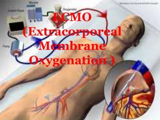
6. ECMO is instituted for the management of
life threatening pulmonary or cardiac failure
(or both), when no other form of treatment
has been or is likely to be successful.
ECMO is used as temporary support, usually
awaiting recovery of organs.
ECMO is essentially a modification of the
cardiopulmonary bypass circuit which is used
routinely in cardiac surgery.
7. MECCANISMO ECMO
Blood is removed from the venous system
either peripherally via cannulation of a
femoral vein or centrally via cannulation
of the right atrium,
Oxygenate
Extract carbon dioxide
Blood is then returned back to the body
either peripherally via a femoral artery or
centrally via the ascending aorta.
9. CONFIGURAZIONE VA
VENO-ARTERIOSA
Blood being drained
from the venous
system and returned
to the arterial system.
Provides both cardiac
and respiratory
support.
Achieved by either
peripheral or central
cannulation
10.
11. CONFIGURAZIONE VV
VENO-VENOSA
Provides oxygenation
Blood being drained from venous system and
returned to venous system.
Only provides respiratory support
Achieved by peripheral cannulation, usually
of both femoral veins.
12.
13. Central vs. Peripheral
VANTAGGI
Cannulation
Flow from Central ECMO is directly from
the outflow cannula into the aorta provides
antegrade flow to the arch vessels,
coronaries and the rest of the body
In contrast, the retrograde aortic flow
provided by peripheral leads to mixing in
the arch.
14. Central vs. Peripheral Cannulation
SVANTAGGI
Previously insertion of central ECMO required
leaving chest open to allow the cannulae to exit.
Increased the risk of bleeding and infection
Newer cannulae are designed to be tunneled
through the subcostal abdominal wall allowing
the chest to be completely closed.
Central cannula are costly (approximately 4
times as much as peripheral)
15. CONSIDERAZIONI
Mechanical ventilation must be continued during
ECMO support to try to maintain oxygen saturation
of blood ejected from the left ventricle to at least
above 90%.
ECMO flow can be very volume dependent
ECMO flow will drop:
Hypovolemia
Cannula malposition
Pneumothorax
Pericardial tamponade.
17. INSUFFICIENZA
CARDIACA
• Cardiomiopatia end-stage in terapia medica massimale, in
attesa di trapianto cardiaco;
• Infarto miocardico acuto complicato da shock cardiogeno
refrattario
• Miocardite acuta con severa insufficienza d’organo o aritmie
ventricolari subentranti non controllabili con terapia medica e
IABP;
• Embolia polmonare massiva con grave compromissione della
funzionalità ventricolare destra e shock cardiogeno o ACR
• Arresto cardiaco nel paziente giovane adulto con precoce
rianimazione cardiopolmonare (con verosimile ottima
prognosi neurologica) refrattario a terapia rianimatoria
medica ed elettrica. La durata del supporto con ECMO dovrà
essere il più breve possibile (sconsigliato per più di 3 giorni).
[Da associare ad altre strategie di neuroprotezione come
l’ipotermia terapeutica]
• Grave depressione della funzione cardiaca da intossicazione di
farmaci o sepsi;
18. Indicazioni ECMO
in UTIC
Si deve tener conto:
• della prognosi, in particolare ripresa della
funzionalità dell’organo [bridge-to-recovery],
• dell’eleggibilità per un trapianto
cardiaco [bridge-to-transplantation],
• della possibilità di posizionamento di
assistenze meccaniche più o meno a
lunga durata (come Levitronix
CentriMag, Jarvik 2000, Cardiowest)
[bridge-to-bridge].
• Ma anche ……….. bridge to decision.
19. INSUFFICIENZA
RESPIRATORIA
• Adult respiratory distress syndrome
(ARDS)
• POLMONITE
• TRAUMA
• Primary graft failure following lung
transplantation.
ECMO is also used for neonatal and
pediatric respiratory support
This is where most of the
research on ECMO has
been done
21. Possibilità di recupero d'organo:. Appropriato solo se il
processo della malattia è reversibile con la terapia e riposo su
Recupero cardiaco: permette un ulteriore recupero cardiaco per
consentire l'impianto del dispositivo (LVAD) o alla lista per il trapianto
CONTROINDICAZIONI
neoplasia disseminata
L'età avanzata
Graft vs host disease
Gravi lesioni cerebrali
DECISIONE DI
ATTUARE
ECMO
ECMO
Arresto cardiaco non testimoniato o di durata prolungata.
Controindicazioni tecniche da considerare: la dissezione
aortica o dell'aorta incompetenza
24. SVEZZAMENTO DA ECMO
QUANDO?
Lo scambio di gas può essere
mantenuto a valori adeguati con una
bassa FiO2 (<30%)
RR e PEEP impostato sul ventilatore
non sono troppo alti (ad esempio <25
atti / min e <15cmH2O,
rispettivamente).
26. COAGULOPATIA
• Continuous activation of
contact and fibrinolytic
systems by the circuit
TROMBOCITOPENIA
• Consumption and dilution of
factors within minutes of
initiation of ECMO
• Platelets adhere
to surface
fibrinogen and
are activated
27. Kidneys
Non-pulsatile
perfusion
to end organs
Splanchnic circulation seems to be particularly
susceptible
GI bleeding, ulceration and perforation
Liver impairment
28. COMPLICANZE
ECMO
Air embolism/
Thromboembolism
Leg
ischemia
• Particularly at peripheral
insertion site of VA
Intracerebral
bleeds
• Largely associated with
sepsis
• Manifest as seizures or
brain death
Mechanical
Complications
• Tubing rupture
• Pump malfunction
• Cannula related problems
29. REGOLARI CONTROLLI DI TESTS
EMATOCHIMICI (ogni 6-8h)
PREVENZIONE
DELLE
COMPLICANZE
• Coagulation Profile
• Platelet Count
• Hemoglobin
• Creatinine to evaluate
for renal insufficiency
AGGRESSIVO REINTEGRO DEI
FATTORI DI COAGULAZIONE
ELETTROLITI, GRC
30. ECMO
• Multidisciplinary team evaluation
(Cardiologist, Anesthesiologist, Cardiac
Surgeon)
• Multi-system work-up
Heart disease (Echo, Cath, etc.)
Respiratory function
Renal function
Liver function
Coagulation cascade and platelet
function
31. CONCLUSIONE
Mechanical circulatory support represents
a concrete and reliable option in different
scenarios of severe acute and chronic
heart failure
Encouraging results related to
• new devices and proper management
• increasing center-related experience
• adequate decision making
• implantation before onset of overt
cardiogenic shock
In severely ill pts, rapid implantation of simple
support (i.e. ECMO) as bridge to decision.
32. ECMO: PROPOSTA DI RETE
HUB di riferimento UTIC SPOKE
Necessità di:
Numero telefonico dedicato
Equipe impianto ECMO H 24
Disponibilità posto letto dedicato
Necessità di:
valutazione indicazioni
stabilizzazione e preparazione del
paziente in accordo con centro HUB
Mezzo di trasporto dedicato H 24
Priorità:
Equipe ECMO in centro SPOKE
Valutazione se impianto ECMO in
centro SPOKE e in quale sede
(UTIC/emodinamica/sala operatoria)
Stabilizzazione e trasporto protetto
(con o senza ECMO) del paziente
Priorità:
Contattare HUB di riferimento
Concordare modalità di
stabilizzazione con protocolli
condivisi
Stabilire sede di impianto
programmare e concordare
trasporto
33. ATTUALMENTE ,IN CASI
PARTICOLARI, SI PUO’ PORRE LA
NECESSITA’ DI TRASFERIRE
PAZIENTI IN ECMO