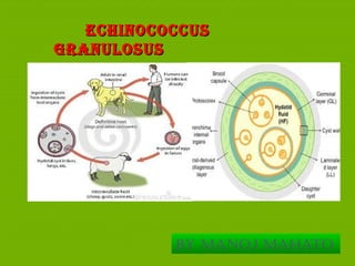
ECHINOCOCCUSECHINOCOCCUS
GRANULOSUSGRANULOSUS
By Manoj Mahato
EPIDEMIOLOGY
 Echinococcus granulosusEchinococcus granulosus is most common in temperateis most common in temperate
sheep-raising areas: South America, East Africa andsheep-raising areas: South America, East Africa and
central Russia, East Africacentral Russia, East Africa
 The highest prevalence is seen in East Africa, KenyaThe highest prevalence is seen in East Africa, Kenya
 The source of most human infections is sheep dog fecesThe source of most human infections is sheep dog feces
containingcontaining E granulosusE granulosus eggseggs
 E. granulosusE. granulosus is prevalent in areas where livestock isis prevalent in areas where livestock is
raised in association with dogs.raised in association with dogs.
INTRODUCTOIINTRODUCTOI
NN
 Echinococcus granulosus is a cestode, and causesEchinococcus granulosus is a cestode, and causes
hydadid cysthydadid cyst
 Hidadid cystHidadid cyst is an accidental human infection, humanis an accidental human infection, human
serving as intermediate host in a life-cycleserving as intermediate host in a life-cycle
 Echinococcosis of humans is caused by the larval stage ofEchinococcosis of humans is caused by the larval stage of
EchinococcusEchinococcus granulosusgranulosus
MORPHOLOGY OFMORPHOLOGY OF
ADULTADULT
 Size: 3-9 mm , smallest of the with proglottids.Size: 3-9 mm , smallest of the with proglottids.
 It has scolex, neck and strobilla.It has scolex, neck and strobilla.
 Scolex is globular in shape and has a prominent rostelluScolex is globular in shape and has a prominent rostellumm
 Aadult worm lives in the jejunum and duodenum ofAadult worm lives in the jejunum and duodenum of
definitive hostdefinitive host
 Buried in the mucosa, between the villi of smallBuried in the mucosa, between the villi of small
intestine with the help of its scolexintestine with the help of its scolex
MORPHOLOGY OF EGGSMORPHOLOGY OF EGGS
 Size: 32-36/25-30 micrometersSize: 32-36/25-30 micrometers
 Shape: OvalarShape: Ovalar
 EmbrionatedEmbrionated
 Six-hooked embriophoreSix-hooked embriophore
MORPHOLOGY OFMORPHOLOGY OF LARVAELARVAE
Size: varySize: vary
Shape: sphericalShape: spherical
New larvae, calledNew larvae, called scolicesscolices, develop in large numbers within, develop in large numbers within
the broodcapsule.the broodcapsule.
Fluid filled hollow bladders containing numerous protoscolicesFluid filled hollow bladders containing numerous protoscolices..
The liver and The lungs aretheMost commonsites for larvaeThe liver and The lungs aretheMost commonsites for larvae
development.development.
LifeLife
CyCLeCyCLe
Mode of Transmission:Mode of Transmission: fecal-oralfecal-oral
Echinococcus granulosusEchinococcus granulosus has 2 hosts:has 2 hosts:
IntermediateIntermediate
Definitive hostsDefinitive hosts
A .A .Definitive hostsDefinitive hosts B.B. Intermediate HostsIntermediate Hosts
DogsDogs SheepSheep
Life CyCLe
LifeLife
CyCLeCyCLe
 The adult worm lives in the smll intestine of the definitive host dogThe adult worm lives in the smll intestine of the definitive host dog..
 Gravid proglottidsGravid proglottids E. granulosusE. granulosus release eggs that are passed in therelease eggs that are passed in the
fecesfeces
 When eggs of EchinococcusWhen eggs of Echinococcus granulosusgranulosus swallowed by man,swallowed by man,
produce embryos that penetrate the small intestine, enter theproduce embryos that penetrate the small intestine, enter the
circulation and form cysts in liver, lung, bones, and sometimes,circulation and form cysts in liver, lung, bones, and sometimes,
brain.brain.
 In the organs they develop into cysts and enlarge graduallyIn the organs they develop into cysts and enlarge gradually
 When Definitive host eats the infected organs and becomes infectedWhen Definitive host eats the infected organs and becomes infected
 After ingestion, the protoscolices evaginate, attach to the intestinalAfter ingestion, the protoscolices evaginate, attach to the intestinal
mucosa and develop into adult stagesmucosa and develop into adult stages
SIGNSANDSIGNSAND
SYMPTOMSSYMPTOMS
Lab.DiagnosiLab.Diagnosi
ss
A specific diagnosis can be made by: the examination ofA specific diagnosis can be made by: the examination of
aspirated fluids for scoliceal hooklets,aspirated fluids for scoliceal hooklets,
ImmunodiagnoImmunodiagno
sissis
(a) Intradermal test (Casoni test): 0.2 ml hydatid fluid (sterilized by(a) Intradermal test (Casoni test): 0.2 ml hydatid fluid (sterilized by
filtration) injected intradermally produces a wheal in about 15 mnfiltration) injected intradermally produces a wheal in about 15 mn
(b) Serological methods:(b) Serological methods:
Precipitin:reaction: equal parts of hydatid fluid and patient's serumPrecipitin:reaction: equal parts of hydatid fluid and patient's serum
incubated at 37°C for 1 hour show flocculation in 36 hours.incubated at 37°C for 1 hour show flocculation in 36 hours.
IHA (Indirect haemagglutination test).IHA (Indirect haemagglutination test).
LA (Latex agglutination test).LA (Latex agglutination test).
IEPIEP
(Immunoelectrophoresis test(Immunoelectrophoresis test
RadioRadio
DiagnosisDiagnosis
--X-ray will define pulmonary cysts:X-ray will define pulmonary cysts:
usually as rounded, uniform densityusually as rounded, uniform density
-CT, is indicative-CT, is indicative ofof E.G.E.G.invasion and helps toinvasion and helps to
distinguish fromcarcinomas, bacterial ordistinguish fromcarcinomas, bacterial or
amebic liveramebic liver
TREATMENTTREATMENT
 Therapy for echinococcosis is based on considerations of the size,Therapy for echinococcosis is based on considerations of the size,
location, and manifestations of cysts and the overall health of thelocation, and manifestations of cysts and the overall health of the
patient.patient.
 Albendazole, given at a dose of:10-15 mg/kg/day for 30 days, withAlbendazole, given at a dose of:10-15 mg/kg/day for 30 days, with
15 days intervals or 400 mg twice a day for 12 weeks,15 days intervals or 400 mg twice a day for 12 weeks,
 Mebendazole can be taken over a long period of time at low dosagesMebendazole can be taken over a long period of time at low dosages
Prevention andPrevention and
ControlControl
 The best method is to disrupt the lifecycleThe best method is to disrupt the lifecycle
 Basic hygiene practicesBasic hygiene practices
 Avoid feeding off raw meat of internal organs of infectedAvoid feeding off raw meat of internal organs of infected
animal to dogsanimal to dogs
 administering praziquantel to infected dogs every 3 monthsadministering praziquantel to infected dogs every 3 months
 Reduce the number of stray dogs THANK YOUReduce the number of stray dogs THANK YOU
1616
THANK YOUTHANK YOU

Echinococcusgranulosusandmultilocularis

  • 1.
  • 2.
    EPIDEMIOLOGY  Echinococcus granulosusEchinococcusgranulosus is most common in temperateis most common in temperate sheep-raising areas: South America, East Africa andsheep-raising areas: South America, East Africa and central Russia, East Africacentral Russia, East Africa  The highest prevalence is seen in East Africa, KenyaThe highest prevalence is seen in East Africa, Kenya  The source of most human infections is sheep dog fecesThe source of most human infections is sheep dog feces containingcontaining E granulosusE granulosus eggseggs  E. granulosusE. granulosus is prevalent in areas where livestock isis prevalent in areas where livestock is raised in association with dogs.raised in association with dogs.
  • 3.
    INTRODUCTOIINTRODUCTOI NN  Echinococcus granulosusis a cestode, and causesEchinococcus granulosus is a cestode, and causes hydadid cysthydadid cyst  Hidadid cystHidadid cyst is an accidental human infection, humanis an accidental human infection, human serving as intermediate host in a life-cycleserving as intermediate host in a life-cycle  Echinococcosis of humans is caused by the larval stage ofEchinococcosis of humans is caused by the larval stage of EchinococcusEchinococcus granulosusgranulosus
  • 4.
    MORPHOLOGY OFMORPHOLOGY OF ADULTADULT Size: 3-9 mm , smallest of the with proglottids.Size: 3-9 mm , smallest of the with proglottids.  It has scolex, neck and strobilla.It has scolex, neck and strobilla.  Scolex is globular in shape and has a prominent rostelluScolex is globular in shape and has a prominent rostellumm  Aadult worm lives in the jejunum and duodenum ofAadult worm lives in the jejunum and duodenum of definitive hostdefinitive host  Buried in the mucosa, between the villi of smallBuried in the mucosa, between the villi of small intestine with the help of its scolexintestine with the help of its scolex
  • 5.
    MORPHOLOGY OF EGGSMORPHOLOGYOF EGGS  Size: 32-36/25-30 micrometersSize: 32-36/25-30 micrometers  Shape: OvalarShape: Ovalar  EmbrionatedEmbrionated  Six-hooked embriophoreSix-hooked embriophore
  • 6.
    MORPHOLOGY OFMORPHOLOGY OFLARVAELARVAE Size: varySize: vary Shape: sphericalShape: spherical New larvae, calledNew larvae, called scolicesscolices, develop in large numbers within, develop in large numbers within the broodcapsule.the broodcapsule. Fluid filled hollow bladders containing numerous protoscolicesFluid filled hollow bladders containing numerous protoscolices.. The liver and The lungs aretheMost commonsites for larvaeThe liver and The lungs aretheMost commonsites for larvae development.development.
  • 7.
    LifeLife CyCLeCyCLe Mode of Transmission:Modeof Transmission: fecal-oralfecal-oral Echinococcus granulosusEchinococcus granulosus has 2 hosts:has 2 hosts: IntermediateIntermediate Definitive hostsDefinitive hosts A .A .Definitive hostsDefinitive hosts B.B. Intermediate HostsIntermediate Hosts DogsDogs SheepSheep
  • 8.
  • 9.
    LifeLife CyCLeCyCLe  The adultworm lives in the smll intestine of the definitive host dogThe adult worm lives in the smll intestine of the definitive host dog..  Gravid proglottidsGravid proglottids E. granulosusE. granulosus release eggs that are passed in therelease eggs that are passed in the fecesfeces  When eggs of EchinococcusWhen eggs of Echinococcus granulosusgranulosus swallowed by man,swallowed by man, produce embryos that penetrate the small intestine, enter theproduce embryos that penetrate the small intestine, enter the circulation and form cysts in liver, lung, bones, and sometimes,circulation and form cysts in liver, lung, bones, and sometimes, brain.brain.  In the organs they develop into cysts and enlarge graduallyIn the organs they develop into cysts and enlarge gradually  When Definitive host eats the infected organs and becomes infectedWhen Definitive host eats the infected organs and becomes infected  After ingestion, the protoscolices evaginate, attach to the intestinalAfter ingestion, the protoscolices evaginate, attach to the intestinal mucosa and develop into adult stagesmucosa and develop into adult stages
  • 10.
  • 11.
    Lab.DiagnosiLab.Diagnosi ss A specific diagnosiscan be made by: the examination ofA specific diagnosis can be made by: the examination of aspirated fluids for scoliceal hooklets,aspirated fluids for scoliceal hooklets,
  • 12.
    ImmunodiagnoImmunodiagno sissis (a) Intradermal test(Casoni test): 0.2 ml hydatid fluid (sterilized by(a) Intradermal test (Casoni test): 0.2 ml hydatid fluid (sterilized by filtration) injected intradermally produces a wheal in about 15 mnfiltration) injected intradermally produces a wheal in about 15 mn (b) Serological methods:(b) Serological methods: Precipitin:reaction: equal parts of hydatid fluid and patient's serumPrecipitin:reaction: equal parts of hydatid fluid and patient's serum incubated at 37°C for 1 hour show flocculation in 36 hours.incubated at 37°C for 1 hour show flocculation in 36 hours. IHA (Indirect haemagglutination test).IHA (Indirect haemagglutination test). LA (Latex agglutination test).LA (Latex agglutination test). IEPIEP (Immunoelectrophoresis test(Immunoelectrophoresis test
  • 13.
    RadioRadio DiagnosisDiagnosis --X-ray will definepulmonary cysts:X-ray will define pulmonary cysts: usually as rounded, uniform densityusually as rounded, uniform density -CT, is indicative-CT, is indicative ofof E.G.E.G.invasion and helps toinvasion and helps to distinguish fromcarcinomas, bacterial ordistinguish fromcarcinomas, bacterial or amebic liveramebic liver
  • 14.
    TREATMENTTREATMENT  Therapy forechinococcosis is based on considerations of the size,Therapy for echinococcosis is based on considerations of the size, location, and manifestations of cysts and the overall health of thelocation, and manifestations of cysts and the overall health of the patient.patient.  Albendazole, given at a dose of:10-15 mg/kg/day for 30 days, withAlbendazole, given at a dose of:10-15 mg/kg/day for 30 days, with 15 days intervals or 400 mg twice a day for 12 weeks,15 days intervals or 400 mg twice a day for 12 weeks,  Mebendazole can be taken over a long period of time at low dosagesMebendazole can be taken over a long period of time at low dosages
  • 15.
    Prevention andPrevention and ControlControl The best method is to disrupt the lifecycleThe best method is to disrupt the lifecycle  Basic hygiene practicesBasic hygiene practices  Avoid feeding off raw meat of internal organs of infectedAvoid feeding off raw meat of internal organs of infected animal to dogsanimal to dogs  administering praziquantel to infected dogs every 3 monthsadministering praziquantel to infected dogs every 3 months  Reduce the number of stray dogs THANK YOUReduce the number of stray dogs THANK YOU
  • 16.