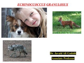
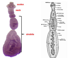
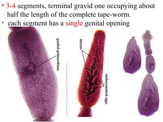
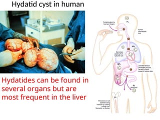
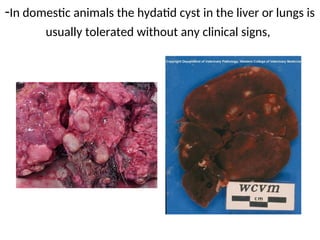
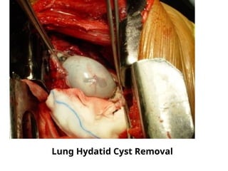
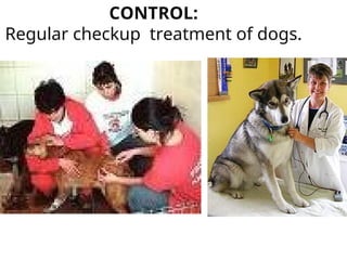
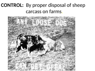
ECHINOCCOCCUS GRANULOSUS
Kingdom Animalia
Phylum Platyhelminthes
Class Cestoda
Order Cyclophyllidea
Family Taeniidae
Genus Echinococcus
Species E. granulosus
Echinococcus granulosus
Echinoccoccus:
• Smallest cestodes of domestic animals.
• Two species, E. granulosus and E. multilocularis are
important in vet: medicine.
• Larval stage, hydatid (G. hydatis means drop of
water) develops in a wide range of intermediate
hosts including man.
scolex
neck
strobila
Echinoccoccus granulosus:
Two major strains of domestic animals are:
E. granulosus granulosus E. granulosus equinus.
Def:Host: Dog, wild canids. Dog, red fox.
Int: Host: Domesticated ruminants, Horse,Donkey.
Man, Wild ruminants, Pig.
E.granulosus granulosus E.granulosus equinus.
Distribution: World wide. Europe.
Echinococcus granulosus: ( Dog tapeworm, Hydatid worm )
Disease: Hydatidosis, Hydatid
disease, Echinococosis ( is a
Zoonosis of great medical,
veterinary and economic
importance).
Habitat: Adults in the small intestine
and hydatid cysts
mainly in liver and lungs.
MORPHOLOGY
• 3.0 - 6.0 mm long and is difficult
to find in freshly opened intestine.
• Scolex is typical taeniid
scolex
neck
strobila
• 3-4 segments, terminal gravid one occupying about
half the length of the complete tape-worm.
• each segment has a single genital opening
Echinococcus granulosus: cystic echinococcosis.
Produces cystic lesions
• Adult worms live in the intestine of dogs.
• produce eggs which are shed with the feces.
•Eggs are infective to herbivores (and humans)
• cyst is lined by a multilayer
parasite tissue with the
innermost layer being the
germinal layer
This layer is a undifferentiated
“stem cell” layer that can
spawn the formation of “brood
capsules” which are themselves
lined by GL
•The daughter cysts "bud" into
the center of the fluid-filled
cyst.
This is a very small portion of
the cyst which may become
quite large.
•Each of the smaller bodies will
develop into tapeworms when
eaten by a definitive or final
Hydatid cyst
Hydatid cyst
(A) Spheric bud.
(B) Early elongated bud showing an anterior and a posterior region.
(C) Late elongated bud with presumptive scolex and body.
(D) Protoscolex in development showing rostellum formation.
(E) Protoscolex in development showing suckers formation
(F) Fully developed protoscolex attached to the germinal layer.
(G) Protoscolex free in the cyst cavity.
laminated layer
germinal layer
tegument
Development of E. granulosus protoscolex in natural infection.
•Thousands of protoscolices
can fill the hydatid
(hydatide sand)
• Protoscolices are the
infective stage for dogs
Hydatides usually grow
slowly but steadily (1-5 cm
per year)
•usually well tolerated until
their size becomes a problem
or they rupture
•Cyst rupture or leakage can
result in allergic reactions
Hydatid Cyst
A wonderful
livestock
guardian dog,
and "her" kids.
Fly on red meat
Life-cycle
• Growth of hydatid cyst is slow, maturity being reached in
6-12 months.
• In the lungs and liver the cyst may have a diameter of up
to 20 cm.
Hydatid cyst in human
Hydatides can be found in
several organs but are
most frequent in the liver
Occurrence and percentage of the cyst
Liver Lungs Other organs.
------ ------ -------------
Sheep 25% 70% 5%
Horse, Cattle 90%
• Fertile cysts when ingested by the dog are capable
of growing into the adult tape-worm in about 6-7
weeks.
• Life span of adult worm in the Canine host is short
(about 6 months)
• Life span of larval worm is considerable and may
continue to develop for several years.
Economic losses
• US$ 212.35 million in India
• US$ 232.3 million in Iran
• US$ 7.708 in turkey
Pathology
• Adult tape worm
non pathogenic,
•Thousands present in
a dog without clinical
sings
-In domestic animals the hydatid cyst in the liver or lungs is
usually tolerated without any clinical signs,
Hydatid cysts in bovine liver
Hydatid cysts in bovine heart. Note the detached germinal
layer
Human, liver. Multiple thin-walled hydatid cysts project from the capsular
surface of the liver.
Left kidney
showing open
hydatid cyst
(black arrows);
fragments of
germinal
membranes
with daughter
cysts are also
visible (white
arrow).
Lung Hydatid Cyst Removal
In contrast when man is involved as intermediate host, the hydatid
in its pulmonary and hepatic site is often of pathogenic significance
Cyst in brain
Even if it does not occlude a vital organ , it can
still cause sudden death if it ruptures
DIAGNOSIS:
• Identification of the gravid segment.
• Dog must be purged orally with arecoline
hydrobromid (12mg/kg) to expel the adult tape-
worm.
• No specific diagnosis in domestic animals
• In man by serological tests.
Treatment:
• No specific drug.
• Treatment of
infected dogs with
praziquantel.
• Surgical removal of
cysts
• Albendazole
therapy
CONTROL:
Regular checkup treatment of dogs.
CONTROL: By proper disposal of sheep
carcass on farms.
By denying dogs access to abattoirs.
wild dogs feeding on carcass
Echinococcus-lec.ppt For every science students

Echinococcus-lec.ppt For every science students

  • 1.
  • 2.
    Kingdom Animalia Phylum Platyhelminthes ClassCestoda Order Cyclophyllidea Family Taeniidae Genus Echinococcus Species E. granulosus Echinococcus granulosus
  • 3.
    Echinoccoccus: • Smallest cestodesof domestic animals. • Two species, E. granulosus and E. multilocularis are important in vet: medicine. • Larval stage, hydatid (G. hydatis means drop of water) develops in a wide range of intermediate hosts including man.
  • 4.
  • 5.
    Echinoccoccus granulosus: Two majorstrains of domestic animals are: E. granulosus granulosus E. granulosus equinus. Def:Host: Dog, wild canids. Dog, red fox.
  • 6.
    Int: Host: Domesticatedruminants, Horse,Donkey. Man, Wild ruminants, Pig. E.granulosus granulosus E.granulosus equinus. Distribution: World wide. Europe.
  • 7.
    Echinococcus granulosus: (Dog tapeworm, Hydatid worm ) Disease: Hydatidosis, Hydatid disease, Echinococosis ( is a Zoonosis of great medical, veterinary and economic importance). Habitat: Adults in the small intestine and hydatid cysts mainly in liver and lungs.
  • 8.
    MORPHOLOGY • 3.0 -6.0 mm long and is difficult to find in freshly opened intestine. • Scolex is typical taeniid scolex neck strobila
  • 9.
    • 3-4 segments,terminal gravid one occupying about half the length of the complete tape-worm. • each segment has a single genital opening
  • 10.
    Echinococcus granulosus: cysticechinococcosis. Produces cystic lesions • Adult worms live in the intestine of dogs. • produce eggs which are shed with the feces. •Eggs are infective to herbivores (and humans)
  • 12.
    • cyst islined by a multilayer parasite tissue with the innermost layer being the germinal layer This layer is a undifferentiated “stem cell” layer that can spawn the formation of “brood capsules” which are themselves lined by GL •The daughter cysts "bud" into the center of the fluid-filled cyst. This is a very small portion of the cyst which may become quite large. •Each of the smaller bodies will develop into tapeworms when eaten by a definitive or final
  • 13.
  • 14.
    (A) Spheric bud. (B)Early elongated bud showing an anterior and a posterior region. (C) Late elongated bud with presumptive scolex and body. (D) Protoscolex in development showing rostellum formation. (E) Protoscolex in development showing suckers formation (F) Fully developed protoscolex attached to the germinal layer. (G) Protoscolex free in the cyst cavity. laminated layer germinal layer tegument Development of E. granulosus protoscolex in natural infection.
  • 15.
    •Thousands of protoscolices canfill the hydatid (hydatide sand) • Protoscolices are the infective stage for dogs Hydatides usually grow slowly but steadily (1-5 cm per year) •usually well tolerated until their size becomes a problem or they rupture •Cyst rupture or leakage can result in allergic reactions Hydatid Cyst
  • 19.
  • 21.
  • 23.
    Life-cycle • Growth ofhydatid cyst is slow, maturity being reached in 6-12 months. • In the lungs and liver the cyst may have a diameter of up to 20 cm.
  • 24.
    Hydatid cyst inhuman Hydatides can be found in several organs but are most frequent in the liver
  • 25.
    Occurrence and percentageof the cyst Liver Lungs Other organs. ------ ------ ------------- Sheep 25% 70% 5% Horse, Cattle 90% • Fertile cysts when ingested by the dog are capable of growing into the adult tape-worm in about 6-7 weeks.
  • 26.
    • Life spanof adult worm in the Canine host is short (about 6 months) • Life span of larval worm is considerable and may continue to develop for several years.
  • 27.
    Economic losses • US$212.35 million in India • US$ 232.3 million in Iran • US$ 7.708 in turkey
  • 28.
    Pathology • Adult tapeworm non pathogenic, •Thousands present in a dog without clinical sings
  • 29.
    -In domestic animalsthe hydatid cyst in the liver or lungs is usually tolerated without any clinical signs,
  • 30.
    Hydatid cysts inbovine liver
  • 32.
    Hydatid cysts inbovine heart. Note the detached germinal layer
  • 33.
    Human, liver. Multiplethin-walled hydatid cysts project from the capsular surface of the liver.
  • 34.
    Left kidney showing open hydatidcyst (black arrows); fragments of germinal membranes with daughter cysts are also visible (white arrow).
  • 35.
  • 37.
    In contrast whenman is involved as intermediate host, the hydatid in its pulmonary and hepatic site is often of pathogenic significance
  • 38.
  • 39.
    Even if itdoes not occlude a vital organ , it can still cause sudden death if it ruptures
  • 40.
    DIAGNOSIS: • Identification ofthe gravid segment. • Dog must be purged orally with arecoline hydrobromid (12mg/kg) to expel the adult tape- worm. • No specific diagnosis in domestic animals • In man by serological tests.
  • 41.
    Treatment: • No specificdrug. • Treatment of infected dogs with praziquantel. • Surgical removal of cysts • Albendazole therapy
  • 42.
  • 43.
    CONTROL: By properdisposal of sheep carcass on farms.
  • 44.
    By denying dogsaccess to abattoirs.
  • 46.