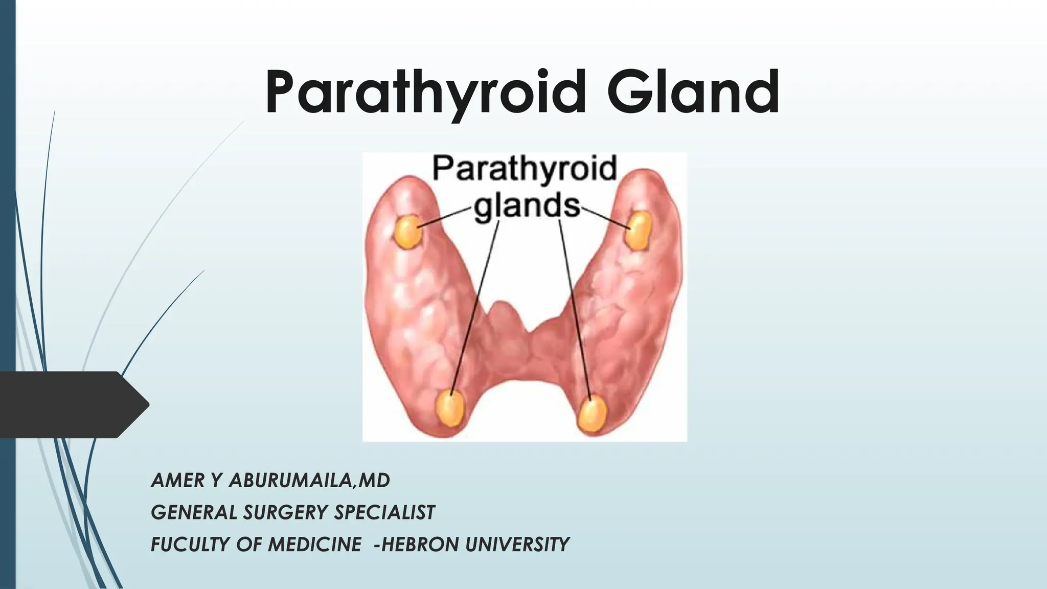
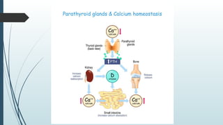
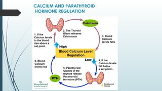
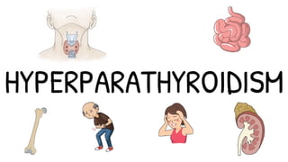
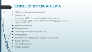
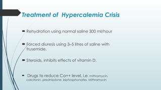
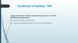
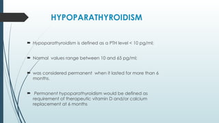
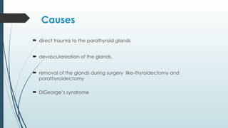
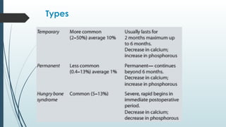
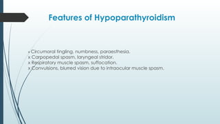
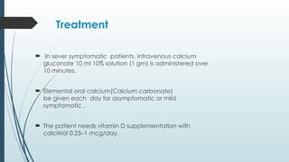
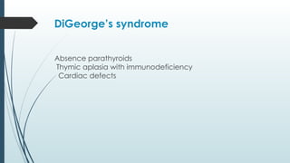
Parathyroid Gland
AMER Y ABURUMAILA,MD
GENERAL SURGERY SPECIALIST
FUCULTY OF MEDICINE -HEBRON UNIVERSITY
Objectives
Anatomy
Embryology
Physiology
Hyperparathyroidism
Hypoparathyroidism
Anatomy
 There are four glands
 5% having a supernumerary gland.
 Develop from the third and fourth
pharyngeal pouches
 They are typically (yellow/brown) in color
 Weigh approximately 30 mg.
 The blood supply of both from the inferior
thyroidal artery.
 The inferior parathyroid gland and the thymus
arise from the third pharyngeal pouch. they are
located at the inferior pole of the thyroid gland
 The superior parathyroid glands arise from fourth
pharyngeal pouch the located at the posterior
aspect of the thyroid lobe in the junction of the
inferior thyroid artery and the recurrent laryngeal
nerve
Anatomy
CALCIUM AND PARATHYROID
HORMONE REGULATION
Hyperparathyroidism
 Primary hyperparathyroidism
 Secondary hyperparathyroidism
 Tertiary hyperparathyroidism
Primary hyperparathyroidism
Adenoma (75–90%)
™
Hyperplasia (20–24%)
™
Carcinoma, rare (1%)
™
Etiology of primary
hyperparathyroidism
 familial or genetic causes are common; MEN I syndrome is often
associated.
 Therapeutic ionizing radiation can cause hyperparathyroidism.
 Lithium can cause parathyroid hyperplasia and HPT but without
causing bone or renal problems.
Primary hyperparathyroidism
 Primary HPT is commonly sporadic than familial
 MEN1 AND MEN IIA
 Adenoma is usually single.
 Multiple adenomas occur in elderly (5%)
 Hyperplasia involves all four parathyroid
Secondary hyperparathyroidism
 due to chronic renal failure
 due to malabsorption, vitamin D deficiency rickets, i.e. due
to chronic hypocalcaemia.
 There is always parathyroid hyperplasia.
 CRF causes hyperphosphataemia and reduced
conversion of 25 DHCC to 1, 25 DHCC.
 absorption of calcium from GIT is reduced causing chronic
hypocalcaemia which stimulates PTH secretion and
parathyroid hyperplasia.
 Serum calcium is normal or low
Tertiary hyperparathyroidism
 is due to autonomous reactive
hyperplasia of parathyroids often seen
like after renal transplantation.
Clinical Picture
 asymptomatic >50% cases
Bones, stones, abdominal graons and psychiatric
moans’
 As palpable neck swelling is uncommon,
 It is common in middle-aged women (3:1).
Incidence is 1:1000 patients.
Bones
:
 Bone pain.
 Subperiosteal erosions on the radial aspect
of the middle phalanges and clavicle.
 Osteitis fibrosa cystica (Brown tumour ).
Stones
:
 renal stones due to hypercalcaemia,
 3% of all renal patient stones is due to primary PTH.
 Recurrent stones are commonly observed in PTH.
 Calculi are usually calcium phosphate or oxalate types.
 calcification of the renal parenchyma resulting in nephrocalcinosis
resulting into renal failure.
 Calcification occurs in the renal vessels leading into renal
hypertension.
Abdominal groans
:
 Hypercalcaemia stimulates gastrin release which causes
peptic ulceration.
 Hypercalcaemia can precipitate acute pancreatitis.
 Gallstone disease is increased due to hypercalcaemia;
Psychic moans:
 Behavioural and neurotic problems with
 depression, anxiety;
Other features:
skin necrosis,
corneal ulceration,
pseudogout,
myalgia, joint pain, back ache,polyuria,polydypsia, constipation,
glycosuria and hypertension.
Investigations for HPT
 High serum calcium level more than 10 mg%.
 Serum albumin level also should be assessed to identify accurate
ionic calcium level.
 Increased serum PTH level is specific and diagnostic—
Assay of ‘Intact PTH’ is more relevant
 Serum phosphate level will be decreased in primary HPT;
but in secondary HPT serum phosphate level is increased.
 Bone density assessment.
 Vitamin D estimation.
 Increased urinary calcium level >250 mg/24 hours
 Raised serum alkaline phosphatase level.
Investigations for HPT
 X-ray skull shows salt and pepper appearance and brown
tumour
 Ultrasound abdomen to find out problems in kidney,
pancreas.
 Ultrasound neck and CT/MRI scan neck and mediastinum.
 Selective venous sampling for PTH is also very useful.
 Thallium-Technetium scan shows hot spots which is
diagnostic of parathyroid adenoma.
 Technetium-99m labelled Sestamibi isotope scan is better
and sensitive (80%)
 single photon emission computerized tomography (SPECT)
CAUSES OF HYPERCALCEMIA
 90% from hyperparathyroidism or CA
 Malignancy :
• Hematologic (25%) – lytic bone lesions (eg multiple myeloma)
• Nonhematologic (75%) – cancers that release PTHrP (eg squamous cell
lung CA, breast CA)
 Hyperparathyroidism
 Hyperthyroidism
 Familial hypercalcemic hypocalciuria
 Immobilization
 Granulomatous disease (sarcoidosis or tuberculosis)
 Excess vitamin D
 Milk–alkali syndrome
 Thiazide diuretics
Treatment of Hypercalemia Crisis
 Rehydration using normal saline 300 ml/hour
 Forced diuresis using 3–5 litres of saline with
frusemide.
 Steroids, inhibits effects of vitamin D.
 Drugs to reduce Ca++ level, i.e. mithramycin,
calcitonin, prednisolone, biphosphonates. Mithramycin
Treatment of Primary HPT
 Indications for parathyroidectomy:
Severe symptoms
™
Young age group
™
Markedly reduced bone density
™
Serum calcium more than 11 mg%
™
Urinary calculi
™
Neuromuscular presentations
™
Urinary calcium more than 400 mg /24 hours
™
Treatment of Primary HPT
 In adenoma surgical removal of the affected glands .
 If it is carcinoma, additional hemithyroidectomy with
postoperative radiotherapy is required.
 When all four glands are diseased,
totalparathyroidectomy with autotransplantation of glands
tissue
 In familial and MEN syndromes, total parathyroidectomy
is better.
Treatment of secondary HPT
 Medical treatment : indicated in most of patient
 Phosphate binder
 Vit D analogues
 Calcemimetics such as cinacalcet
 Surgical treatment : indicated in patient who is refractory
to medical treatment or who have intractable symptoms
 Subtatal parathyroidectomy
 tatal parathyroidectomy with autotransplantation of
parathyroid tissue
Treatment of tertiary HPT
Surgical treatment: When persistent longer than 12 months
following transplantation,
 subtotal parathyroidectomy
 total parathyroidectomy with autotransplantation.
HYPOPARATHYROIDISM
 Hypoparathyroidism is defined as a PTH level < 10 pg/ml;
 Normal values range between 10 and 65 pg/ml;
 was considered permanent when it lasted for more than 6
months.
 Permanent hypoparathyroidism would be defined as
requirement of therapeutic vitamin D and/or calcium
replacement at 6 months
Causes
 direct trauma to the parathyroid glands
 devascularization of the glands,
 removal of the glands during surgery like-thyroidectomy and
parathyroidectomy
 DiGeorge’s syndrome
Types
Features of Hypoparathyroidism
x Circumoral tingling, numbness, paraesthesia.
x Carpopedal spasm, laryngeal stridor.
x Respiratory muscle spasm, suffocation.
x Convulsions, blurred vision due to intraocular muscle spasm.
Treatment
 In sever symptomatic patients, intravenous calcium
gluconate 10 ml 10% solution (1 gm) is administered over
10 minutes.
 Elemental oral calcium(Calcium carbonate)
be given each day for asymptomatic or mild
symptomatic .
 The patient needs vitamin D supplementation with
calcitriol 0.25–1 mcg/day.
DiGeorge’s syndrome
™ Absence parathyroids
Thymic aplasia with immunodeficiency
™
Cardiac defects
™
Any Question
????
parathyroid final  6  year 2024.pptx fff

parathyroid final 6 year 2024.pptx fff

  • 1.
    Parathyroid Gland AMER YABURUMAILA,MD GENERAL SURGERY SPECIALIST FUCULTY OF MEDICINE -HEBRON UNIVERSITY
  • 2.
  • 3.
    Anatomy  There arefour glands  5% having a supernumerary gland.  Develop from the third and fourth pharyngeal pouches  They are typically (yellow/brown) in color  Weigh approximately 30 mg.  The blood supply of both from the inferior thyroidal artery.
  • 4.
     The inferiorparathyroid gland and the thymus arise from the third pharyngeal pouch. they are located at the inferior pole of the thyroid gland  The superior parathyroid glands arise from fourth pharyngeal pouch the located at the posterior aspect of the thyroid lobe in the junction of the inferior thyroid artery and the recurrent laryngeal nerve Anatomy
  • 6.
  • 8.
    Hyperparathyroidism  Primary hyperparathyroidism Secondary hyperparathyroidism  Tertiary hyperparathyroidism
  • 9.
  • 10.
    Etiology of primary hyperparathyroidism familial or genetic causes are common; MEN I syndrome is often associated.  Therapeutic ionizing radiation can cause hyperparathyroidism.  Lithium can cause parathyroid hyperplasia and HPT but without causing bone or renal problems.
  • 11.
    Primary hyperparathyroidism  PrimaryHPT is commonly sporadic than familial  MEN1 AND MEN IIA  Adenoma is usually single.  Multiple adenomas occur in elderly (5%)  Hyperplasia involves all four parathyroid
  • 12.
    Secondary hyperparathyroidism  dueto chronic renal failure  due to malabsorption, vitamin D deficiency rickets, i.e. due to chronic hypocalcaemia.  There is always parathyroid hyperplasia.  CRF causes hyperphosphataemia and reduced conversion of 25 DHCC to 1, 25 DHCC.  absorption of calcium from GIT is reduced causing chronic hypocalcaemia which stimulates PTH secretion and parathyroid hyperplasia.  Serum calcium is normal or low
  • 13.
    Tertiary hyperparathyroidism  isdue to autonomous reactive hyperplasia of parathyroids often seen like after renal transplantation.
  • 14.
    Clinical Picture  asymptomatic>50% cases Bones, stones, abdominal graons and psychiatric moans’  As palpable neck swelling is uncommon,  It is common in middle-aged women (3:1). Incidence is 1:1000 patients.
  • 15.
    Bones :  Bone pain. Subperiosteal erosions on the radial aspect of the middle phalanges and clavicle.  Osteitis fibrosa cystica (Brown tumour ).
  • 16.
    Stones :  renal stonesdue to hypercalcaemia,  3% of all renal patient stones is due to primary PTH.  Recurrent stones are commonly observed in PTH.  Calculi are usually calcium phosphate or oxalate types.  calcification of the renal parenchyma resulting in nephrocalcinosis resulting into renal failure.  Calcification occurs in the renal vessels leading into renal hypertension.
  • 17.
    Abdominal groans :  Hypercalcaemiastimulates gastrin release which causes peptic ulceration.  Hypercalcaemia can precipitate acute pancreatitis.  Gallstone disease is increased due to hypercalcaemia;
  • 18.
    Psychic moans:  Behaviouraland neurotic problems with  depression, anxiety; Other features: skin necrosis, corneal ulceration, pseudogout, myalgia, joint pain, back ache,polyuria,polydypsia, constipation, glycosuria and hypertension.
  • 19.
    Investigations for HPT High serum calcium level more than 10 mg%.  Serum albumin level also should be assessed to identify accurate ionic calcium level.  Increased serum PTH level is specific and diagnostic— Assay of ‘Intact PTH’ is more relevant  Serum phosphate level will be decreased in primary HPT; but in secondary HPT serum phosphate level is increased.  Bone density assessment.  Vitamin D estimation.  Increased urinary calcium level >250 mg/24 hours  Raised serum alkaline phosphatase level.
  • 21.
    Investigations for HPT X-ray skull shows salt and pepper appearance and brown tumour  Ultrasound abdomen to find out problems in kidney, pancreas.  Ultrasound neck and CT/MRI scan neck and mediastinum.  Selective venous sampling for PTH is also very useful.  Thallium-Technetium scan shows hot spots which is diagnostic of parathyroid adenoma.  Technetium-99m labelled Sestamibi isotope scan is better and sensitive (80%)  single photon emission computerized tomography (SPECT)
  • 24.
    CAUSES OF HYPERCALCEMIA 90% from hyperparathyroidism or CA  Malignancy : • Hematologic (25%) – lytic bone lesions (eg multiple myeloma) • Nonhematologic (75%) – cancers that release PTHrP (eg squamous cell lung CA, breast CA)  Hyperparathyroidism  Hyperthyroidism  Familial hypercalcemic hypocalciuria  Immobilization  Granulomatous disease (sarcoidosis or tuberculosis)  Excess vitamin D  Milk–alkali syndrome  Thiazide diuretics
  • 25.
    Treatment of HypercalemiaCrisis  Rehydration using normal saline 300 ml/hour  Forced diuresis using 3–5 litres of saline with frusemide.  Steroids, inhibits effects of vitamin D.  Drugs to reduce Ca++ level, i.e. mithramycin, calcitonin, prednisolone, biphosphonates. Mithramycin
  • 26.
    Treatment of PrimaryHPT  Indications for parathyroidectomy: Severe symptoms ™ Young age group ™ Markedly reduced bone density ™ Serum calcium more than 11 mg% ™ Urinary calculi ™ Neuromuscular presentations ™ Urinary calcium more than 400 mg /24 hours ™
  • 28.
    Treatment of PrimaryHPT  In adenoma surgical removal of the affected glands .  If it is carcinoma, additional hemithyroidectomy with postoperative radiotherapy is required.  When all four glands are diseased, totalparathyroidectomy with autotransplantation of glands tissue  In familial and MEN syndromes, total parathyroidectomy is better.
  • 29.
    Treatment of secondaryHPT  Medical treatment : indicated in most of patient  Phosphate binder  Vit D analogues  Calcemimetics such as cinacalcet  Surgical treatment : indicated in patient who is refractory to medical treatment or who have intractable symptoms  Subtatal parathyroidectomy  tatal parathyroidectomy with autotransplantation of parathyroid tissue
  • 30.
    Treatment of tertiaryHPT Surgical treatment: When persistent longer than 12 months following transplantation,  subtotal parathyroidectomy  total parathyroidectomy with autotransplantation.
  • 31.
    HYPOPARATHYROIDISM  Hypoparathyroidism isdefined as a PTH level < 10 pg/ml;  Normal values range between 10 and 65 pg/ml;  was considered permanent when it lasted for more than 6 months.  Permanent hypoparathyroidism would be defined as requirement of therapeutic vitamin D and/or calcium replacement at 6 months
  • 32.
    Causes  direct traumato the parathyroid glands  devascularization of the glands,  removal of the glands during surgery like-thyroidectomy and parathyroidectomy  DiGeorge’s syndrome
  • 33.
  • 34.
    Features of Hypoparathyroidism xCircumoral tingling, numbness, paraesthesia. x Carpopedal spasm, laryngeal stridor. x Respiratory muscle spasm, suffocation. x Convulsions, blurred vision due to intraocular muscle spasm.
  • 35.
    Treatment  In seversymptomatic patients, intravenous calcium gluconate 10 ml 10% solution (1 gm) is administered over 10 minutes.  Elemental oral calcium(Calcium carbonate) be given each day for asymptomatic or mild symptomatic .  The patient needs vitamin D supplementation with calcitriol 0.25–1 mcg/day.
  • 36.
    DiGeorge’s syndrome ™ Absenceparathyroids Thymic aplasia with immunodeficiency ™ Cardiac defects ™
  • 37.