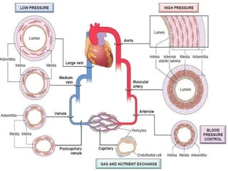
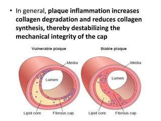
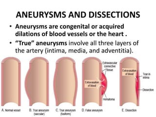
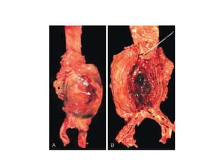
Blood Vessels
Dr. Maria Idrees: PT
MS NMPT; RIU
DPT; TUF
• Vascular disease develops through two
principal mechanisms:
• Narrowing or complete obstruction of vessel
lumina, occurring either progressively (e.g., by
atherosclerosis) or acutely (e.g., by
thrombosis or embolism)
• Weakening of vessel walls, causing dilation
and/or rupture
STRUCTURE AND FUNCTION OF
BLOOD VESSELS
• Blood vessels are fundamentally all tubes
composed of smooth muscle cells (SMCs) and
extracellular matrix (ECM), with an inner
luminal face covered by a continuous lining of
endothelial cells (ECs).
• However, the relative amounts of SMCs and
ECM, and the properties of the ECs, vary
throughout the vasculature depending on
unique functional needs.
• To accommodate pulsatile flow and higher
blood pressures, arterial walls are thicker than
veins and invested with several reinforcing
layers of SMCs.
• To facilitate maximal diffusion, capillaries are
essentially single-cell linings of ECs lying on a
basement membrane
Vascular Organization
• Arteries are divided into three types based on their size
and structure:
Large elastic arteries (e.g., aorta, arch vessels, iliac
and pulmonary arteries). In these vessels, elastic fibers
alternate with SMCs throughout the media, which
expands during systole (storing some of the energy of
each cardiac contraction), and recoils during diastole to
propel blood distally. With age and/or diseases, such as
diabetes and hypertension, the elasticity is lost; the
vessels become “stiff pipes” that transmit high arterial
pressures to distal organs, or become dilated and
tortuous (ectatic) and prone to rupture.
Medium-sized muscular arteries (e.g.,
coronary and renal arteries). Here, the media
is composed primarily of SMCs, with elastin
limited to the internal and external elastic
lamina.
• The medial SMCs are circularly or spirally
arranged around the lumen, and regional
blood flow is regulated by SMC contraction
(vasoconstriction) and relaxation
(vasodilation) controlled by the autonomic
nervous system and local metabolic factors
(e.g., acidosis)
Small arteries (2 mm or less in diameter) and arterioles
(20 to 100 μm in diameter) lie within the connective
tissue of organs. The media in these vessels is mostly
composed of SMCs. Arterioles are where blood flow
resistance is regulated. As pressures drop during
passage through arterioles, the velocity of blood flow is
sharply reduced, and flow becomes steady rather than
pulsatile. Because the resistance to fluid flow is
inversely proportional to the fourth power of the
diameter (i.e., halving the diameter increases
resistance 16-fold), small changes in arteriolar lumen
size have profound effects on blood pressure.
• Capillaries have lumen diameters slightly smaller
than those of red blood cells (7 to 8 μm). These
vessels are lined by ECs and partially surrounded
by SMC-like cells called pericytes. Collectively,
capillary beds have a very large total cross-
sectional area and a low rate of blood flow. With
their thin walls and slow flow, capillaries are
ideally suited to the rapid exchange of diffusible
substances between blood and tissue.
• Lymphatics are thin-walled, endothelium-lined
channels that drain lymph (water, electrolytes,
glucose, fat, proteins, and inflammatory cells)
from the interstitium of tissues, eventually
reconnecting with the blood stream via the
thoracic duct
Endothelial Cells
• Endothelium is a continuous sheet of cells
lining the entire vascular tree that regulates
many aspects of blood and blood vessel
function.
smooth muscle
tone
smooth muscle
contraction
Vascular Smooth Muscle Cells
• SMCs participate in both normal vascular repair
and pathologic processes such as atherosclerosis.
When stimulated by various factors, SMCs can do
the following:
• Proliferate
• Upregulate ECM collagen, elastin, and
proteoglycan production
• Elaborate growth factors and cytokines
• SMCs also mediate the vasoconstriction or
vasodilation that occurs in response to
physiologic or pharmacologic stimuli.
• The migratory and proliferative activities of
SMCs are regulated by numerous factors.
Among the most important pro–growth
factors are platelet-derived growth factor
(PDGF), endothelin, thrombin, fibroblast
growth factors, and inflammatory mediators
such as interferon-γ (IFN-γ) and interleukin-1
(IL-1). Factors that maintain SMCs in a
quiescent state include heparan sulfate, NO,
and transforming growth factor-α (TGF-α).
CONGENITAL ANOMALIES
• Among the other congenital vascular
anomalies, three deserve further mention:
Berry aneurysms are thin-walled arterial
outpouchings in cerebral vessels, classically at
branch points around the circle of Willis; they
occur where the arterial media is congenitally
attenuated and can spontaneously rupture
causing fatal intracerebral hemorrhage
Arteriovenous (AV) fistulas are abnormal
connections between arteries and veins without
an intervening capillary bed.
• They occur most commonly as developmental
defects but can also result from rupture of
arterial aneurysms into adjacent veins, from
penetrating injuries that pierce arteries and veins,
or from inflammatory necrosis of adjacent
vessels.
• Extensive AV fistulas can cause high-output
cardiac failure by shunting large volumes of blood
from the arterial to the venous circulation.
Fibromuscular dysplasia is a focal irregular
thickening of the walls of medium- and large-
sized muscular arteries due to a combination
of medial and intimal hyperplasia and fibrosis.
It can manifest at any age but occurs most
frequently in young women.
• The focal wall thickening results in luminal
stenosis or can be associated with vessel
spasm that reduces vascular flow; in the renal
arteries, it can lead to renovascular
hypertension.
BLOOD PRESSURE REGULATION
• Low blood pressure (hypotension) results in
inadequate organ perfusion, organ
dysfunction, and sometimes tissue death.
• Conversely, high blood pressure (hypertension)
causes vessel and end-organ damage and is
one of the major risk factors for
atherosclerosis.
• Blood pressure is a function of cardiac output
and peripheral vascular resistance, both of which
are influenced by multiple genetic and
environmental factors.
• Cardiac output is a function of stroke volume and
heart rate. The most important determinant of
stroke volume is the filling pressure, which is
regulated through sodium homeostasis and its
effect on blood volume.
• Heart rate and myocardial contractility (a second
factor affecting stroke volume) are both regulated
by the α- and β-adrenergic systems (in addition to
their effects on vascular tone).
• Peripheral resistance is regulated
predominantly at the level of the arterioles by
neural and humoral inputs.
• Vascular tone reflects a balance between the
actions of vasoconstrictors (including
angiotensin II, catecholamines, and
endothelin) and vasodilators (including kinins,
prostaglandins, and NO).
HYPERTENSIVE VASCULAR DISEASE
• This insidious condition has been dubbed
essential hypertension, so called because the
gradual age-associated rise in blood pressure was
considered “essential” for normal perfusion of
endorgans such as the brain.
• However, such increases are neither essential
nor benign; besides increasing the risk for stroke
and atherosclerotic coronary heart disease,
hypertension can lead to cardiac hypertrophy and
heart failure
Epidemiology of Hypertension
• Nevertheless, sustained diastolic pressures
greater than 90 mm Hg or sustained systolic
pressures in excess of 140 mm Hg are reliably
associated with an increased risk for
atherosclerosis and are therefore used as
cutoffs in diagnosing hypertension in clinical
practice.
Pathogenesis
• Hypertension may be primary (idiopathic) or
less commonly secondary to an identifiable
underlying condition. In close to 95% of cases
hypertension is idiopathic or “essential”.
Most of the remaining cases (secondary
hypertension) are due to primary renal
disease, renal artery narrowing (renovascular
hypertension), or adrenal disorders
VASCULAR WALL RESPONSE
TO INJURY
• Injury to the vessel wall—and in particular to
ECs—is the fundamental basis for the vast
majority of vascular disorders.
Intimal Thickening: A Stereotypical Response to Vascular Injury
• Vascular injury leading to EC loss or dysfunction stimulates
SMC growth, ECM synthesis, and thickening of the vascular
wall.
ARTERIOSCLEROSIS
• Arteriosclerosis literally means “hardening of
the arteries”; it is a generic term reflecting
arterial wall thickening and loss of elasticity.
Four distinct types are recognized, each with
different clinical and pathologic causes and
consequences:
• Arteriolosclerosis affects small arteries and
arterioles and may cause downstream ischemic
injury. The two variants, hyaline and hyperplastic
arteriolosclerosis.
• Mönckeberg medial sclerosis is characterized by
the presence of calcific deposits in muscular
arteries, usually centered on the internal elastic
lamina, and typically in individuals older than 50
years of age. The lesions do not encroach on the
vessel lumen and usually are not clinically
significant.
• Fibromuscular intimal hyperplasia is a non-
atherosclerotic process that occurs in muscular
arteries larger than arterioles. This is
predominantly an SMC- and ECM-rich lesion
driven by inflammation or by mechanical injury
• Atherosclerosis
ATHEROSCLEROSIS
• Atherosclerosis is characterized by intimal
lesions called atheromas (or atheromatous or
atherosclerotic plaques) that impinge on the
vascular lumen and can rupture to cause
sudden occlusion.
Pathogenesis
Atherosclerosis results from the following pathogenic events:
• EC injury—and resultant endothelial dysfunction— leading to
increased permeability, leukocyte adhesion, and thrombosis
• Accumulation of lipoproteins (mainly oxidized LDL and
cholesterol crystals) in the vessel wall
• Platelet adhesion
• Monocyte adhesion to the endothelium, migration into the
intima, and differentiation into macrophages and foam cells
• Lipid accumulation within macrophages, which respond by
releasing inflammatory cytokines
• SMC recruitment due to factors released from activated
platelets, macrophages, and vascular wall cells
• SMC proliferation and ECM production
• Atherosclerotic plaques have three principal
components:
• (1) cells, including SMCs, macrophages, and T
cells; (2) ECM, including collagen, elastic
fibers, and proteoglycans; and (3)
intracellular and extracellular lipid.
• Atherosclerotic plaques are susceptible to
several clinically important changes:
• Rupture, ulceration
• Hemorrhage into a plaque
• Atheroembolism. Ruptured plaque can
discharge debris into the blood, producing
microemboli composed of plaque contents.
• Aneurysm formation.
Clinicopathologic Consequences of
Atherosclerosis
• In general, plaque inflammation increases
collagen degradation and reduces collagen
synthesis, thereby destabilizing the
mechanical integrity of the cap
ANEURYSMS AND DISSECTIONS
• Aneurysms are congenital or acquired
dilations of blood vessels or the heart .
• “True” aneurysms involve all three layers of
the artery (intima, media, and adventitia).
Abdominal Aortic Aneurysm
• Aneurysms occurring as a consequence of
atherosclerosis form most commonly in the
abdominal aorta and common iliac arteries, and
may also involve the aortic arch and descending
thoracic aorta.
• Abdominal aortic aneurysms (AAAs) occur more
frequently in men and in smokers and rarely
develop before 50 years of age. Atherosclerosis is
a major cause of AAA, but other factors clearly
contribute,
Clinical Consequences
The clinical consequences of AAA include the following:
• Obstruction of a vessel branching off the aorta (e.g.,
the renal, iliac, vertebral, or mesenteric arteries),
resulting in ischemia of the kidneys, legs, spinal cord,
or gastrointestinal tract, respectively
• Embolism of atheromatous material (e.g., cholesterol
crystals) or mural thrombus
• Impingement on adjacent structures (e.g., compression
of a ureter or erosion of vertebrae by the expanding
aneurysm)
• An abdominal mass (often palpably pulsating) that
simulates a tumor
• Rupture into the peritoneal cavity or retroperitoneal
tissues, leading to massive, often fatal hemorrhage
Thoracic Aortic Aneurysm
These aneurysms manifest with the following signs and
symptoms:
• Respiratory or feeding difficulties due to airway or
esophageal compression, respectively, because of
encroachment on mediastinal structures
• Persistent cough from irritation of the recurrent
laryngeal nerves
• Pain caused by erosion of bone (i.e., ribs and vertebral
bodies)
• Cardiac disease due to valvular insufficiency or
narrowing of the coronary ostia; heart failure induced
by aortic valvular incompetence
• Aortic dissection or rupture
Aortic Dissection
• Aortic dissection occurs when blood splays
apart the laminar planes of the media to
form a blood-filled channel within the aortic
wall.
Clinical Consequences
• Thus, aortic dissections generally are classified
into two types
• Proximal lesions (type A dissections), involving
the ascending aorta, with or without
involvement of the descending aorta (DeBakey
type I or II, respectively)
• Distal lesions (type B dissections), usually
beginning beyond the subclavian artery
(DeBakey type III)
• The classic clinical symptom of aortic
dissection is the sudden onset of excruciating
tearing or stabbing pain, usually beginning in
the anterior chest, radiating to the back
between the scapulae, and moving downward
as the dissection progresses.
• The most common cause of death is rupture
of the dissection into the pericardial, pleural,
or peritoneal cavity.
VASCULITIS
• Vasculitis is a general term for vessel wall
inflammation.
• The two most common pathogenic
mechanisms of vasculitis are immune-
mediated inflammation and direct vascular
invasion by infectious pathogens
Noninfectious Vasculitis
• The main immunologic mechanisms
underlying noninfectious vasculitis are as
follows:
• Immune complex deposition
• Anti-neutrophil cytoplasmic antibodies
• Anti-EC antibodies
• Autoreactive T cells
Immune Complex–Associated
Vasculitis
• This form of vasculitis is seen in immunologic
disorders such as systemic lupus
erythematosus that are associated with
autoantibody production.
Anti-Neutrophil Cytoplasmic
Antibodies
• Many patients with vasculitis have circulating
antibodies that react with neutrophil
cytoplasmic antigens, so-called “anti-
neutrophil cytoplasmic antibodies (ANCAs).”
• Although a number of ANCAs have been
described, two are most important. These are
classified according to their antigen specificity:
Anti-proteinase-3 (PR3-ANCA)
Anti-myeloperoxidase (MPO-ANCA)
Anti-Endothelial Cell Antibodies and
Autoreactive T Cells
• Antibodies to ECs underlie certain vasculitides,
such as Kawasaki disease. Autoreactive T cells
cause injury in some forms of vasculitides
characterized by formation of granulomas.
Giant Cell (Temporal) Arteritis
• Giant cell (temporal) arteritis is a chronic
inflammatory disorder, typically with
granulomatous inflammation, that principally
affects large- to small-sized arteries in the
head. The temporal arteries are not more
vulnerable than other.
• Vertebral and ophthalmic arteries, as well as
the aorta (giant cell aortitis), are other
common sites of involvement.
Clinical Features
• Signs and symptoms may be vague and
constitutional (e.g., fever, fatigue, weight loss) or
take the form of facial pain or headache, most
intense along the course of the superficial
temporal artery, which is painful to palpation.
• Ocular symptoms (associated with involvement of
the ophthalmic artery) abruptly appear in about
50% of patients; these range from diplopia to
complete vision loss.
Takayasu Arteritis
• Takayasu arteritis is a granulomatous vasculitis of
medium- and large-sized arteries characterized
principally by ocular disturbances and marked
weakening of the pulses in the upper extremities
(hence the alternate name, pulseless disease).
• This disorder manifests with transmural scarring
and thickening of the aorta—particularly the
aortic arch and great vessels—with severe
luminal narrowing of the major branch vessels
Differential Diagnosis
• Aortic lesions share many of the clinical and
histologic features of giant cell aortitis.
• Indeed, the distinction between the two
entities is made largely on the basis of a
patient’s age; in those older than 50 years of
age are said to have giant cell aortitis, and
lesions that occur in those younger than 50
years of age are designated Takayasu aortitis
Clinical Features
• Initial signs and symptoms usually are
nonspecific, including fatigue, weight loss, and
fever.
• With progression, reduced upper-extremity blood
pressure and pulse strength; neurologic deficits;
and ocular disturbances, including visual field
defects, retinal hemorrhages, and total blindness.
Distal aorta disease can manifest as leg
claudication, and pulmonary artery involvement
can cause pulmonary hypertension
Polyarteritis Nodosa
• Polyarteritis nodosa (PAN) is a systemic
vasculitis of small- or medium-sized muscular
arteries; it typically involves the renal and
visceral vessels and spares the pulmonary
circulation.
Clinical Features
• A “classic” presentation involves involve some
combination of rapidly accelerating
hypertension due to renal artery involvement;
abdominal pain and bloody stools caused by
gastrointestinal lesions; diffuse muscular
aches and pains; and peripheral neuritis,
predominantly affecting motor nerves.
• Renal involvement often is prominent and is a
major cause of death.
Kawasaki Disease
• Kawasaki disease is an acute, febrile, usually
self-limited illness of infancy and childhood
associated with an arteritis of mainly large- to
medium-sized vessels.
• Approximately 80% of patients are younger
than 4 years of age. Its clinical significance
stems from the involvement of coronary
arteries
Clinical Features
• Kawasaki disease typically manifests with
conjunctival and oral erythema and blistering,
edema of the hands and feet, erythema of the
palms and soles, a desquamative rash, and
cervical lymph node enlargement (hence its
other name, mucocutaneous lymph node
syndrome).
Microscopic Polyangiitis
• Microscopic polyangiitis is a necrotizing
vasculitis that generally affects capillaries, as
well as small arterioles and venules.
• It also is called hypersensitivity vasculitis or
leukocytoclastic vasculitis.
• The skin, mucous membranes, lungs, brain, heart,
gastrointestinal tract, kidneys, and muscle all can
be involved; necrotizing glomerulonephritis and
pulmonary capillaritis are particularly common.
Clinical Features
• Depending on the vascular bed involved,
major features include hemoptysis,
hematuria, proteinuria, abdominal pain or
bleeding, muscle pain or weakness, and
palpable cutaneous purpura.
• Renal or CNS involvement,
immunosuppression and removal of the
offending agent induce durable remissions.
Granulomatosis With Polyangiitis
• Previously called Wegener granulomatosis,
granulomatosis with polyangiitis (GPA) is a necrotizing
vasculitis characterized by a triad of the following:
• Necrotizing granulomas of the upper-respiratory tract
(ear, nose, sinuses, throat) or the lower-respiratory
tract (lung) or both
• Necrotizing or granulomatous vasculitis affecting
small- to medium-sized vessels (e.g., capillaries,
venules, arterioles, and arteries), most prominently the
lungs and upper airways but other sites as well
• Focal necrotizing, often crescentic, glomerulonephritis
Clinical Features
• Classic presentations include
• Bilateral pneumonitis with nodules and
cavitary lesions
• Chronic sinusitis
• Mucosal ulcerations of the nasopharynx
• Renal disease
Churg-Strauss Syndrome
• Churg-Strauss syndrome (also called allergic
granulomatosis and angiitis) is a small-vessel
necrotizing vasculitis classically associated with
asthma, allergic rhinitis, lung infiltrates,
peripheral eosinophilia, extravascular necrotizing
granulomas, and a striking infiltration of vessels
and perivascular tissues by eosinophils.
• It is a rare disorder, affecting 1 in 1 million
individuals.
Thromboangiitis Obliterans (Buerger
Disease)
• Thromboangiitis obliterans (Buerger disease) is
characterized by segmental, thrombosing, acute
and chronic inflammation of medium- and small-
sized arteries, principally the tibial and radial
arteries, with occasional secondary extension
into the veins and nerves of the extremities.
• Buerger disease occurs almost exclusively in
heavy tobacco smokers and usually develops
before 35 years of age.
Clinical Features
• Early manifestations include cold-induced
Raynaud phenomenon
• instep foot pain induced by exercise (instep
claudication),
• superficial nodular phlebitis (venous
inflammation)
• The vascular insufficiency of Buerger disease
tends to be accompanied by severe pain—even at
rest—undoubtedly from the neural involvement
Vasculitis Associated With Other
Noninfectious Disorders
• Rheumatoid vasculitis may occur in patients
with severe, long-standing rheumatoid
arthritis; it can cause a clinically significant
aortitis but more typically affects small- and
medium-sized arteries, leading to visceral
infarction.
• Systemic lupus erythematosus,
• Anti-phospholipid antibody syndrome
Infectious Vasculitis
• Localized arteritis may be caused by the direct
invasion of arteries by infectious agents,
usually bacteria or fungi, and in particular
Aspergillus and Mucor spp.
• May arise from hematogenous spread of
bacteria

5. Blood Vessels.pptx

  • 1.
    Blood Vessels Dr. MariaIdrees: PT MS NMPT; RIU DPT; TUF
  • 2.
    • Vascular diseasedevelops through two principal mechanisms: • Narrowing or complete obstruction of vessel lumina, occurring either progressively (e.g., by atherosclerosis) or acutely (e.g., by thrombosis or embolism) • Weakening of vessel walls, causing dilation and/or rupture
  • 3.
    STRUCTURE AND FUNCTIONOF BLOOD VESSELS • Blood vessels are fundamentally all tubes composed of smooth muscle cells (SMCs) and extracellular matrix (ECM), with an inner luminal face covered by a continuous lining of endothelial cells (ECs). • However, the relative amounts of SMCs and ECM, and the properties of the ECs, vary throughout the vasculature depending on unique functional needs.
  • 4.
    • To accommodatepulsatile flow and higher blood pressures, arterial walls are thicker than veins and invested with several reinforcing layers of SMCs. • To facilitate maximal diffusion, capillaries are essentially single-cell linings of ECs lying on a basement membrane
  • 6.
    Vascular Organization • Arteriesare divided into three types based on their size and structure: Large elastic arteries (e.g., aorta, arch vessels, iliac and pulmonary arteries). In these vessels, elastic fibers alternate with SMCs throughout the media, which expands during systole (storing some of the energy of each cardiac contraction), and recoils during diastole to propel blood distally. With age and/or diseases, such as diabetes and hypertension, the elasticity is lost; the vessels become “stiff pipes” that transmit high arterial pressures to distal organs, or become dilated and tortuous (ectatic) and prone to rupture.
  • 7.
    Medium-sized muscular arteries(e.g., coronary and renal arteries). Here, the media is composed primarily of SMCs, with elastin limited to the internal and external elastic lamina. • The medial SMCs are circularly or spirally arranged around the lumen, and regional blood flow is regulated by SMC contraction (vasoconstriction) and relaxation (vasodilation) controlled by the autonomic nervous system and local metabolic factors (e.g., acidosis)
  • 8.
    Small arteries (2mm or less in diameter) and arterioles (20 to 100 μm in diameter) lie within the connective tissue of organs. The media in these vessels is mostly composed of SMCs. Arterioles are where blood flow resistance is regulated. As pressures drop during passage through arterioles, the velocity of blood flow is sharply reduced, and flow becomes steady rather than pulsatile. Because the resistance to fluid flow is inversely proportional to the fourth power of the diameter (i.e., halving the diameter increases resistance 16-fold), small changes in arteriolar lumen size have profound effects on blood pressure.
  • 9.
    • Capillaries havelumen diameters slightly smaller than those of red blood cells (7 to 8 μm). These vessels are lined by ECs and partially surrounded by SMC-like cells called pericytes. Collectively, capillary beds have a very large total cross- sectional area and a low rate of blood flow. With their thin walls and slow flow, capillaries are ideally suited to the rapid exchange of diffusible substances between blood and tissue. • Lymphatics are thin-walled, endothelium-lined channels that drain lymph (water, electrolytes, glucose, fat, proteins, and inflammatory cells) from the interstitium of tissues, eventually reconnecting with the blood stream via the thoracic duct
  • 10.
    Endothelial Cells • Endotheliumis a continuous sheet of cells lining the entire vascular tree that regulates many aspects of blood and blood vessel function.
  • 12.
  • 13.
    Vascular Smooth MuscleCells • SMCs participate in both normal vascular repair and pathologic processes such as atherosclerosis. When stimulated by various factors, SMCs can do the following: • Proliferate • Upregulate ECM collagen, elastin, and proteoglycan production • Elaborate growth factors and cytokines • SMCs also mediate the vasoconstriction or vasodilation that occurs in response to physiologic or pharmacologic stimuli.
  • 14.
    • The migratoryand proliferative activities of SMCs are regulated by numerous factors. Among the most important pro–growth factors are platelet-derived growth factor (PDGF), endothelin, thrombin, fibroblast growth factors, and inflammatory mediators such as interferon-γ (IFN-γ) and interleukin-1 (IL-1). Factors that maintain SMCs in a quiescent state include heparan sulfate, NO, and transforming growth factor-α (TGF-α).
  • 15.
    CONGENITAL ANOMALIES • Amongthe other congenital vascular anomalies, three deserve further mention: Berry aneurysms are thin-walled arterial outpouchings in cerebral vessels, classically at branch points around the circle of Willis; they occur where the arterial media is congenitally attenuated and can spontaneously rupture causing fatal intracerebral hemorrhage
  • 16.
    Arteriovenous (AV) fistulasare abnormal connections between arteries and veins without an intervening capillary bed. • They occur most commonly as developmental defects but can also result from rupture of arterial aneurysms into adjacent veins, from penetrating injuries that pierce arteries and veins, or from inflammatory necrosis of adjacent vessels. • Extensive AV fistulas can cause high-output cardiac failure by shunting large volumes of blood from the arterial to the venous circulation.
  • 17.
    Fibromuscular dysplasia isa focal irregular thickening of the walls of medium- and large- sized muscular arteries due to a combination of medial and intimal hyperplasia and fibrosis. It can manifest at any age but occurs most frequently in young women. • The focal wall thickening results in luminal stenosis or can be associated with vessel spasm that reduces vascular flow; in the renal arteries, it can lead to renovascular hypertension.
  • 18.
    BLOOD PRESSURE REGULATION •Low blood pressure (hypotension) results in inadequate organ perfusion, organ dysfunction, and sometimes tissue death. • Conversely, high blood pressure (hypertension) causes vessel and end-organ damage and is one of the major risk factors for atherosclerosis.
  • 19.
    • Blood pressureis a function of cardiac output and peripheral vascular resistance, both of which are influenced by multiple genetic and environmental factors. • Cardiac output is a function of stroke volume and heart rate. The most important determinant of stroke volume is the filling pressure, which is regulated through sodium homeostasis and its effect on blood volume. • Heart rate and myocardial contractility (a second factor affecting stroke volume) are both regulated by the α- and β-adrenergic systems (in addition to their effects on vascular tone).
  • 20.
    • Peripheral resistanceis regulated predominantly at the level of the arterioles by neural and humoral inputs. • Vascular tone reflects a balance between the actions of vasoconstrictors (including angiotensin II, catecholamines, and endothelin) and vasodilators (including kinins, prostaglandins, and NO).
  • 23.
    HYPERTENSIVE VASCULAR DISEASE •This insidious condition has been dubbed essential hypertension, so called because the gradual age-associated rise in blood pressure was considered “essential” for normal perfusion of endorgans such as the brain. • However, such increases are neither essential nor benign; besides increasing the risk for stroke and atherosclerotic coronary heart disease, hypertension can lead to cardiac hypertrophy and heart failure
  • 24.
    Epidemiology of Hypertension •Nevertheless, sustained diastolic pressures greater than 90 mm Hg or sustained systolic pressures in excess of 140 mm Hg are reliably associated with an increased risk for atherosclerosis and are therefore used as cutoffs in diagnosing hypertension in clinical practice.
  • 25.
    Pathogenesis • Hypertension maybe primary (idiopathic) or less commonly secondary to an identifiable underlying condition. In close to 95% of cases hypertension is idiopathic or “essential”. Most of the remaining cases (secondary hypertension) are due to primary renal disease, renal artery narrowing (renovascular hypertension), or adrenal disorders
  • 26.
    VASCULAR WALL RESPONSE TOINJURY • Injury to the vessel wall—and in particular to ECs—is the fundamental basis for the vast majority of vascular disorders.
  • 27.
    Intimal Thickening: AStereotypical Response to Vascular Injury • Vascular injury leading to EC loss or dysfunction stimulates SMC growth, ECM synthesis, and thickening of the vascular wall.
  • 28.
    ARTERIOSCLEROSIS • Arteriosclerosis literallymeans “hardening of the arteries”; it is a generic term reflecting arterial wall thickening and loss of elasticity. Four distinct types are recognized, each with different clinical and pathologic causes and consequences: • Arteriolosclerosis affects small arteries and arterioles and may cause downstream ischemic injury. The two variants, hyaline and hyperplastic arteriolosclerosis.
  • 29.
    • Mönckeberg medialsclerosis is characterized by the presence of calcific deposits in muscular arteries, usually centered on the internal elastic lamina, and typically in individuals older than 50 years of age. The lesions do not encroach on the vessel lumen and usually are not clinically significant. • Fibromuscular intimal hyperplasia is a non- atherosclerotic process that occurs in muscular arteries larger than arterioles. This is predominantly an SMC- and ECM-rich lesion driven by inflammation or by mechanical injury • Atherosclerosis
  • 30.
    ATHEROSCLEROSIS • Atherosclerosis ischaracterized by intimal lesions called atheromas (or atheromatous or atherosclerotic plaques) that impinge on the vascular lumen and can rupture to cause sudden occlusion.
  • 32.
    Pathogenesis Atherosclerosis results fromthe following pathogenic events: • EC injury—and resultant endothelial dysfunction— leading to increased permeability, leukocyte adhesion, and thrombosis • Accumulation of lipoproteins (mainly oxidized LDL and cholesterol crystals) in the vessel wall • Platelet adhesion • Monocyte adhesion to the endothelium, migration into the intima, and differentiation into macrophages and foam cells • Lipid accumulation within macrophages, which respond by releasing inflammatory cytokines • SMC recruitment due to factors released from activated platelets, macrophages, and vascular wall cells • SMC proliferation and ECM production
  • 34.
    • Atherosclerotic plaqueshave three principal components: • (1) cells, including SMCs, macrophages, and T cells; (2) ECM, including collagen, elastic fibers, and proteoglycans; and (3) intracellular and extracellular lipid.
  • 35.
    • Atherosclerotic plaquesare susceptible to several clinically important changes: • Rupture, ulceration • Hemorrhage into a plaque • Atheroembolism. Ruptured plaque can discharge debris into the blood, producing microemboli composed of plaque contents. • Aneurysm formation.
  • 36.
  • 37.
    • In general,plaque inflammation increases collagen degradation and reduces collagen synthesis, thereby destabilizing the mechanical integrity of the cap
  • 38.
    ANEURYSMS AND DISSECTIONS •Aneurysms are congenital or acquired dilations of blood vessels or the heart . • “True” aneurysms involve all three layers of the artery (intima, media, and adventitia).
  • 39.
    Abdominal Aortic Aneurysm •Aneurysms occurring as a consequence of atherosclerosis form most commonly in the abdominal aorta and common iliac arteries, and may also involve the aortic arch and descending thoracic aorta. • Abdominal aortic aneurysms (AAAs) occur more frequently in men and in smokers and rarely develop before 50 years of age. Atherosclerosis is a major cause of AAA, but other factors clearly contribute,
  • 41.
    Clinical Consequences The clinicalconsequences of AAA include the following: • Obstruction of a vessel branching off the aorta (e.g., the renal, iliac, vertebral, or mesenteric arteries), resulting in ischemia of the kidneys, legs, spinal cord, or gastrointestinal tract, respectively • Embolism of atheromatous material (e.g., cholesterol crystals) or mural thrombus • Impingement on adjacent structures (e.g., compression of a ureter or erosion of vertebrae by the expanding aneurysm) • An abdominal mass (often palpably pulsating) that simulates a tumor • Rupture into the peritoneal cavity or retroperitoneal tissues, leading to massive, often fatal hemorrhage
  • 42.
    Thoracic Aortic Aneurysm Theseaneurysms manifest with the following signs and symptoms: • Respiratory or feeding difficulties due to airway or esophageal compression, respectively, because of encroachment on mediastinal structures • Persistent cough from irritation of the recurrent laryngeal nerves • Pain caused by erosion of bone (i.e., ribs and vertebral bodies) • Cardiac disease due to valvular insufficiency or narrowing of the coronary ostia; heart failure induced by aortic valvular incompetence • Aortic dissection or rupture
  • 43.
    Aortic Dissection • Aorticdissection occurs when blood splays apart the laminar planes of the media to form a blood-filled channel within the aortic wall.
  • 44.
    Clinical Consequences • Thus,aortic dissections generally are classified into two types • Proximal lesions (type A dissections), involving the ascending aorta, with or without involvement of the descending aorta (DeBakey type I or II, respectively) • Distal lesions (type B dissections), usually beginning beyond the subclavian artery (DeBakey type III)
  • 46.
    • The classicclinical symptom of aortic dissection is the sudden onset of excruciating tearing or stabbing pain, usually beginning in the anterior chest, radiating to the back between the scapulae, and moving downward as the dissection progresses. • The most common cause of death is rupture of the dissection into the pericardial, pleural, or peritoneal cavity.
  • 47.
    VASCULITIS • Vasculitis isa general term for vessel wall inflammation. • The two most common pathogenic mechanisms of vasculitis are immune- mediated inflammation and direct vascular invasion by infectious pathogens
  • 48.
    Noninfectious Vasculitis • Themain immunologic mechanisms underlying noninfectious vasculitis are as follows: • Immune complex deposition • Anti-neutrophil cytoplasmic antibodies • Anti-EC antibodies • Autoreactive T cells
  • 49.
    Immune Complex–Associated Vasculitis • Thisform of vasculitis is seen in immunologic disorders such as systemic lupus erythematosus that are associated with autoantibody production.
  • 50.
    Anti-Neutrophil Cytoplasmic Antibodies • Manypatients with vasculitis have circulating antibodies that react with neutrophil cytoplasmic antigens, so-called “anti- neutrophil cytoplasmic antibodies (ANCAs).” • Although a number of ANCAs have been described, two are most important. These are classified according to their antigen specificity: Anti-proteinase-3 (PR3-ANCA) Anti-myeloperoxidase (MPO-ANCA)
  • 51.
    Anti-Endothelial Cell Antibodiesand Autoreactive T Cells • Antibodies to ECs underlie certain vasculitides, such as Kawasaki disease. Autoreactive T cells cause injury in some forms of vasculitides characterized by formation of granulomas.
  • 52.
    Giant Cell (Temporal)Arteritis • Giant cell (temporal) arteritis is a chronic inflammatory disorder, typically with granulomatous inflammation, that principally affects large- to small-sized arteries in the head. The temporal arteries are not more vulnerable than other. • Vertebral and ophthalmic arteries, as well as the aorta (giant cell aortitis), are other common sites of involvement.
  • 53.
    Clinical Features • Signsand symptoms may be vague and constitutional (e.g., fever, fatigue, weight loss) or take the form of facial pain or headache, most intense along the course of the superficial temporal artery, which is painful to palpation. • Ocular symptoms (associated with involvement of the ophthalmic artery) abruptly appear in about 50% of patients; these range from diplopia to complete vision loss.
  • 55.
    Takayasu Arteritis • Takayasuarteritis is a granulomatous vasculitis of medium- and large-sized arteries characterized principally by ocular disturbances and marked weakening of the pulses in the upper extremities (hence the alternate name, pulseless disease). • This disorder manifests with transmural scarring and thickening of the aorta—particularly the aortic arch and great vessels—with severe luminal narrowing of the major branch vessels
  • 56.
    Differential Diagnosis • Aorticlesions share many of the clinical and histologic features of giant cell aortitis. • Indeed, the distinction between the two entities is made largely on the basis of a patient’s age; in those older than 50 years of age are said to have giant cell aortitis, and lesions that occur in those younger than 50 years of age are designated Takayasu aortitis
  • 57.
    Clinical Features • Initialsigns and symptoms usually are nonspecific, including fatigue, weight loss, and fever. • With progression, reduced upper-extremity blood pressure and pulse strength; neurologic deficits; and ocular disturbances, including visual field defects, retinal hemorrhages, and total blindness. Distal aorta disease can manifest as leg claudication, and pulmonary artery involvement can cause pulmonary hypertension
  • 58.
    Polyarteritis Nodosa • Polyarteritisnodosa (PAN) is a systemic vasculitis of small- or medium-sized muscular arteries; it typically involves the renal and visceral vessels and spares the pulmonary circulation.
  • 59.
    Clinical Features • A“classic” presentation involves involve some combination of rapidly accelerating hypertension due to renal artery involvement; abdominal pain and bloody stools caused by gastrointestinal lesions; diffuse muscular aches and pains; and peripheral neuritis, predominantly affecting motor nerves. • Renal involvement often is prominent and is a major cause of death.
  • 60.
    Kawasaki Disease • Kawasakidisease is an acute, febrile, usually self-limited illness of infancy and childhood associated with an arteritis of mainly large- to medium-sized vessels. • Approximately 80% of patients are younger than 4 years of age. Its clinical significance stems from the involvement of coronary arteries
  • 61.
    Clinical Features • Kawasakidisease typically manifests with conjunctival and oral erythema and blistering, edema of the hands and feet, erythema of the palms and soles, a desquamative rash, and cervical lymph node enlargement (hence its other name, mucocutaneous lymph node syndrome).
  • 62.
    Microscopic Polyangiitis • Microscopicpolyangiitis is a necrotizing vasculitis that generally affects capillaries, as well as small arterioles and venules. • It also is called hypersensitivity vasculitis or leukocytoclastic vasculitis. • The skin, mucous membranes, lungs, brain, heart, gastrointestinal tract, kidneys, and muscle all can be involved; necrotizing glomerulonephritis and pulmonary capillaritis are particularly common.
  • 63.
    Clinical Features • Dependingon the vascular bed involved, major features include hemoptysis, hematuria, proteinuria, abdominal pain or bleeding, muscle pain or weakness, and palpable cutaneous purpura. • Renal or CNS involvement, immunosuppression and removal of the offending agent induce durable remissions.
  • 64.
    Granulomatosis With Polyangiitis •Previously called Wegener granulomatosis, granulomatosis with polyangiitis (GPA) is a necrotizing vasculitis characterized by a triad of the following: • Necrotizing granulomas of the upper-respiratory tract (ear, nose, sinuses, throat) or the lower-respiratory tract (lung) or both • Necrotizing or granulomatous vasculitis affecting small- to medium-sized vessels (e.g., capillaries, venules, arterioles, and arteries), most prominently the lungs and upper airways but other sites as well • Focal necrotizing, often crescentic, glomerulonephritis
  • 65.
    Clinical Features • Classicpresentations include • Bilateral pneumonitis with nodules and cavitary lesions • Chronic sinusitis • Mucosal ulcerations of the nasopharynx • Renal disease
  • 66.
    Churg-Strauss Syndrome • Churg-Strausssyndrome (also called allergic granulomatosis and angiitis) is a small-vessel necrotizing vasculitis classically associated with asthma, allergic rhinitis, lung infiltrates, peripheral eosinophilia, extravascular necrotizing granulomas, and a striking infiltration of vessels and perivascular tissues by eosinophils. • It is a rare disorder, affecting 1 in 1 million individuals.
  • 67.
    Thromboangiitis Obliterans (Buerger Disease) •Thromboangiitis obliterans (Buerger disease) is characterized by segmental, thrombosing, acute and chronic inflammation of medium- and small- sized arteries, principally the tibial and radial arteries, with occasional secondary extension into the veins and nerves of the extremities. • Buerger disease occurs almost exclusively in heavy tobacco smokers and usually develops before 35 years of age.
  • 68.
    Clinical Features • Earlymanifestations include cold-induced Raynaud phenomenon • instep foot pain induced by exercise (instep claudication), • superficial nodular phlebitis (venous inflammation) • The vascular insufficiency of Buerger disease tends to be accompanied by severe pain—even at rest—undoubtedly from the neural involvement
  • 69.
    Vasculitis Associated WithOther Noninfectious Disorders • Rheumatoid vasculitis may occur in patients with severe, long-standing rheumatoid arthritis; it can cause a clinically significant aortitis but more typically affects small- and medium-sized arteries, leading to visceral infarction. • Systemic lupus erythematosus, • Anti-phospholipid antibody syndrome
  • 70.
    Infectious Vasculitis • Localizedarteritis may be caused by the direct invasion of arteries by infectious agents, usually bacteria or fungi, and in particular Aspergillus and Mucor spp. • May arise from hematogenous spread of bacteria

Editor's Notes

  • #9 vasa vasorum — literally, “vessels of the vessels