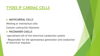
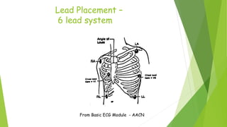
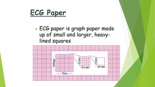
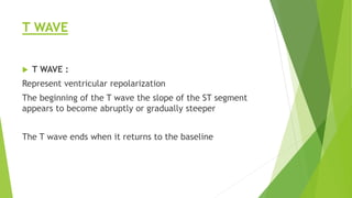
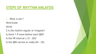
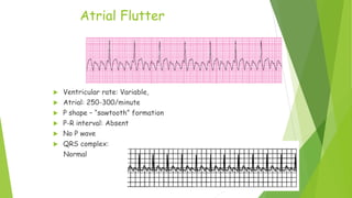
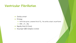
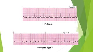
ECG
PATEL.YASH.GIRISHBHAI (KAPC)
TYOES IF CARDIAC CELLS
 MAYOCARDIAL CELLS
Working or mechanical cells
Contain contractile filaments
 PACEMAKER CAELLS
-specialized cell of the electrical conduction system
- Responsible for the spontaneous generation and conduction
of electrical impulses
Cardiac
Conduction
From Basic ECG Module - AACN
SA Node
Cardiac Cycle & the ECG
Isometric line
ST Segment
Lead Placement –
6 lead system
From Basic ECG Module - AACN
ECG Paper
 ECG paper is graph paper made
up of small and larger, heavy-
lined squares
 Smallest squares are 1 mm wide and 1 mm high
 5 small squares between the heavier black lines
 25 small squares within each large square
What Does the ECG Measure?
V
O
L
T
A
G
E
T I M E
PR INTERVAL
 Normal P wave – small, round,
Upright
PR INTERVAL:
Begins with the onset of the P
Wave and ends with the inset of QRS
Complex normally measures 0.12
To 0.20 seconds
5 small boxes
QRS COMPLEX
 A QRS complex normally
Follows each P wave
Consists of Q wave, R wave,
And S wave
Represents the spread of electrical
Impulse through the ventricles
normal – 0.04- 0.12 seconds
ST SEGMENT
 ST – segment
Begins with the end of the QRS complex and ends with the of
the onset of the T wave and is on the same line as the PR
interval
ST segment depression of more than 1 mm is suggestive of
myocardial ischemia
ST segment elevation of more then 1 mm is suggestive of
myocardial injury or pericarditis.
T WAVE
 T WAVE :
Represent ventricular repolarization
The beginning of the T wave the slope of the ST segment
appears to become abruptly or gradually steeper
The T wave ends when it returns to the baseline
ST Segment
 The ST segment is considered:
 “Elevated” if the segment deviates above the baseline of the
PR segment
 “Depressed” if the segment deviates below it
STEPS OF RHYTHM ANLAYSIS
1. What is ate ?
Ventricular
Atrial
2 is the rhythm regular or irregular?
Is there 1 P wave before each QRS?
Is the PR interval (.12- .20)?
Is the QRS narrow or wide(.04- .10)
Determining the Rate
 1500 ÷ No of small boxes within an RR interval (regular rhythms)
 300 ÷ No of Large boxes within an RR interval (regular rhythms)
 10 x No of R complexes in 6 seconds(30 large box)
 10 x No of R complexes in 6 seconds
Rhythm: Regular / Irregular
 Distance between the ‘R’ waves
Normal Sinus Rhythm
 Ventricular rate: 60-100; Regular rhythm
 Atrial: Same as ventricular
 P consistent shape – always positive
 P-R interval: 0.12-0.20
 QRS complex: 0.04-0.10
 1 P wave for every QRS
DYSRHYTHMIAS
 Disorders of electrical impulse:
Formation
Conduction
 Named by
Site of origin of impulse
Mechanism of formation or conduction involved
DYSRTHMIAS
 Site of origin
SA node
Bradycardia, tachycardia
AYRIAL tissue
Flutter, fibrillation
AV node
blocks
SINUS TACHYCARDIA
 ETIOLOGY
↑ CNS response : anxiety; pain; fever; anemia
INTERVANTION
Identify cause, select best treatment
Goal : ↓ HR to normal levels
β-blockers, ACE Inhibitors
Sinus Tachycardia
 Ventricular rate: > 100 (up to180);
Regular rhythm
 Atrial: Same as ventricular
 P consistent shape
 P wave for every QRS
 QRS complex: Normal
Sinus Tachycardia
Sinus Bradycardia
 Ventricular rate:< 60; regular rhythm
 Atrial: same as ventricular
 P consistent shape
 P wave for every QRS
 QRS complex: Normal
Sinus Bradycardia
 Etiology
 PNS dominant; Excessive vagal (Valsalva) stimulation to the heart (↓ SA node
discharge = ↓ HR, ↓ conduction)
 Interventions
 Atropine = Tx of choice
 Pacemaker placement
PAC (premature atraial
contractions):
Irregular P-R rhythms
 Premature, irregular P waves (sometimes “lost” in the T
Wave.
Premature Atrial Contractions:
 Triggered by: Alcohol, nicotine, anxiety, fatigue, fever, and
infections
 Clinical Manifestations: Palpitations or “skipped beats”
Flutter and Fibrillation
Atrial Flutter
 Etiology
 AV node selectively blocks # impulses that reach
ventricles (protective mechanism)
 Rheumatic Heart disease, CHF, AV valve disease, post
cardiac surgery
 Clinical manifestations dependent upon ventricular
response
Atrial Flutter
 Ventricular rate: Variable,
 Atrial: 250-300/minute
 P shape – “sawtooth” formation
 P-R interval: Absent
 No P wave
 QRS complex:
Normal
Atrial Fibrillation
 Etiology
 Most common dysrhythmia in US
 Aging, MI, MS, Cardiomyopathy
 Multiple, rapid impulses many atrial foci;
Atrial depolarization disorganized
Atrial Fibrillation
 Commonly seen after cardiac
surgery (transient)
 Can be intermittent or chronic
 Symptoms:
 SOB
 Fatigue
 Weakness,
 Distended neck veins
 Anxiety
 Palpitations
 Chest discomfort
 Irregular pulse
Atrial Fibrillation
 Ventricular rate: < 100 (controlled)
 Atrial: Unable to determine (>350)
 No P waves (fibrillatory waves)
 P-R interval: Absent
 QRS complex: Normal
Atrial Fibrillation
Premature Ventricular Contractions
 Etiology
 Early ventricular complexes, followed by pause
 Ventricular contraction originating in an ectopic
focus outside ventricles
 Aging, MI, Caffeine, ↓ K+
Premature Ventricular Contractions
 No P wave
 QRS wide and unusual
 ST segment often slops in the opposite direction
PVC
Ventricular Tachycardia
 Etiology
 Repetitive firing of an irritated ventricular ectopic
focus
 Intermittent
 Sustained: > 15-30 sec
Ventricular Tachycardia (V Tach)
 Unable to determine rhythm
 No P waves present
 QRS complex > 0.10 sec
Ventricular Fibrillation
 Cardiac arrest
 Etiology
 Ventricles quiver, consume lots of O2, No cardiac output, no perfusion
 AMI, ↓ K+, ↓ Mg+
 Rapidly fatal (3-5 min)
 No proper QRS Complex is noted
Ventricular Fibrillation
 Assessment
 LOC, Absence of Pulse
 Apnea
 Seizures
 Development of respiratory & metabolic acidosis
 Treatment
 CPR (ACLS)
 Defibrillation
Ventricular Fibrillation (V Fib)
Coarse
Fine
Heart Blocks
 Occur when there is a delay in the conduction of the impulse
through the AV node
 PR is > 0.20 seconds
 SA node function is normal
Heart Block Overview
 1st degree –
 PR interval > 0.20 seconds
 All impulses reach the ventricles
 2nd degree – (2 types)
 Mobitz I – each impulses takes longer to conduct
until 1 is blocked and a QRS complex is dropped and
a pause occurs; then cycle repeats
 Mobitz II – None conduction to ventricles.2-4 p
waves between QRS.
 3rd degree –
 None of the atrial impulses reach the ventricles
 Activity of the atria and ventricles is ‘divorced’
 Results in inadequate cardiac output
 Requires pacemaker
2nd degree Type 1
1st degree
3rd degree
2nd degree Type II
Pacemakers
ECG

ECG

  • 1.
  • 2.
    TYOES IF CARDIACCELLS  MAYOCARDIAL CELLS Working or mechanical cells Contain contractile filaments  PACEMAKER CAELLS -specialized cell of the electrical conduction system - Responsible for the spontaneous generation and conduction of electrical impulses
  • 3.
  • 4.
    From Basic ECGModule - AACN SA Node
  • 5.
    Cardiac Cycle &the ECG Isometric line ST Segment
  • 6.
    Lead Placement – 6lead system From Basic ECG Module - AACN
  • 7.
    ECG Paper  ECGpaper is graph paper made up of small and larger, heavy- lined squares  Smallest squares are 1 mm wide and 1 mm high  5 small squares between the heavier black lines  25 small squares within each large square
  • 8.
    What Does theECG Measure? V O L T A G E T I M E
  • 9.
    PR INTERVAL  NormalP wave – small, round, Upright PR INTERVAL: Begins with the onset of the P Wave and ends with the inset of QRS Complex normally measures 0.12 To 0.20 seconds 5 small boxes
  • 10.
    QRS COMPLEX  AQRS complex normally Follows each P wave Consists of Q wave, R wave, And S wave Represents the spread of electrical Impulse through the ventricles normal – 0.04- 0.12 seconds
  • 11.
    ST SEGMENT  ST– segment Begins with the end of the QRS complex and ends with the of the onset of the T wave and is on the same line as the PR interval ST segment depression of more than 1 mm is suggestive of myocardial ischemia ST segment elevation of more then 1 mm is suggestive of myocardial injury or pericarditis.
  • 12.
    T WAVE  TWAVE : Represent ventricular repolarization The beginning of the T wave the slope of the ST segment appears to become abruptly or gradually steeper The T wave ends when it returns to the baseline
  • 13.
    ST Segment  TheST segment is considered:  “Elevated” if the segment deviates above the baseline of the PR segment  “Depressed” if the segment deviates below it
  • 14.
    STEPS OF RHYTHMANLAYSIS 1. What is ate ? Ventricular Atrial 2 is the rhythm regular or irregular? Is there 1 P wave before each QRS? Is the PR interval (.12- .20)? Is the QRS narrow or wide(.04- .10)
  • 15.
    Determining the Rate 1500 ÷ No of small boxes within an RR interval (regular rhythms)  300 ÷ No of Large boxes within an RR interval (regular rhythms)  10 x No of R complexes in 6 seconds(30 large box)  10 x No of R complexes in 6 seconds
  • 16.
    Rhythm: Regular /Irregular  Distance between the ‘R’ waves
  • 17.
    Normal Sinus Rhythm Ventricular rate: 60-100; Regular rhythm  Atrial: Same as ventricular  P consistent shape – always positive  P-R interval: 0.12-0.20  QRS complex: 0.04-0.10  1 P wave for every QRS
  • 18.
    DYSRHYTHMIAS  Disorders ofelectrical impulse: Formation Conduction  Named by Site of origin of impulse Mechanism of formation or conduction involved
  • 19.
    DYSRTHMIAS  Site oforigin SA node Bradycardia, tachycardia AYRIAL tissue Flutter, fibrillation AV node blocks
  • 20.
    SINUS TACHYCARDIA  ETIOLOGY ↑CNS response : anxiety; pain; fever; anemia INTERVANTION Identify cause, select best treatment Goal : ↓ HR to normal levels β-blockers, ACE Inhibitors
  • 21.
    Sinus Tachycardia  Ventricularrate: > 100 (up to180); Regular rhythm  Atrial: Same as ventricular  P consistent shape  P wave for every QRS  QRS complex: Normal Sinus Tachycardia
  • 22.
    Sinus Bradycardia  Ventricularrate:< 60; regular rhythm  Atrial: same as ventricular  P consistent shape  P wave for every QRS  QRS complex: Normal
  • 23.
    Sinus Bradycardia  Etiology PNS dominant; Excessive vagal (Valsalva) stimulation to the heart (↓ SA node discharge = ↓ HR, ↓ conduction)  Interventions  Atropine = Tx of choice  Pacemaker placement
  • 24.
    PAC (premature atraial contractions): IrregularP-R rhythms  Premature, irregular P waves (sometimes “lost” in the T Wave.
  • 25.
    Premature Atrial Contractions: Triggered by: Alcohol, nicotine, anxiety, fatigue, fever, and infections  Clinical Manifestations: Palpitations or “skipped beats”
  • 26.
  • 27.
    Atrial Flutter  Etiology AV node selectively blocks # impulses that reach ventricles (protective mechanism)  Rheumatic Heart disease, CHF, AV valve disease, post cardiac surgery  Clinical manifestations dependent upon ventricular response
  • 28.
    Atrial Flutter  Ventricularrate: Variable,  Atrial: 250-300/minute  P shape – “sawtooth” formation  P-R interval: Absent  No P wave  QRS complex: Normal
  • 29.
    Atrial Fibrillation  Etiology Most common dysrhythmia in US  Aging, MI, MS, Cardiomyopathy  Multiple, rapid impulses many atrial foci; Atrial depolarization disorganized
  • 30.
    Atrial Fibrillation  Commonlyseen after cardiac surgery (transient)  Can be intermittent or chronic  Symptoms:  SOB  Fatigue  Weakness,  Distended neck veins  Anxiety  Palpitations  Chest discomfort  Irregular pulse
  • 31.
    Atrial Fibrillation  Ventricularrate: < 100 (controlled)  Atrial: Unable to determine (>350)  No P waves (fibrillatory waves)  P-R interval: Absent  QRS complex: Normal
  • 32.
  • 33.
    Premature Ventricular Contractions Etiology  Early ventricular complexes, followed by pause  Ventricular contraction originating in an ectopic focus outside ventricles  Aging, MI, Caffeine, ↓ K+
  • 34.
    Premature Ventricular Contractions No P wave  QRS wide and unusual  ST segment often slops in the opposite direction PVC
  • 35.
    Ventricular Tachycardia  Etiology Repetitive firing of an irritated ventricular ectopic focus  Intermittent  Sustained: > 15-30 sec
  • 36.
    Ventricular Tachycardia (VTach)  Unable to determine rhythm  No P waves present  QRS complex > 0.10 sec
  • 37.
    Ventricular Fibrillation  Cardiacarrest  Etiology  Ventricles quiver, consume lots of O2, No cardiac output, no perfusion  AMI, ↓ K+, ↓ Mg+  Rapidly fatal (3-5 min)  No proper QRS Complex is noted
  • 38.
    Ventricular Fibrillation  Assessment LOC, Absence of Pulse  Apnea  Seizures  Development of respiratory & metabolic acidosis  Treatment  CPR (ACLS)  Defibrillation
  • 39.
  • 40.
    Heart Blocks  Occurwhen there is a delay in the conduction of the impulse through the AV node  PR is > 0.20 seconds  SA node function is normal
  • 41.
    Heart Block Overview 1st degree –  PR interval > 0.20 seconds  All impulses reach the ventricles  2nd degree – (2 types)  Mobitz I – each impulses takes longer to conduct until 1 is blocked and a QRS complex is dropped and a pause occurs; then cycle repeats  Mobitz II – None conduction to ventricles.2-4 p waves between QRS.  3rd degree –  None of the atrial impulses reach the ventricles  Activity of the atria and ventricles is ‘divorced’  Results in inadequate cardiac output  Requires pacemaker
  • 42.
    2nd degree Type1 1st degree
  • 43.
  • 44.