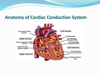
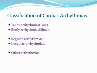
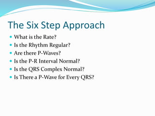
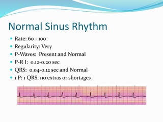
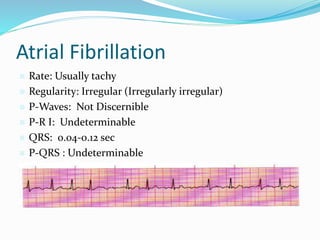
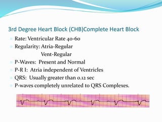
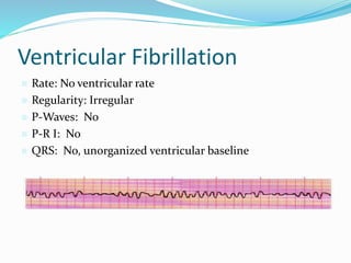
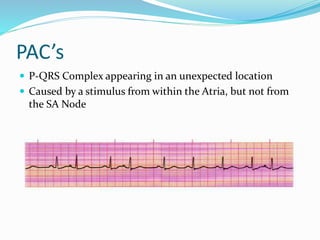
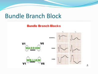
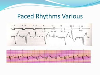
BY
Rizk Elazhary
MD, Anesthesiology
Benha University
Introduction
Anatomy of Cardiac Conduction System
Physiology of the heart
Electrocardiogram  Action potentials
through myocardium
during cardiac cycle
produces electric
currents than can be
measured
 Pattern
 P wave
 Atria depolarization
 QRS complex
 Ventricle depolarization
 Atria repolarization
 T wave:
 Ventricle repolarization
What is Arrhythmias?
Classification of Cardiac Arrhythmias
 Tachy arrhythmias(Fast).
 Brady arrhythmias(Slow).
 Regular-arrhythmias.
 Irregular-arrhythmias.
 Other arrhythmias.
Approach to Arrhythmias
The Six Step Approach
 What is the Rate?
 Is the Rhythm Regular?
 Are there P-Waves?
 Is the P-R Interval Normal?
 Is the QRS Complex Normal?
 Is There a P-Wave for Every QRS?
Normal Sinus Rhythm
 Rate: 60 - 100
 Regularity: Very
 P-Waves: Present and Normal
 P-R I: 0.12-0.20 sec
 QRS: 0.04-0.12 sec and Normal
 1 P: 1 QRS, no extras or shortages
Sinus Tachycardia
 Rate: Over 100
 Regularity: Regular
 P-Waves: Present and Normal
 P-R I: 0.12-0.20 sec
 QRS: 0.04-0.12 sec and Normal
 1 P: 1 QRS, no extras or shortages
Sinus Bradycardia
 Rate: Less than 60
 Regularity: Regular
 P-Waves: Present and Normal
 P-R I: 0.12-0.20 sec
 QRS: 0.04-0.12 sec and Normal
 1 P: 1 QRS, no extras or shortages
Atrial Fibrillation
 Rate: Usually tachy
 Regularity: Irregular (Irregularly irregular)
 P-Waves: Not Discernible
 P-R I: Undeterminable
 QRS: 0.04-0.12 sec
 P-QRS : Undeterminable
Atrial Flutter
 Rate: Usually tachy
 Regularity: Atria Regular
• Ventricles May be Irregular
 P-Waves: Saw tooth Pattern 2:1, 3:1, 4:1...
 P-R I: 0.12-0.20 sec on conducting beat
 QRS: 0.04-0.12 sec
 P-waves outnumber QRS
(Paroxysmal) Supra Ventricular Tach
 Rate: 140-220
 Regularity: Regular
 P-Waves: Usually falls within the QRS-T complex (
sometimes not visible)
 P-R I: Shorter than 0.12, or absent
 QRS: 0.04-0.12 sec and Normal
 P-QRS :Undeterminable
SVT
 WPW
 Usually based on Hx.
 Delta wave on Q
 Shortened PR-I
 No Verapamil – Accessory Path use increase
1st Degree Heart Block
 Rate: 60 - 100
 Regularity: Very
 P-Waves: Present and Normal
 P-R I: Longer than 0.20 sec
 QRS: 0.04-0.12 sec and Normal
 1 P: 1 QRS, no extras or shortages
2nd Degree Heart Block (Type 1) Wenkebach
 Rate: Can be Normal, or usually brady
 Regularity: Irregular
 P-Waves: Present and Normal
 P-R I: Lengthens until beat is dropped
 QRS: 0.04-0.12 sec and Normal
 P-wave present on conducting beats, increased delay
causes missed QRS
2nd Degree Heart Block (Type 2) Mobitz II
 Rate: Less than 60
 Regularity: Irregular
 P-Waves: Present, 2:1, 3:1, 4:1
 P-R I: 0.12-0.20 sec on conducting beat
 QRS: 0.04-0.12 sec, may begin to widen
 P-wave for every QRS and extras depending on conduction
ratio
3rd Degree Heart Block (CHB)Complete Heart Block
 Rate: Ventricular Rate 40-60
 Regularity: Atria-Regular
• Vent-Regular
 P-Waves: Present and Normal
 P-R I: Atria independent of Ventricles
 QRS: Usually greater than 0.12 sec
 P-waves completely unrelated to QRS Complexes.
Junctional Rhythm
 Rate: 40-60
 Regularity: Regular
 P-Waves: Inverted, Retrograde or Absent
 P-R I: Shortened or absent
 QRS: 0.04-0.12 sec
 P-wave for every QRS, sometimes not visible
Ventricular Tachycardia
 Rate: 100-220
 Regularity: Regular
 P-Waves: None
 P-R I: None
 QRS: Greater than 0.12 sec
Ventricular Fibrillation
 Rate: No ventricular rate
 Regularity: Irregular
 P-Waves: No
 P-R I: No
 QRS: No, unorganized ventricular baseline
Asystole
 Rate: 0
 Regularity: N/A
 P-Waves: None
 P-R I: N/A
 QRS: None
(check another lead)
Agonal / Idioventricular
 Rate: 20-40
 Regularity: Irregular
 P-Waves: None
 P-R I: N/A
 QRS: Wider than 0.12 sec
(a dying heart)
Other Arrhythmias
 Premature Ventricular Contractions
 Premature Atrial Contractions
 Bundle Branch Blocks
 Pacer Considerations (Atrial, Ventricular or Both)
Premature Ventricular Contractions
 Wide, Bizarre QRS Complex
 Always identify the underlying rhythm first
 Can appear in couplets, triplets, short runs of V-Tach,
bigeminy and trigeminy
 Can be uni-focal or multi-focal
 Caused by random firing within the ventricles
 Not accompanied by a P-wave
PAC’s
 P-QRS Complex appearing in an unexpected location
 Caused by a stimulus from within the Atria, but not from
the SA Node
Bundle Branch Block
 Any rhythm having a BBB will have a widened twin
peaked R-Wave
Bundle Branch Block
Paced Rhythms Various
QT Prolongation
 QT represents the ventricular refractory period
 Normal
 Men 450ms
 Women 460ms
 Corrected QT (QTc)
 QTm/√(R-R)
 Causes
 Drugs (Na channel blockers)
 Hypocalcemia, hypomagnesemia, hypokalemia
 Hypothermia
 AMI
 Congenital
 Increased ICP
Torsade de pointes
 Torsades de pointes is a polymorphic ventricular
tachycardia . The phrase “torsades de pointes” means
“twisting on a point” which explains the action of the QRS
complex and how it varies from beat to beat. It is a
shockable rhythm.
Normal Variants
 Always normal:
 Sinus Arrhythmia
 Supraventricular Extrasystoles
 Partial RBBB
 Often normal:
 Sinus Bradycardia (and pauses in athletes)
 First Degree Heart Block
 Ventricular Extrasystoles
 Left/Right Axis Deviation
 RBBB
Special Situations
 Dextrocardia – reverse precordial leads
 Large breasts – don't place electrodes on top of breast
 Bilateral breast implants you should apply V4, V5, and V6
close to the midaxillary line.
 Note patient abnormalities on ECG
 Do not place electrodes on open wounds, burns, or clear
dressings
 Do not allow electrodes to touch one another
References
 Pictures and info from:
 Flip and See ECG, 2nd Edition
 Cohn/Gilroy-Doohan
 A great resource
 Paramedic Paramedic Textbook, Revised 2nd Edition
 Mick J. Sanders, Mosby
 ECG’s Made Easy, 2nd Edition
 Barbara Aehlert, RN, Mosby
 Basic Dysrhythmias, Interpretation and Management,
3rd Edition
 Robert J. Huszar, Mosby
Rizk  ecg

Rizk ecg

  • 1.
  • 2.
  • 3.
    Anatomy of CardiacConduction System
  • 4.
  • 5.
    Electrocardiogram  Actionpotentials through myocardium during cardiac cycle produces electric currents than can be measured  Pattern  P wave  Atria depolarization  QRS complex  Ventricle depolarization  Atria repolarization  T wave:  Ventricle repolarization
  • 8.
  • 9.
    Classification of CardiacArrhythmias  Tachy arrhythmias(Fast).  Brady arrhythmias(Slow).  Regular-arrhythmias.  Irregular-arrhythmias.  Other arrhythmias.
  • 10.
  • 11.
    The Six StepApproach  What is the Rate?  Is the Rhythm Regular?  Are there P-Waves?  Is the P-R Interval Normal?  Is the QRS Complex Normal?  Is There a P-Wave for Every QRS?
  • 12.
    Normal Sinus Rhythm Rate: 60 - 100  Regularity: Very  P-Waves: Present and Normal  P-R I: 0.12-0.20 sec  QRS: 0.04-0.12 sec and Normal  1 P: 1 QRS, no extras or shortages
  • 13.
    Sinus Tachycardia  Rate:Over 100  Regularity: Regular  P-Waves: Present and Normal  P-R I: 0.12-0.20 sec  QRS: 0.04-0.12 sec and Normal  1 P: 1 QRS, no extras or shortages
  • 14.
    Sinus Bradycardia  Rate:Less than 60  Regularity: Regular  P-Waves: Present and Normal  P-R I: 0.12-0.20 sec  QRS: 0.04-0.12 sec and Normal  1 P: 1 QRS, no extras or shortages
  • 15.
    Atrial Fibrillation  Rate:Usually tachy  Regularity: Irregular (Irregularly irregular)  P-Waves: Not Discernible  P-R I: Undeterminable  QRS: 0.04-0.12 sec  P-QRS : Undeterminable
  • 16.
    Atrial Flutter  Rate:Usually tachy  Regularity: Atria Regular • Ventricles May be Irregular  P-Waves: Saw tooth Pattern 2:1, 3:1, 4:1...  P-R I: 0.12-0.20 sec on conducting beat  QRS: 0.04-0.12 sec  P-waves outnumber QRS
  • 17.
    (Paroxysmal) Supra VentricularTach  Rate: 140-220  Regularity: Regular  P-Waves: Usually falls within the QRS-T complex ( sometimes not visible)  P-R I: Shorter than 0.12, or absent  QRS: 0.04-0.12 sec and Normal  P-QRS :Undeterminable
  • 18.
    SVT  WPW  Usuallybased on Hx.  Delta wave on Q  Shortened PR-I  No Verapamil – Accessory Path use increase
  • 19.
    1st Degree HeartBlock  Rate: 60 - 100  Regularity: Very  P-Waves: Present and Normal  P-R I: Longer than 0.20 sec  QRS: 0.04-0.12 sec and Normal  1 P: 1 QRS, no extras or shortages
  • 20.
    2nd Degree HeartBlock (Type 1) Wenkebach  Rate: Can be Normal, or usually brady  Regularity: Irregular  P-Waves: Present and Normal  P-R I: Lengthens until beat is dropped  QRS: 0.04-0.12 sec and Normal  P-wave present on conducting beats, increased delay causes missed QRS
  • 21.
    2nd Degree HeartBlock (Type 2) Mobitz II  Rate: Less than 60  Regularity: Irregular  P-Waves: Present, 2:1, 3:1, 4:1  P-R I: 0.12-0.20 sec on conducting beat  QRS: 0.04-0.12 sec, may begin to widen  P-wave for every QRS and extras depending on conduction ratio
  • 22.
    3rd Degree HeartBlock (CHB)Complete Heart Block  Rate: Ventricular Rate 40-60  Regularity: Atria-Regular • Vent-Regular  P-Waves: Present and Normal  P-R I: Atria independent of Ventricles  QRS: Usually greater than 0.12 sec  P-waves completely unrelated to QRS Complexes.
  • 23.
    Junctional Rhythm  Rate:40-60  Regularity: Regular  P-Waves: Inverted, Retrograde or Absent  P-R I: Shortened or absent  QRS: 0.04-0.12 sec  P-wave for every QRS, sometimes not visible
  • 24.
    Ventricular Tachycardia  Rate:100-220  Regularity: Regular  P-Waves: None  P-R I: None  QRS: Greater than 0.12 sec
  • 25.
    Ventricular Fibrillation  Rate:No ventricular rate  Regularity: Irregular  P-Waves: No  P-R I: No  QRS: No, unorganized ventricular baseline
  • 26.
    Asystole  Rate: 0 Regularity: N/A  P-Waves: None  P-R I: N/A  QRS: None (check another lead)
  • 27.
    Agonal / Idioventricular Rate: 20-40  Regularity: Irregular  P-Waves: None  P-R I: N/A  QRS: Wider than 0.12 sec (a dying heart)
  • 28.
    Other Arrhythmias  PrematureVentricular Contractions  Premature Atrial Contractions  Bundle Branch Blocks  Pacer Considerations (Atrial, Ventricular or Both)
  • 29.
    Premature Ventricular Contractions Wide, Bizarre QRS Complex  Always identify the underlying rhythm first  Can appear in couplets, triplets, short runs of V-Tach, bigeminy and trigeminy  Can be uni-focal or multi-focal  Caused by random firing within the ventricles  Not accompanied by a P-wave
  • 30.
    PAC’s  P-QRS Complexappearing in an unexpected location  Caused by a stimulus from within the Atria, but not from the SA Node
  • 31.
    Bundle Branch Block Any rhythm having a BBB will have a widened twin peaked R-Wave
  • 32.
  • 33.
  • 34.
    QT Prolongation  QTrepresents the ventricular refractory period  Normal  Men 450ms  Women 460ms  Corrected QT (QTc)  QTm/√(R-R)  Causes  Drugs (Na channel blockers)  Hypocalcemia, hypomagnesemia, hypokalemia  Hypothermia  AMI  Congenital  Increased ICP
  • 35.
    Torsade de pointes Torsades de pointes is a polymorphic ventricular tachycardia . The phrase “torsades de pointes” means “twisting on a point” which explains the action of the QRS complex and how it varies from beat to beat. It is a shockable rhythm.
  • 36.
    Normal Variants  Alwaysnormal:  Sinus Arrhythmia  Supraventricular Extrasystoles  Partial RBBB  Often normal:  Sinus Bradycardia (and pauses in athletes)  First Degree Heart Block  Ventricular Extrasystoles  Left/Right Axis Deviation  RBBB
  • 37.
    Special Situations  Dextrocardia– reverse precordial leads  Large breasts – don't place electrodes on top of breast  Bilateral breast implants you should apply V4, V5, and V6 close to the midaxillary line.  Note patient abnormalities on ECG  Do not place electrodes on open wounds, burns, or clear dressings  Do not allow electrodes to touch one another
  • 38.
    References  Pictures andinfo from:  Flip and See ECG, 2nd Edition  Cohn/Gilroy-Doohan  A great resource  Paramedic Paramedic Textbook, Revised 2nd Edition  Mick J. Sanders, Mosby  ECG’s Made Easy, 2nd Edition  Barbara Aehlert, RN, Mosby  Basic Dysrhythmias, Interpretation and Management, 3rd Edition  Robert J. Huszar, Mosby