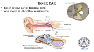
Ear
Submitted by :
•Amit Kumar Shah
• Deepashree sah
• Kiran Gautam
• Kiran Kathayat
• Neelam Bist
• Nischala Raut
• Sangita Bhajagain
• SimRan Shrestha
• Sudhakar tharu
Submitted TO :
Department of
anatomy
Madan Bhandari
Academy of Health
Sciences
2.
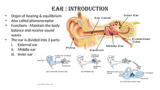
• Organ ofhearing & equilibrium
• Also called phonoreceptor
• Functions : Maintain the body
balance and receive sound
waves
• The ear is divided into 3 parts:
i. External ear
ii. Middle ear
iii. Inner ear
Ear : Introduction
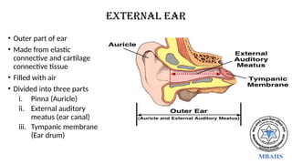
4.
• Outer partof ear
• Made from elastic
connective and cartilage
connective tissue
• Filled with air
• Divided into three parts
i. Pinna (Auricle)
ii. External auditory
meatus (ear canal)
iii. Tympanic membrane
(Ear drum)
External ear
5.
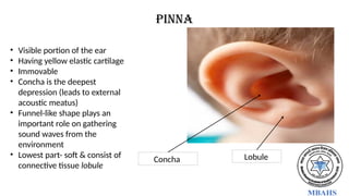
Pinna
• Visible portionof the ear
• Having yellow elastic cartilage
• Immovable
• Concha is the deepest
depression (leads to external
acoustic meatus)
• Funnel-like shape plays an
important role on gathering
sound waves from the
environment
• Lowest part- soft & consist of
connective tissue lobule
Concha Lobule
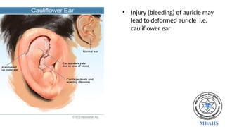
7.
• Injury (bleeding)of auricle may
lead to deformed auricle i.e.
cauliflower ear
8.
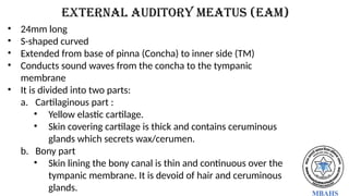
External auditory meatus(EAM)
• 24mm long
• S-shaped curved
• Extended from base of pinna (Concha) to inner side (TM)
• Conducts sound waves from the concha to the tympanic
membrane
• It is divided into two parts:
a. Cartilaginous part :
• Yellow elastic cartilage.
• Skin covering cartilage is thick and contains ceruminous
glands which secrets wax/cerumen.
b. Bony part
• Skin lining the bony canal is thin and continuous over the
tympanic membrane. It is devoid of hair and ceruminous
glands.
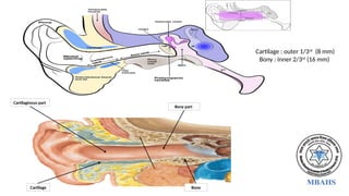
9.
Cartilage : outer1/3rd
(8 mm)
Bony : Inner 2/3rd
(16 mm)
Cartilaginous part
Cartilage
Bony part
Bone
10.
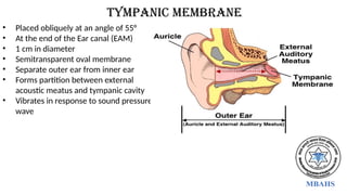
Tympanic membrane
• Placedobliquely at an angle of 55°
• At the end of the Ear canal (EAM)
• 1 cm in diameter
• Semitransparent oval membrane
• Separate outer ear from inner ear
• Forms partition between external
acoustic meatus and tympanic cavity
• Vibrates in response to sound pressure
wave
11.
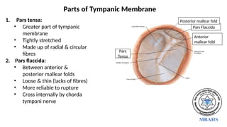
1. Pars tensa:
•Greater part of tympanic
membrane
• Tightly stretched
• Made up of radial & circular
fibres
2. Pars flaccida:
• Between anterior &
posterior mallear folds
• Loose & thin (lacks of fibres)
• More reliable to rupture
• Cross internally by chorda
tympani nerve
Parts of Tympanic Membrane
Pars
Tensa
Pars Flaccida
Anterior
mallear fold
Posterior mallear fold
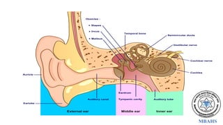
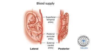
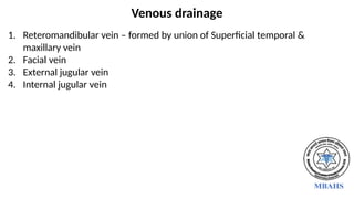
Venous drainage
1. Reteromandibularvein – formed by union of Superficial temporal &
maxillary vein
2. Facial vein
3. External jugular vein
4. Internal jugular vein
15.
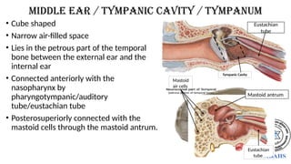
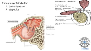
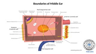
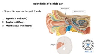
• Cube shaped
•Narrow air-filled space
• Lies in the petrous part of the temporal
bone between the external ear and the
internal ear
• Connected anteriorly with the
nasopharynx by
pharyngotympanic/auditory
tube/eustachian tube
• Posterosuperiorly connected with the
mastoid cells through the mastoid antrum.
middle ear / Tympanic cavity / tympanum
Tympanic Cavity
Eustachian
tube
Mastoid
air cells
Mastoid antrum
Eustachian
tube
16.
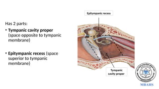
Has 2 parts:
•Tympanic cavity proper
(space opposite to tympanic
membrane)
• Epitympanic recess (space
superior to tympanic
membrane)
Tympanic
cavity proper
Epitympanic recess
17.
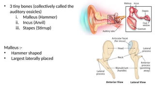
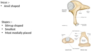
• 3 tinybones (collectively called the
auditory ossicles)
i. Malleus (Hammer)
ii. Incus (Anvil)
iii. Stapes (Stirrup)
Malleus :-
• Hammer shaped
• Largest laterally placed
inner ear
• Liesin petrous part of temporal bone
• Also known as Labrynth or Auris Interna
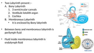
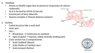
25.
• Two Labyrinthpresent :-
A. Bony Labyrinth
1. 3 semicircular cannals
2. Vestibule lotolith organ
3. Cochlea
B. Membranous Labyrinth
• It is enclosed by Bony labyrinth
• Between bony and membranous labyrinth is
perilymph fluid
• Fluid inside membranous labyrinth is
endolymph fluid
26.
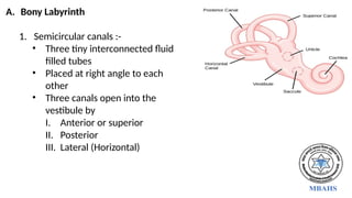
A. Bony Labyrinth
1.Semicircular canals :-
• Three tiny interconnected fluid
filled tubes
• Placed at right angle to each
other
• Three canals open into the
vestibule by
I. Anterior or superior
II. Posterior
III. Lateral (Horizontal)
27.
2. Vestibule
• Knownas Otolith organ (due to presence of granules of calcium
carbonate)
• As it is formed by Utricle & Saccule
• Central part of bony labyrinth
• Sensory receptor is Macula (balance posture)
3. Cochlea
• Coiled structure like a snail shell
• Inner part
• Has :-
Broad base → Continuous to vestibule
Apex (Cupala) → Narrow, coiled centrally (ending part)
• Cross section has 3 compartments
Scala Vestibuli (Above)
Scala Media or Cochlear duct
Scala tympani (below)
28.
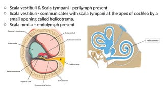
o Scala vestibuli& Scala tympani - perilymph present.
o Scala vestibuli - communicates with scala tympani at the apex of cochlea by a
small opening called helicotrema.
o Scala media – endolymph present
Helicotrema
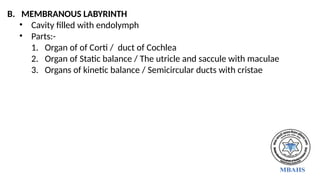
B. MEMBRANOUS LABYRINTH
•Cavity filled with endolymph
• Parts:-
1. Organ of of Corti / duct of Cochlea
2. Organ of Static balance / The utricle and saccule with maculae
3. Organs of kinetic balance / Semicircular ducts with cristae
31.
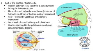
1. Duct ofthe Cochlea / Scala Media
• Present between scala vestibule & scala tympani
• Triangular in cross-section
• Floor – formed by basilar membrane (presence of
hair cells i.e. Organ of Corti or auditory receptors)
• Roof – formed by vestibular or Reissner’s
membrane
• Outer wall – formed by bony wall of cochlea
• Hair is embedded in the gelatinous membrane
called membrane tectoria
32.
2. Saccule &Utricle
• Saccule lies anteroinferiorly of
vestibule
• Utricle lies posterosuperiorly
of vestibule
• Utricle is larger than saccule
• Utricle receives the ends of 3
semicircular ducts through 5
openings
• They are static balance
receptors
• Maculae are receptors that
give information about
position of head
33.
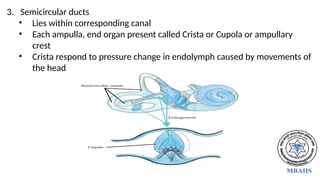
3. Semicircular ducts
•Lies within corresponding canal
• Each ampulla, end organ present called Crista or Cupola or ampullary
crest
• Crista respond to pressure change in endolymph caused by movements of
the head


![CTEV [ clubfoot] DR ARUN LAL ,DR MOHAMED ASHRAF travancore medical college k...](https://cdn.slidesharecdn.com/ss_thumbnails/ctevclubfootdrarunlaldrmohamedashraftravancoremedicalcollegekollamkeralaindia-260208063247-18fc466c-thumbnail.jpg?width=640&height=640&fit=bounds)


![PERI-PROSTHETIC FRACTURE NAIL-PLATE CONSTRUCT [NPC].pptx](https://cdn.slidesharecdn.com/ss_thumbnails/drarunkumardrmohamedashrafperiprostheticfrasturenail-plateconstructnpc-260209164459-7e9d15a1-thumbnail.jpg?width=640&height=640&fit=bounds)
