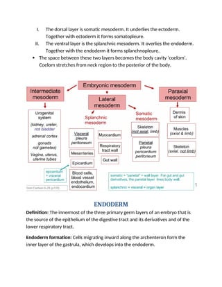
Early Vertebrate Development
NEURULATION
Neurulation is a folding process in vertebrates’ embryo which includes the
transformation of the neural plate into the neural tube.
Neurula: At this stage the embryo is called Neurula.
Types:
There are two types of Neurulation.
1. Primary Neurulation:
Primary neurulation leads to the development of brain and spinal cord.
2. Secondary Neurulation:
Secondary neurulation leads to the formation of lower sacral and coccygeal
cord.
Occurance:
Neurulation occur after gastrulation.
In human it occurs in two distinct phases.
• Phase l:
This is the phase of 3rd
and 4th
week of gestation. During this phase primary
neurulation occur.
• Phase ll:
This is the phase of 5th
and 6th
week of gestation. During this phase secondary
neurulation occur.
Process of Neurulation
Process of neurulation complete in the following steps:
1. Neuroectodermal tissue differentiates from the
ectoderm and thickens into the neural plate. The neural
plate borders separate the ectoderm from the neural
plate.
2. The neural plate bends dorsally, with the two end
eventually joining at the neural plate borders, which
are now referred to as to the neural crest.
3. The closer of the neural tube disconnect the neural
crest from the epidermis. Neural crest cells
differentiate to form most of the peripheral nervous
system.
4. The notochord degenerates and only persists as the
nucleus pulposus of the intervertebral discs. Other
mesoderm cells differentiate into the somites, the
precursors of the axial skeleton and skeletal muscle.
ECTODERM
The word ectoderm comes from the Greek ektos meaning "outside", and derma
meaning "skin".
Definition: The ectoderm is one of the three primary germ layers formed in early
embryonic development. It is the outermost layer, and is superficial to the
mesoderm (the middle layer) and endoderm (the innermost layer).
Origination: It emerges and originates from the outer layer of germ cells.
Gastrulation process occurs in third week of pregnancy in which the outer germ
cells form the ectoderm.
Differentiation of Ectoderm into epidermis and neural tube
Derivatives of Ectoderm:
THE ECTODERM gives rise to the central nervous system (the brain and spinal
cord); the peripheral nervous system; the sensory epithelia of the eye, ear, and
nose; the epidermis and its appendages (the nails and hair); the mammary glands;
the hypophysis; the subcutaneous glands; and the enamel of the teeth.
MESODERM
The mesoderm is the middle layer of the three germ layers that develops during
gastrulation in the very early development of the embryo of most animals.
Mesoderm Formation
In the early blastula, the future mesoderm cells are found on the surface. During
gastrulation the cells will rearrange until the mesoderm (and endoderm) are on
the inside of the embryo, and the ectoderm is on the outside surface. This occurs
by invagination of the mesoderm and endoderm cells; they migrate to the interior
while the ectoderm spreads to cover the exterior.
Derivatives of Mesoderm
The trunk mesoderm of a neurula stage embryo is divided into 4 regions.
1. Chordamesoderm: This forms notochord. Its main function is to establish
anterior posterior axis.
2. Paraxial mesoderm: Both sides on the notochord is the paraxial mesoderm.
The cells from this region will form somites.
3. Intermediate mesoderm: Forms urinogenital system.
4. Lateral plate mesoderm: forms heart, blood vessels and blood cells, lining of
the body cavities, extraembryonic membranes. The lateral plate mesoderm
splits horizontally into two layers.
I. The dorsal layer is somatic mesoderm. It underlies the ectoderm.
Together with ectoderm it forms somatopleure.
II. The ventral layer is the splanchnic mesoderm. It overlies the endoderm.
Together with the endoderm it forms splanchnopleure.
 The space between these two layers becomes the body cavity ‘coelom’.
Coelom stretches from neck region to the posterior of the body.
ENDODERM
Definition: The innermost of the three primary germ layers of an embryo that is
the source of the epithelium of the digestive tract and its derivatives and of the
lower respiratory tract.
Endoderm formation: Cells migrating inward along the archenteron form the
inner layer of the gastrula, which develops into the endoderm.
Endoderm Function
The two main functions of the endoderm are:
1. Induce the formation of several mesodermal organs.
2. Construct the lining of the digestive and respiratory tubes.
Derivatives of Endoderm
The endoderm will become the digestive tract (or gut), as well as a number of
associated organs and glands. It will give rise to the lungs, liver, and pancreas, as
well as the thymus, thyroid, and parathyroid glands. In addition, endoderm cells
will form the lining of many of the body’s organ systems including the respiratory
system, the digestive system, auditory system and the urinary system.
Gastrointestinal
tract
the entire alimentary canal except part of the mouth,
pharynx and the terminal part of the rectum (which are
lined by involutions of the ectoderm), the lining cells of all
the glands which open into the digestive tube, including
those of the liver and pancreas
Respiratory tract the trachea, bronchi, and alveoli of the lungs
Endocrine glands
and organs
the lining of the follicles of the thyroid gland and the
epithelial component of the thymus (i.e. thymic epithelial
cells).
Auditory system the epithelium of the auditory tube and tympanic cavity
Urinary system the urinary bladder and part of the urethra

Early Vertebrate Development, NEURULATION, ECTODERM, MESODERM, ENDODERM.docx

  • 1.
    Early Vertebrate Development NEURULATION Neurulationis a folding process in vertebrates’ embryo which includes the transformation of the neural plate into the neural tube. Neurula: At this stage the embryo is called Neurula. Types: There are two types of Neurulation. 1. Primary Neurulation: Primary neurulation leads to the development of brain and spinal cord. 2. Secondary Neurulation: Secondary neurulation leads to the formation of lower sacral and coccygeal cord. Occurance: Neurulation occur after gastrulation. In human it occurs in two distinct phases. • Phase l: This is the phase of 3rd and 4th week of gestation. During this phase primary neurulation occur. • Phase ll: This is the phase of 5th and 6th week of gestation. During this phase secondary neurulation occur.
  • 2.
    Process of Neurulation Processof neurulation complete in the following steps: 1. Neuroectodermal tissue differentiates from the ectoderm and thickens into the neural plate. The neural plate borders separate the ectoderm from the neural plate. 2. The neural plate bends dorsally, with the two end eventually joining at the neural plate borders, which are now referred to as to the neural crest. 3. The closer of the neural tube disconnect the neural crest from the epidermis. Neural crest cells differentiate to form most of the peripheral nervous system. 4. The notochord degenerates and only persists as the nucleus pulposus of the intervertebral discs. Other mesoderm cells differentiate into the somites, the precursors of the axial skeleton and skeletal muscle. ECTODERM The word ectoderm comes from the Greek ektos meaning "outside", and derma meaning "skin". Definition: The ectoderm is one of the three primary germ layers formed in early embryonic development. It is the outermost layer, and is superficial to the mesoderm (the middle layer) and endoderm (the innermost layer).
  • 3.
    Origination: It emergesand originates from the outer layer of germ cells. Gastrulation process occurs in third week of pregnancy in which the outer germ cells form the ectoderm. Differentiation of Ectoderm into epidermis and neural tube Derivatives of Ectoderm: THE ECTODERM gives rise to the central nervous system (the brain and spinal cord); the peripheral nervous system; the sensory epithelia of the eye, ear, and nose; the epidermis and its appendages (the nails and hair); the mammary glands; the hypophysis; the subcutaneous glands; and the enamel of the teeth.
  • 4.
    MESODERM The mesoderm isthe middle layer of the three germ layers that develops during gastrulation in the very early development of the embryo of most animals. Mesoderm Formation In the early blastula, the future mesoderm cells are found on the surface. During gastrulation the cells will rearrange until the mesoderm (and endoderm) are on the inside of the embryo, and the ectoderm is on the outside surface. This occurs by invagination of the mesoderm and endoderm cells; they migrate to the interior while the ectoderm spreads to cover the exterior. Derivatives of Mesoderm The trunk mesoderm of a neurula stage embryo is divided into 4 regions. 1. Chordamesoderm: This forms notochord. Its main function is to establish anterior posterior axis. 2. Paraxial mesoderm: Both sides on the notochord is the paraxial mesoderm. The cells from this region will form somites. 3. Intermediate mesoderm: Forms urinogenital system. 4. Lateral plate mesoderm: forms heart, blood vessels and blood cells, lining of the body cavities, extraembryonic membranes. The lateral plate mesoderm splits horizontally into two layers.
  • 5.
    I. The dorsallayer is somatic mesoderm. It underlies the ectoderm. Together with ectoderm it forms somatopleure. II. The ventral layer is the splanchnic mesoderm. It overlies the endoderm. Together with the endoderm it forms splanchnopleure.  The space between these two layers becomes the body cavity ‘coelom’. Coelom stretches from neck region to the posterior of the body. ENDODERM Definition: The innermost of the three primary germ layers of an embryo that is the source of the epithelium of the digestive tract and its derivatives and of the lower respiratory tract. Endoderm formation: Cells migrating inward along the archenteron form the inner layer of the gastrula, which develops into the endoderm.
  • 6.
    Endoderm Function The twomain functions of the endoderm are: 1. Induce the formation of several mesodermal organs. 2. Construct the lining of the digestive and respiratory tubes. Derivatives of Endoderm The endoderm will become the digestive tract (or gut), as well as a number of associated organs and glands. It will give rise to the lungs, liver, and pancreas, as well as the thymus, thyroid, and parathyroid glands. In addition, endoderm cells will form the lining of many of the body’s organ systems including the respiratory system, the digestive system, auditory system and the urinary system. Gastrointestinal tract the entire alimentary canal except part of the mouth, pharynx and the terminal part of the rectum (which are lined by involutions of the ectoderm), the lining cells of all the glands which open into the digestive tube, including those of the liver and pancreas Respiratory tract the trachea, bronchi, and alveoli of the lungs Endocrine glands and organs the lining of the follicles of the thyroid gland and the epithelial component of the thymus (i.e. thymic epithelial cells). Auditory system the epithelium of the auditory tube and tympanic cavity Urinary system the urinary bladder and part of the urethra