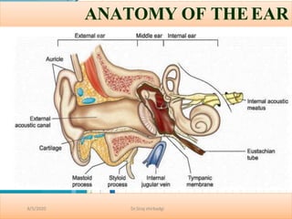
6. Pinna(auricle)
The visible portion that is commonly referredto
as "the ear" ,It consists of cartilege and skin
Helps localize sound sources and directs them
towards the external auditory meatus and on to
the tympanic membrane
• Lymphatics;drain into parotid group,upper deep
cervical and mastoid lymphnodes
• Veins;dain into into external jugular,common
facial vein
• Arteries;posterior auricular branch of external
carotid artery,ant.auricular branch of superficial
2
/
1
9
/
t2
e0
1
m9poral and a branch of occD
ir
pN
id
ta
ay
li
s
a
ab
ra
tC
eo
rr
n
ye
i
l
l
e
Dr.Siraj shirbadgi
8/5/2020
7. Auriculotemporal nerve (CN V3): It is a
branch of mandibular division of
trigeminal nerve and supplies
anterosuperior part of lateral surface of
pinna including tragusand crus of helix.
CN VII (facial nerve): It innervates the
skin of lateral concha and antihelix,
lobule and mastoid.
CN X (vagus nerve): Its auricular
branch (Arnold’s nerve) supplies to
concha and post auricular skin.
Greater auricular nerve (C2,3): This
nerve of cervical plexus supplies most of
the medial surface of auricle and
posterior part of lateral surface and the
postauricular region.
Lesser occipital nerve (C2): This nerve
of cervical plexus supplies upper part
of medial surface of auricle and
postauricular region.
Nerve supply
8/5/2020 Dr.Siraj shirbadgi
8. Is a curved tube about 2.5cm that lies in the temporal bone and leads fromthe
auricle to the eardrum (tympanic memb)
Near to the exterior of its opening there is a few hairs and specialized
sebaceous(oil)glands called ceruminous glands that secrete cerumen(earwax)
The combination of hairs and cerumen help prevent dust and foreign objects
from entering the ear and cleaning mechanism of ear
NERVE SUPLY
(i)Anterior wall and roof: auriculotemporal (V3) supplies anterosuperior wall
of external auditory canal
(ii)Posterior wall and floor: auricular branch of vagus nerve(CNX) suppliesto
inferoposterior external auditory canal.
Posterior wall of the auditory canal also receives sensory fibres of cranial NVII
(facial nerve) through auricular branch of vagus.
8/5/2020 Dr.Siraj shirbadgi
External Auditory Meatus
10. Eardrum(tympanic membrane)
It is a thin,semitransparent partiton between the external auditory canal and
middle ear.
It has two parts :(a) Pars Tensa:
It forms most of tympanic membrane..
Its periphery is thickened to form a fibro cartilaginous ring calledthe
annulus tympanicus which fits in the tympanicsulcus.
The central part is tented inwards at the level of the tip of malleus and is
called the umbo.
(b) Pars Flaccida (Shrapnel's Membrane)
• This is situated above the lateral process of malleus between the notch of
Rivinus and the anterior and posterior malleolar folds.
It has three layers:
(i) Outer epithelial layer, which is continuous with the skin liningthe
meatus. no hairs and glands
(ii)Middle fibrous layer, which encloses the handle of malleus and has three
types of fibres-the radial, circular and the parabolic.
(2
ii/
1
i9
)/
2
I0
n1
9
nermucosal layer, whichD
r
isN
d
ca
y
oi
s
na
b
ta
inC
o
ur
n
oe
ui
l
l
e
swith the mucosa of the middle
ear
Dr.Siraj shirbadgi
8/5/2020
11. Tympanic membrane
1. Malleus 2,6
2. Anterior mallear fold
3. Post mallear fold
4. Pars flaccida
5. projection of long process of
incus.
6. Pars tensa
7. Annular ligament.
NERVE SUPPLY
(i) Anterior half of lateral surface:
auriculotemporal(V 3)
(ii)Posterior half of lateral surface: auricular
branch of vagus nerve (CN X) (Arnold’s nerve)
(iii)Medial surface: Tympanic branch of CN
IX glossopharyngeal nerve (Jacobson's nerve).
8/5/2020 Dr.Siraj shirbadgi
14. MIDDLE EAR
It has two main parts:
Auditory ossicles
(transmit and mplify sound
from the tympanic membrane
to the oval window)
Auditory(eustachian)tube(e
qualizes pressure on both
sides of the tympanic
membrane
8/5/2020 Dr.Siraj shirbadgi
15. MIDDLE EAR
The middle ear together with the Eustachian tube,
aditus,antrum and mastoid air cells is called the
middle ear cleft.
its lined with mucous membrane and filled with
air.
Aditus and Antrum: Aditus is an opening through
which the attic communicates with the antrum.
The Mastoid and its Air Cell System:The
mastoid consists of bone cortex with a
"honeycomb“ of air cells underneath.
Depending on development of air cell, three types
of mastoid have been described: Well-pneumatised
or cellular; Diploetic; Sclerotic or acellular
It is divided into:
I. Mesotympanum(lying opposite to parsa
tensa).
II. Epitmpanum or attic( lying above parsa
tensa but medial to shrapnell’s membrane
and the bony lateral attic wall)
III. Hypotympanum( lying below the level of
2/19/201p9arca tensa). Dr NdayisabaCorneille
Dr.Siraj shirbadgi
8/5/2020
17. BOUNDARIES OF MIDDLE EAR
8/5/2020 Dr.Siraj shirbadgi
Roof (Tegmental wall): is formed by a thin plate of bone called tegmen tympani.
Floor (Jugular wall):is also thin plate of bone which separates tympanic cavityfrom
the jagular bulb
Anterior (Carotid wall):has a thin plate of bone which separates the cavity from
internal carotid artery. has following features: Eustachian tube, Canal of tensor
tympani muscle, Canal for chorda tympani nerve, Attachment of anterior malleolar
ligament
Posterior (Mastoid wall):lies close to the mastoid air cells. Has:
Pyramid, Aditus ad antrum, Facial nerve
Medial (Labyrinthine wall):is formed by labyrinth( including promontory, Round
window, oval window.)
Lateral wall: is formed largely by tympanic membrane.
18. Auditory ossicles
n
– Malleus
• Attaches to ear drum
• Articulates with incus
– Incus
• Articulates with stapes
– Stapes (stirrup)
• Footplate of stapes fits into oval
window
Ossicles Allows communication bt
the external and internal ear and
amplification
8/5/2020 Dr.Siraj shirbadgi
19. MIDDLE EAR MUSCLES
There are two middle ear muscles also
called intratympanic muscle : tensor
tympani and the stapedius.
1.Tensor tympani: It runs above the
eustachian tube. Originate from Bony
tunnel above the osseous part of
eustachian tube.inserted Just below the
neck of malleus and is supplied by a
branch of mandibular division of
trigeminal nerve (CN V3)
2.Stapedius: On contraction it reduces
the loud sounds and
prevents noise trauma to the inner ear.
Originate from Conical cavity and canal
within pyramid.it insert to the neck stapes
and supplied by a branch of CN VII
(nerve to stapedius of facial nerve).
8/5/2020 Dr.Siraj shirbadgi
20. Protection by Two Tiny Muscles
• Tensor Tympani
– Attaches to Malleus to
increase tension on ear
drum & prevent damage
to inner ear.
• Stapedius
– Smallest skeletal muscle
– Dampens large vibrations
of stapes to protect oval
window.
stapedius
21. Auditory(eustachian)tube
• It consists of both of both bone and hyaline cartilage
and connects the middle ear with the nasopharynx.itis
normally closed at its medial(pharyngeal)end;during
swallowing and yawning,it opens,then atmospheric
pressure from throat enters or leaves the middle ear
until int.pressure is =to external pressure.
• When the pressures are balanced,the eardrumvibrates
freely as soundwaves strike it
• It is also a route where pathogens can travel from throat
and nose to the middle ear
8/5/2020 Dr.Siraj shirbadgi
22. SUMMARY
8/5/2020 Dr.Siraj shirbadgi
Middle ear contains:
1. Air
2. Two muscles ( Tensor tympani and stapedius)
3. Two nerves( corda tympani and tympanic plexus on the promontory)
4. 3 bone (maleus, incus, stapes)
Mucosa of middle ear is stratified columnar with goblet and seromucinous glands
Its function:
Conduction
– Conduct sound from the outer ear to the inner ear
Protection
– Creates a barrier that protects the middle and inner areas from foreign objects
– Middle ear muscles may provide protection from loud sounds
Transducer
– Converts acoustic energy to mechanical energy
– Converts mechanical energy to hydraulic energy
Amplifier
– Transformer action of the middle ear
– only about 1/1000 of the acoustic energy in air would be transmitted to the inner-ear
fluids (about 30 dB hearing loss)
25. INNER EAR
The internal ear or the labyrinth is an important
organ of hearing and balance. It consists of a
bony and a membranous labyrinth.
The membranous labyrinth is filled with a clear
fluid called endolymph while the space between
membranous and bony labyrinths is filled with
perilymph.
Bony labyrinth: It consists of
i. the vestibule,
ii. the semicircular canals and
iii. the cochlea.
Membranous labyrinth: consists of
i. The membranous cochlear duct
ii. The membranous semicircular canals.
iii. The utricle and saccule (that lie within the
vestibule)
iv. Th2e/19e/2n0d19olymphaticduct andsac Dr NdayisabaCorneille
Dr.Siraj shirbadgi
8/5/2020
26. A. Cochlear part
it is fluid filled organ.
it is bony coiled up on axis like
a snail’s shell (central pyramid called
modiolus)
Its basal turn forms the promontory.
It has three compartment
a. Scala vestibule
b. scala tympani
Above 2 are filled with perilymph and
communicate with each other at apex of cochlea through Helicotrema
c.scala media(cochlear duct): its blind coiled tube and it appears triangular oncross-
section and its three wall are formed by:
i. basilar membrane which supports the organ of corti( inner, outer hair cellsand
tectorial mem)
ii. The Reissner’s membrane which separate it from scalavestibuli,
iii. The stria vasculars which contains vascular epithelium and is concernedwith
secretion of endolymph.
8/5/2020 Dr.Siraj shirbadgi
28. B) Vestibular part:
- its bony cavity that lodges the utricle and saccule.
for equilibrium ( responsible for linear
- it contains the sensory organs responsible
acceleration)
C) Semicircular canals(SCC):
they are not complete circles, the have one ampulary and one non-ampulary ends.
Oriented as superior, lateral, posterior canals with 90 degree relation to each other.
Sensory organs are crista ampularis(which is responsible for angular acceleration).
the vestibular nerve:
Superior and inferior vestibular nerve arise from the sensory organs of SCC and utricle and
saccule to enter the internal acoustic canal.
8/5/2020 Dr.Siraj shirbadgi
29. Membranous labyrinth
8/5/2020 Dr.Siraj shirbadgi
Cochlear Duct (Membranous Cochlea or Scala Media) This blind coiledtube,
which appears triangular on cross section, is connected to the saccule through
ductus reunions
Saccule: The saccule lies anterior to the utricle opposite the stapes footplate in
the bony vestibule. Its sensory epithelium, macula responds to linear
acceleration and deceleration. The saccule is connected to the cochleathrough
the thin reunion duct
Semicircular Ducts: The three semicircular ducts, which open in theutricle,
correspond exactly to the three bony canals
Endolymphatic Duct and Sac: The ducts from utricle and saccule unite andform
utriculo saccular duct, which continues as endolymphatic duct that passes
through thevestibular aqueduct
Endolymphatic sac is thought to regulate pressure of membranouslabyrinth.
30. INNER EAR FLUIDS
8/5/2020 Dr.Siraj shirbadgi
Perilymph fills the space between bony and membranous labyrinth while endolymph
fills the entire membranous labyrinth
Perilymph
It resembles extracellular fluid and is rich in sodium ions
Originate from Filtrate of blood serum from the capillaries of spiral ligament and CSF
reaching labyrinth via aqueduct of cochlea.
Endolymph
It resembles intracellular fluid and is rich in potassium ions Protein and glucose
contents are less than in perilymph.
„originate from Stria vascularis and Dark cells of utricle and ampullated ends of
semicircular ducts.
31. This sensory organ of the hearing, is situated on the basilar membrane. It is spread like a ribbon
along the entire length of basilar membrane. It consists of: 1. Tunnel of Corti: This tunnel, which
is situated between the inner and outer rods, contains a fluid called cortilymph. The functions of
the rods and cortilymph are yet not clear.
2. Hair Cells: hese important receptor cells of hearing transduce sound energy into electrical
energy. There are two types of hair cells—inner and outer.
ORGAN OF CORTI
8/5/2020 Dr.Siraj shirbadgi
32. Organ of Corti
• The end organ of
hearing
– Contains stereocilia
& receptor hair cells
– Tectorial and Basilar
Membranes
– Cochlear fluids
– Fluid movement
causes deflection of
nerve endings
– Nerve impulses
(electrical energy)
are generated and
sent to the brain
34. Summary of How We Hear
Acoustic energy, in the form of
sound waves, is channeled into the
ear canal by the pinna. Sound
waves hit the tympanic membrane
and cause it to vibrate, like a
drum, changing it into mechanical
energy. The malleus, which is
attached to the tympanic membrane,
starts the ossicles into motion. The
stapes moves in and out of the oval
window of the cochlea creating a
fluid motion, or hydraulic energy.
The fluid movement causes
membranes in the Organ of Corti to
shear against the hair cells. This
creates an electrical signal which is
sent up the Auditory Nerve
(cochlear nerve) to the brain.The
brain interprets it as sound!
Dr.Siraj shirbadgi
8/5/2020
40. COCHLEA
Derived from greek word (cochlos for snail)
5mm from base to apex & 9mm around its base
2.5 to 2.75 turns around a central pyramid of bone = MODIOLOUS.
The base of MODIOLUS is directed towards IAM & transmits nerves to
cochlea.
PROMONTORY =Basal turn of cochlea produces a bulge in medial
wall of middle ear
Dr.Siraj shirbadgi
8/5/2020
41. Thin plate of bone winding spirally around MODIOLUS like a
thread of screw k/a BONY SPIRALLAMINA.
ROSENTHAL’S CANAL: spiral ganglion are situated in this
canal which runs along the osseous spiral lamina
Dr.Siraj shirbadgi
8/5/2020
43. Bony cochlea is divivded into :
1. SCALA VESTIBULI : closed by stapes footplate and contains perilymph
2. SCALA TYMPANI : closed by secondary TM & containsperilymph.
Connected to sub archanoid space through Aqueduct ofcochlea.
3. SCALA MEDIA : also known as COCHLEAR DUCT & containsendolymph.
Dr.Siraj shirbadgi
8/5/2020
45. ENDOLYMPHATIC DUCT & SAC
Formed by the union of two ducts one forms the utricle
& another from the saccule.
Passes through the vestibular aqueduct
Terminal part is dilated to form Endolymphatic sac.
Dr.Siraj shirbadgi
8/5/2020