Best Rate (Hyderabad) Call Girls Jahanuma ⟟ 8250192130 ⟟ High Class Call Girl...
Anatomy Of Ear.pdf
1. 1
Head and Neck - Anatomy DMK @ 19/20
Introduction -----------
▪ The ear is an organ of hearing and maintaining the equilibrium of the body.
▪ It consists of Three parts:
- The External Ear
- Middle Ear / tympanic Membrane
- Internal Ear / inner Ear
External and middle ear is separated by the tympanic membrane
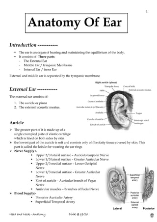
External Ear -----------
The external ear consists of:
1. The auricle or pinna
2. The external acoustic meatus.
Auricle
➢ The greater part of it is made up of a
single crumpled plate of elastic cartilage
which is lined on both sides by skin
➢ the lowest part of the auricle is soft and consists only of fibrofatty tissue covered by skin: This
part is called the lobule for wearing the ear rings
➢ Nerve Supply :-
▪ Upper 2/3 lateral surface – Auriculotemporal Nerve
▪ Lower 1/3 lateral surface – Greater Auricular Nerve
▪ Upper 2/3 medial surface – Lesser Occipital
Nerve
▪ Lower 1/3 medial surface – Greater Auricular
Nerve
▪ Root of auricle – Auricular branch of Vegas
Nerve
▪ Auricular muscles – Branches of Facial Nerve
➢ Blood Supply:-
▪ Posterior Auricular Artery
▪ Superficial Temporal Artery
Anatomy Of Ear
2. 2
Head and Neck - Anatomy DMK @ 19/20
Clinical Relevance of Auricle : Auricular Haematoma
An auricular haematoma refers to a collection of blood between the cartilage of the ear and the
overlying perichondrium. It is usually occurs as a result of trauma, commonly seen in contact
sports (e.g. rugby).
The accumulation of blood can
disrupt the blood supply to the
cartilage, and requires prompt
drainage. Untreated cases can result
in avascular necrosis of the cartilage,
resulting in a ‘cauliflower ear’
deformity.
External Acoustic Meatus
➢ The external acoustic meatus is a
sigmoid shaped tube that
extends from the deep part of
the concha to the tympanic
membrane.
➢ The walls of the external 1/3 are
formed by cartilage, whereas the inner 2/3 are formed by the temporal bone.
➢ The external acoustic meatus does not have a straight path, and instead travels in an S-
shaped curve as follows:
- Initially it travels in a superoanterior direction.
- In then turns slightly to move superoposteriorly.
- It ends by running in an inferoanterior direction.
➢ Ceruminous glands are specialized sudoriferous glands (sweat glands) located subcutaneous
in the external auditory canal, in the outer 1/3.
➢ Ceruminous glands are simple, coiled, tubular glands classed as apocrine glands Which
drain into larger ducts, which then drain into the guard hairs that reside in the the ext aud.
canal.
➢ Here they produce cerumen, or earwax, by mixing their secretion with sebum and dead
epidermal cells.
➢ Cerumen keeps the eardrum pliable, lubricates and cleans the ext aud. canal, waterproofs the
canal, kills bacteria, and serves as a barrier to trap foreign particles (dust, fungal spores, etc.)
by coating the guard hairs of the ear, making them sticky.
➢ Blood Supply :-
The outer part of the canal - superficial temporal and posterior auricular arteries,
The Inner Part - the deep auricular branch of the maxillary artery.
➢ Nerve Supply
The skin lining the anterior half of the meatus is supplied by the auriculotemporal
nerve, and that lining the posterior half, by the auricular branch of the vagus.
3. 3
Head and Neck - Anatomy DMK @ 19/20
Tympanic membrane
•Tympanic membrane has two parts
- pars tensa (bigger and tense lower part)
- pars flaccida (small and flaccid upper part)
• Sound waves cause vibration
• Postero-inferior part is safest for incisions
• Perforation may lead to deafness
Blood Supply :-
The outer surface is supplied by the deep
auricular branch of the maxillary artery.
The inner surface is supplied by the anterior tympanic branch of the maxillary artery
and by the posterior tympanic branch of the stylomastoid branch of the posterior auricular
artery
Venous Drainage :-
Veins from the outer surface drain into the external jugular vein. Those from the inner
surface drain into the transverse sinus and into the venous plexus around the auditory tube.
Middle Ear -----------
Middle ear lies inside the petrous part of
the temporal bone
Has two parts:
• tympanic cavity (proper)
• epitympanic recess (extends
superiorly from the upper border
of tympanic membrane)
Postero-superiorly connects with the
mastoid cells through the mastoid
antrum
Contents of the middle ear are:
• Auditory ossicles(malleus, incus, stapes)
• Stapedius and tensor tympani muscles (related with transmission of sound)
• Chorda tympani (branch of facial nerve) -presynaptic parasympathetic fibers of the facial
nerve to the sublingual and submandibular, and also taste sensation from the anterior part
of the tongue.
• Tympanic nerve plexus
4. 4
Head and Neck - Anatomy DMK @ 19/20
Tympanic Plexus
A plexus that is located on the promontory of the labyrinthine wall of the tympanic cavity, is
formed by the tympanic nerve, by an anastomotic branch of the facial nerve, and by sympathetic
branches coming from the internal carotid plexus, supplies the mucosa of the middle ear, the
mastoid cells, and the auditory tube, and gives off the lesser petrosal nerve to the otic ganglion.
Walls of the tympanic cavity
Lateral wall (membranous)
➢ Tympanic membrane (inf)
➢ Bony wall of the epitympanic
recess (sup)
Medial wall (labyrinthine)
➢ Promontorium (basal turn of
cochlea)
➢ Oval window (base of
stapes)
➢ Round window(closed by
secondary tympanic
membrane)
Roof (tegmental)
➢ Tegmen tympani (thin plate of temporal
bone separating the tympanic cavity from
the middle cranial fossa)
Floor (jugular)
➢ Layer of bone separating from the superior
bulb of internal jugular vein all the veins
from the cranial cavities forms the internal
jugular vein at the bulb.
Anterior wall (carotid)
➢ Tensor tympani muscle (sup) (innervated
by SVE)
➢ Auditory tube (mid)
➢ Plate of bone separating from carotid canal (inf)
Posterior wall (mastoid)
➢ Aditus (entrance) to mastoid antrum (connects to mastoid air cells)
➢ Pyramidal eminence (origin for stapedius muscle)
➢ Stapedius muscle
5. 5
Head and Neck - Anatomy DMK @ 19/20
Auditory tube (pharyngotympanic tube)
▪ Connects the tympanic cavity with nasopharynx
▪ About 3.5-4 cm long
▪ Equalize the pressure inside the tympanic cavity with the outside pressure.
▪ Contraction of tensor tympani muscle and some pharyngeal muscles opens the tube which
is normally closed.
▪ These muscles contract especially during swallowing and yawning–opens the tube!!
Auditory ossicles (malleus, incus, stapes)
These bones conduct the movements of the tympanic
membrane to the inner ear.
✓ Malleus attaches to the tympanic membrane
✓ Base of the stapes fits into the oval window
✓ Incus lies between these two bones.
Muscles controlling the movements of the auditory ossicles
- prevents excessive movements of the bones in middle ear…..if they don’t function we
get hyperacusis.
Tensor tympani muscle
• Inserts to the handle of malleus
• Tenses the tympanic membrane thus reducing the oscillations of the tympanic membrane.
• Prevents damage to the inner ear due to the excessively loud sounds
• Innervated by medial pterygoid nerve (br of V3)
6. 6
Head and Neck - Anatomy DMK @ 19/20
Stapedius muscle
• Extends between the pyramidal eminence and stapes
• Reduces the oscillatory range of the base of the stapes
• Innervated by the nerve to stapedius muscle (branch of facial nerve)
• Lesions of the facial nerve leads to hyperacusis
Facial nerve (CN VII)
• Leaves cranial cavity through the internal acoustic
meatus
• Traverses the petrous part of the temporal bone
within the facial canal
• In the facial canal it is related to the medial and
posterior walls of the tympanic cavity
• Exits the skull through the stylomastoid foramen
•Within the facial canal, the facial nerve gives of three
branches
▪ Greater petrosal nerve –
presynaptic fibers to the
peterygopalatine ganglion.
▪ Nerve to the stapedius –
innervates the stapedius muscle,
damage causes hyperacusis.
▪ Chorda tympani
•Leaves the skull through the
stylomastoid foramen and gives rise to
the
•Posterior auricular nerve(Sensory
branch)
•Then it enters into the tissue of the
parotid gland and here it gives of its
five terminal (muscular) branches:
▪ Temporal
▪ Zygomatic
▪ Buccal
▪ Marginal mandibular
▪ Cervical
7. 7
Head and Neck - Anatomy DMK @ 19/20
• SVE fibers
- All of the muscles of facial expression and stapedius muscle(skeletal)are innervated by
the facial nerve.
• SVA fibers
- Receive taste sensation from the anterior 2/3 of the tongue.
•GVE fibers
- Supply parasympathetic motor fibers to the submandibular and sublingual glands, as
well as the small salivary glands around the mouth
- Supply parasympathetic motor fibers to the lacrimal gland.
•GSA fibers
- Receive sensation around the external acoustic meatus (post auricular nerve .
Inner Ear -----------
• Contains the vestibulocochlear organ
• Vestibulocochlear organ is composed of three parts
• Lies inside the petrous part of the temporal bone
i. Cochlea (receptor for hearing)
ii. Vestibule (receptor for equilibrium)
iii. Labyrinth – semicircular canal (receptor for equilibrium)
Internal Ear
Bony labyrinth
Membranous labyrinth (inside the bony labyrinth)
Between the bony labyrinth and the
membranous labyrinththere is a fluid
called the perilymph
Inside the membranous labyrinththere is a
fluid called the endolymph
8. 8
Head and Neck - Anatomy DMK @ 19/20
Bony labyrinth
• cochlea (ant)
• vestibule (mid)
• semicircular canals (post)
❖ Inner surface of the bony labyrinth is lined with a thin fibrous membrane
❖ Inner surface of this fibrous membrane is lined by a thin layer of epithelium which secretes
the perilymph
Cochlea
✓ Begins at the vestibule and makes 2.5 turns around a bony core called the modiolus(middle
part of the turns).
✓ Upper part is called the cupula
✓ Osseous spiral lamina partially divides the cochlea into two parts
✓ scala vestibuli(upper) –connected directly to the oval window, base of stapes.
✓ scala tympani(lower)
✓ Both scala are filled with perilymph
✓ Just below the cupula at the region called helicotrema both scala are continuous, thus the
fluid reaches the top and returns back to the scala tympani
✓ The two scalae are connected with each other through the helicotremaat the apex
✓ Between the two scala, lies the cochlear ductof the membranous labyrinth –contains
endolymph.
Vestibule
• Lies between the cochlea and the semicircular canals
• Contains the utricle and saccule parts of the membranous
labyrinth.
• Semi cirular canals - utricle.
• Its medial wall bears:
- oval window (base of the stapes
- round window (closed by secondary tympanic membrane)
- absorbs the final movement of the perilymph when it returns back by tympani scala , it is
stopped by this membrane.
Semicircular canals
There are three semicircular canals
➢ anterior (superior), posterior, lateral (horizontal)
➢ Each semicircular canal extends from the vestibule and ends with a swelling called ampulla
➢ Contains the semicircular ducts of the membranous labyrinth
9. 9
Head and Neck - Anatomy DMK @ 19/20
Membranous Labyrinth
✓ Lies inside the bony labyrinth
✓ Contains endolymph
✓ Composed of ducts and sacs that communicate with each other
✓ Membranous labyrinth has two parts
➢ Cochlear duct
➢ Vestibular labyrinth
❖ Utricle and saccule
❖ Semicircular ducts
Cochlear duct
✓ Divides the perilymph filled spiral canal into two channels
- Scala vestibuli
- Scala tympani
✓ Contains endolymph
Pathway
1. Base of the stapes starts waves of hydaulic pressure(base of the stapes fits the oval window)
2. Wave is transmitted through the scala vestibuli
3. Passing through helicotrema waves reach the scala tympani
4. The pressure is finally absorbed by the secondary tympanic membrane of the round window.
Cochlear duct lies between two membranes:
• Vestibular membrane ( forms the roof)
• Basilar membrane ( forms the floor)
Structure of the organ of Corti and the formation of the cochlear nerve
Spiral organ of Corti (receptor organ) is situated on the basillar membrane and it is overlaid by
the tectorial membrane
• There are hair cellsin the organ of Corti
• Tips of thesehair cells are embedded in the tectorial membraneand are stimulated by the
movements of the membrane
• The sensory cells for hearing lie in the spiral ganglion(situated in the middle of modiolus)–a
sensory ganglion.
• The peripheral processes of these neurons terminate at the base of the hair cells in the organ of
corti
•The central processes (8th CN) of these neurons form the cochlear nerve
•8 th CN has 2 parts: cochlear (hearing) & vestibular (equilibrium)
10. 10
Head and Neck - Anatomy DMK @ 19/20
•Internal acoustic meatus: 8 CN & 7 CN pass through it
Mechanism Of Hearing
• Movements of the tectorial membrane stimulate the hair cells, which is transmitted to
theneurons of the spiral ganglion
• Central axons of the neurons of spiral ganglion form the cochlear nerve which is a part of the
CN8
Semicircular ducts, utricle and saccule
• Semicircular ducts open into the utricle through five openings (ant. and post. canals shares a
single canal at one of the limbs)
• Contains endolymph
• Utricle and saccule communicates through the utriculosaccular canal(duct)
• The saccule is continuous with the cochlear duct through ductus reuniens
• Utricle and saccule have regions called macula where the hair cells are located (receptors for
equilibrium)
• Ampulla of the semicircular canals also bear hair
cells
•Peripheral processes of the neurons in the
vestibular ganglion (in the internal acoustic
meatus) has nerve ending that terminate at the base
of the hair cells
•Central processes of the neurons of the vestibular
ganglion constitute the vestibular nerve.
The semicircular canals detect angular
acceleration
11. 11
Head and Neck - Anatomy DMK @ 19/20
The utricle and saccule detect linear acceleration
✓ Cochlear and vestibular nerves constitute the vestibulocochlear nerve (CN VIII), which passes
through the internal acoustic meatus (together with the facial nerve) enters the brainstem.
✓ As the axons enter the brainstem they make synapses with the second order neurons of the
related nuclei.
Clinical Notes
❖ Otitis externa
❖ Otoscopic examination
❖ Otitis media
❖ Perforation of the tympanic membrane
❖ Blockage of the pharygo tympanic tube
❖ Paralysis of the stapedius muscle (lesion to
facial nerve)
❖ Hearing loss (conductive, sensorineural)
❖ Meniere syndrome
❖ High tone deafness
Otitis Externa
• Inflammation of external acoustic meatus.
• Infection develops:
- in swimmers who do not dry their meatus or
because of a bacterial infection of the skin lining
the meatus.
• Symptoms:
- itching, pain in external ear, pulling the auricle or applying pressure on the tragus increases
the pain.
Otoscopic Examination
•Examination of external acoustic meatus and tympanic membrane begins by straightening the
meatus.
•Insertion of otoscope, by pulling the ear.
•Tenderness indicates infection
•Handle of malleus is visible in the center.
•In a healthy ear a bright cone of reflex is visible.
12. 12
Head and Neck - Anatomy DMK @ 19/20
Paralysis of Stapedius Muscle
•Can result from lesion of facial nerve.
•Is associated with excessive acuteness of hearing called hyperacusis.
•This condition results from the uninhibited movements of the stapes.
Meniere’s disease
•Related to the blockage of the cochlear aqueduct and is characterized by recurrent attacks of
tinnitus, hearing loss and vertigo.
•Symptoms are accompanied by a sense of pressure in the ear, distortion of sounds, and
sensitivity to noises.
•Increase in endolymph volume will cause the ballooning of the cochlear duct, utricle, and
saccule.
High Tone Deafness
•Persistent exposure to excessively loud sounds causes degenerative changes in the spiral organ.
•Commonly occurs in workers who are exposed to loud noises and do not wear earmuffs.
Dizziness & Hearing Loss
•Injuries of the peripheral auditory system cause the 3 major symptoms:
- hearing loss
- vertigo (dizziness) –when injury is localized in the semicircular canals.
- tinnitus (buzzing or ringing) –when injury is localized in chochlear duct
2 kinds of hearing loss:
1. Conductive hearing loss–when the round or oval windows are affected.
2. Sensorineural hearing loss: defects in cochlea, cochlear nerve, brainstem or cortical
connections.
Blockage of Pharyngotympanic tube
•Infection passes from the nasopharynx to the tympanic cavity.
•This tube is easily blocked by swelling of its mucous membrane, even because of cold.
•Lower pressure is build in the tympanic cavity.
•Finally hearing is affected.
13. 13
Head and Neck - Anatomy DMK @ 19/20
Perforation of Tympanic Membrane
•May result from otitis media and one of the several causes of the middle ear deafness.
•May also result from foreign bodies in the EAM, trauma, or excessive pressure (scuba diving).
•Minor ruptures – heal itself
•Major ruptures – surgery
•Incision are made postero inferiorly because of less amount of blood vessels in this area.
Otitis Media
•An ear-ache and bulging red tympanic membrane may indicate pus or fluid in the middle ear.
•Infection of the middle ear is often secondary to upper respiratory inflammation.
•Swelling of mucous membrane can cause the blockage of the pharyngotympanic tube.
•If untreated it can produce impaired hearing as the result of the inability of the ossiclesto move
in response to sound.