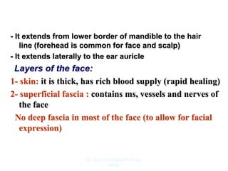
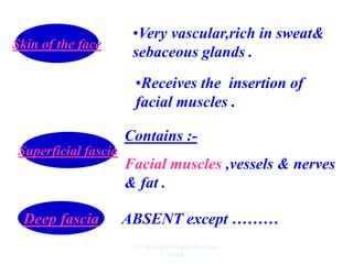
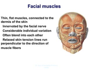
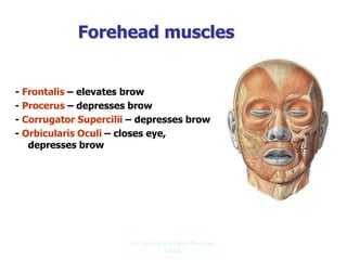
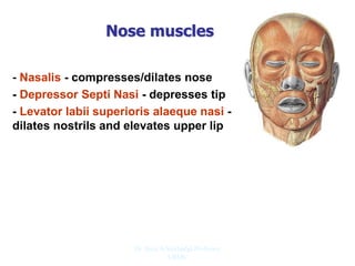
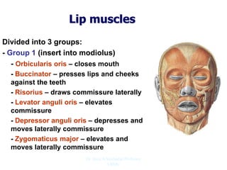
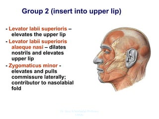
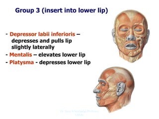
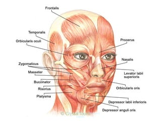
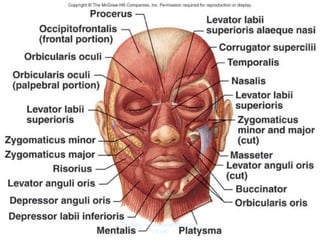
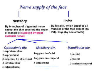
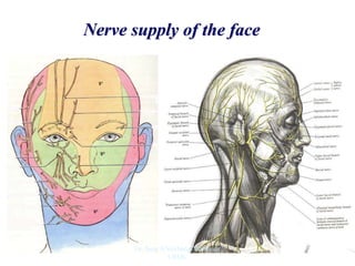
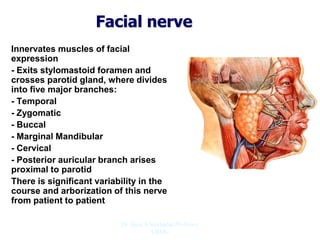
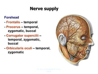
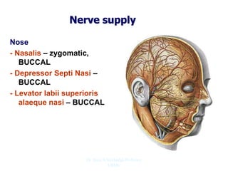
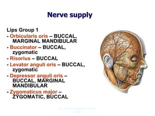
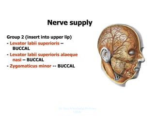
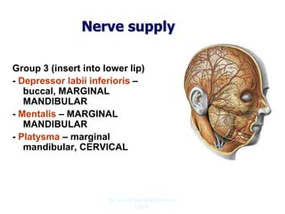
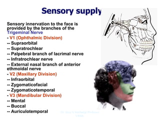
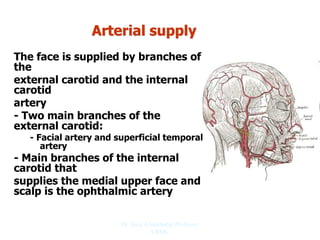
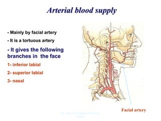
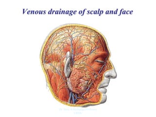
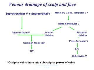
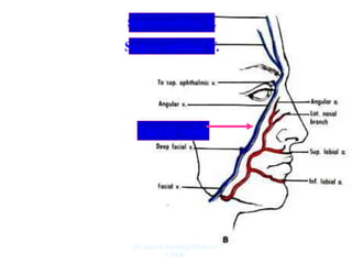
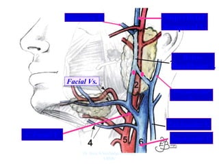
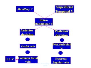
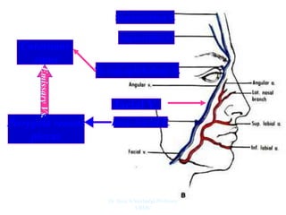
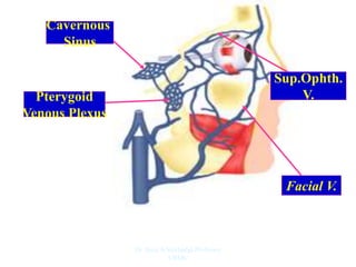
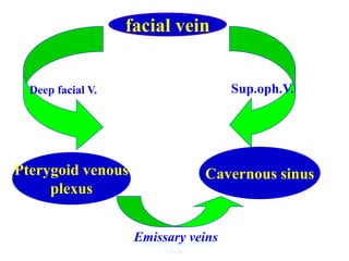
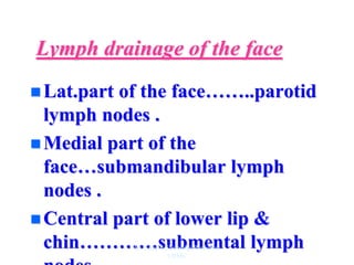
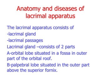
Dr. Siraj A Shirbadgi Professor
VBMC
- It extends from lower border of mandible to the hair
line (forehead is common for face and scalp)
- It extends laterally to the ear auricle
Layers of the face:
1- skin: it is thick, has rich blood supply (rapid healing)
2- superficial fascia : contains ms, vessels and nerves of
the face
No deep fascia in most of the face (to allow for facial
expression)
Dr. Siraj A Shirbadgi Professor
VBMC
Skin of the face
•Very vascular,rich in sweat&
sebaceous glands .
•Receives the insertion of
facial muscles .
Superficial fascia
Contains :-
Facial muscles ,vessels & nerves
& fat .
Deep fascia ABSENT except ………
Dr. Siraj A Shirbadgi Professor
VBMC
Facial muscles
Thin, flat muscles, connected to the
dermis of the skin
Innervated by the facial nerve
Considerable individual variation
Often blend into each other
Relaxed skin tension lines run
perpendicular to the direction of
muscle fibers
Dr. Siraj A Shirbadgi Professor
VBMC
Forehead muscles
- Frontalis – elevates brow
- Procerus – depresses brow
- Corrugator Supercilii – depresses brow
- Orbicularis Oculi – closes eye,
depresses brow
Dr. Siraj A Shirbadgi Professor
VBMC
Nose muscles
- Nasalis - compresses/dilates nose
- Depressor Septi Nasi - depresses tip
- Levator labii superioris alaeque nasi -
dilates nostrils and elevates upper lip
Dr. Siraj A Shirbadgi Professor
VBMC
Lip muscles
Divided into 3 groups:
- Group 1 (insert into modiolus)
- Orbicularis oris – closes mouth
- Buccinator – presses lips and cheeks
against the teeth
- Risorius – draws commissure laterally
- Levator anguli oris – elevates
commissure
- Depressor anguli oris – depresses and
moves laterally commissure
- Zygomaticus major – elevates and
moves laterally commissure
Dr. Siraj A Shirbadgi Professor
VBMC
Group 2 (insert into upper lip)
- Levator labii superioris –
elevates the upper lip
- Levator labii superioris
alaeque nasi – dilates
nostrils and elevates
upper lip
- Zygomaticus minor -
elevates and pulls
commissure laterally;
contributor to nasolabial
fold
Dr. Siraj A Shirbadgi Professor
VBMC
Group 3 (insert into lower lip)
- Depressor labii inferioris –
depresses and pulls lip
slightly laterally
- Mentalis – elevates lower lip
- Platysma - depresses lower lip
Dr. Siraj A Shirbadgi Professor
VBMC
Dr. Siraj A Shirbadgi Professor
VBMC
Dr. Siraj A Shirbadgi Professor
VBMC
Nerve supply of the face
By facial N. which supplies all
muscles of the face except lev.
Palp. Sup. (by oculomotor)
sensory motor
By branches of trigeminal nerve
except the skin covering the angle
of mandible (supplied by great
auricular nerve)
Ophthalmic div. Maxillary div. Mandibular div.
1-supratrrochlear
2-supraorbital
3-palpebral br. of lacrimal
4-infratrochlear
5-external nasal
1-zygomaticofacial
2-zygomaticotemporal
3-infraorbital
1-mental
2-buccal
3-auriculotemporal
Dr. Siraj A Shirbadgi Professor
VBMC
Nerve supply of the face
Dr. Siraj A Shirbadgi Professor
VBMC
Facial nerve
Innervates muscles of facial
expression
- Exits stylomastoid foramen and
crosses parotid gland, where divides
into five major branches:
- Temporal
- Zygomatic
- Buccal
- Marginal Mandibular
- Cervical
- Posterior auricular branch arises
proximal to parotid
There is significant variability in the
course and arborization of this nerve
from patient to patient
Dr. Siraj A Shirbadgi Professor
VBMC
Nerve supply
Forehead
- Frontalis – temporal
- Procerus – temporal,
zygomatic, buccal
- Corrugator supercilii –
temporal, zygomatic,
buccal
- Orbicularis oculi – temporal,
zygomatic
Dr. Siraj A Shirbadgi Professor
VBMC
Nerve supply
Nose
- Nasalis – zygomatic,
BUCCAL
- Depressor Septi Nasi –
BUCCAL
- Levator labii superioris
alaeque nasi – BUCCAL
Dr. Siraj A Shirbadgi Professor
VBMC
Nerve supply
Lips Group 1
- Orbicularis oris – BUCCAL,
MARGINAL MANDIBULAR
- Buccinator – BUCCAL,
zygomatic
- Risorius – BUCCAL
- Levator anguli oris – BUCCAL,
zygomatic
- Depressor anguli oris –
BUCCAL, MARGINAL
MANDIBULAR
- Zygomaticus major –
ZYGOMATIC, BUCCAL
Dr. Siraj A Shirbadgi Professor
VBMC
Nerve supply
Group 2 (insert into upper lip)
- Levator labii superioris –
BUCCAL
- Levator labii superioris alaeque
nasi – BUCCAL
- Zygomaticus minor -- BUCCAL
Dr. Siraj A Shirbadgi Professor
VBMC
Nerve supply
Group 3 (insert into lower lip)
- Depressor labii inferioris –
buccal, MARGINAL
MANDIBULAR
- Mentalis – MARGINAL
MANDIBULAR
- Platysma – marginal
mandibular, CERVICAL
Dr. Siraj A Shirbadgi Professor
VBMC
Sensory supply
Sensory innervation to the face is
provided by the branches of the
Trigeminal Nerve
- V1 (Ophthalmic Division)
-- Supraorbital
-- Supratrochlear
-- Palpebral branch of lacrimal nerve
-- Infratrochlear nerve
-- External nasal branch of anterior
ethmoidal nerve
- V2 (Maxillary Division)
-- Infraorbital
-- Zygomaticofacial
-- Zygomaticotemporal
- V3 (Mandibular Division)
-- Mental
-- Buccal
-- Auriculotemporal Dr. Siraj A Shirbadgi Professor
VBMC
Sensory supply
Sensory branches of the trigeminal nerve
course superficially are prone to damage.
- However, most of the resulting sensory
dysfunction is not debilitating because of
collateral sprouting of nearby sensory nerves.
- Sensation to the posterior scalp and superior
neck is provided by branches of C2 and C3
- Knowledge of sensory nerve anatomy is useful
for regional anesthesia/nerve blocks.
Dr. Siraj A Shirbadgi Professor
VBMC
THANKS
FOR
YOUR
ATTENTION
Dr. Siraj A Shirbadgi Professor
VBMC
Dr. Siraj A Shirbadgi Professor
VBMC
Dr. Siraj A Shirbadgi Professor
VBMC
Arterial supply
The face is supplied by branches of
the
external carotid and the internal
carotid
artery
- Two main branches of the
external carotid:
- Facial artery and superficial temporal
artery
- Main branches of the internal
carotid that
supplies the medial upper face and
scalp is the ophthalmic artery
Dr. Siraj A Shirbadgi Professor
VBMC
Arterial blood supply
- Mainly by facial artery
- It is a tortuous artery
- It gives the following
branches in the face
1- inferior labial
2- superior labial
3- nasal
Facial artery
Dr. Siraj A Shirbadgi Professor
VBMC
Venous drainage of scalp and face
Dr. Siraj A Shirbadgi Professor
VBMC
Venous drainage of scalp and face
Supratrochlear V + Supraorbital V
Anterior facial V
Maxillary V Sup. Temporal V +
Retromandibular V
Anterior
division
Posterior
division
Common facial vein
+
Post. Auricular V
EJV
IJV
Subclavian V
* Occipital veins drain into suboccipital plexus of veins
Dr. Siraj A Shirbadgi Professor
VBMC
Supratrochlear V.
Supra-orbital V.
facial vein
Dr. Siraj A Shirbadgi Professor
VBMC
Retro-
Mandibular V.
Ext.jugular V.
Post.auricular
V.
C.Facial V.
Superficial
Temporal V.
Maxillary V.
Int.jugular V.
Facial Vs.
Dr. Siraj A Shirbadgi Professor
VBMC
Superficial
Temporal V.
Maxillary V.
Retro-
Mandibular V.
Post.auricular
V.
Facial vein
Posterior
devision
Anterior
devision
+
+
Common facial
vein
=
External
Jugular vein
=
I.J.V.
Dr. Siraj A Shirbadgi Professor
VBMC
Supratrochlear V.
Supraorbital V.
facial V.
To sup.ophthalmic V.
Cavernous
sinus
Deep Facial V.
Pterygoid venous
plexus
Emissary
Vs.
Dr. Siraj A Shirbadgi Professor
VBMC
Cavernous
Sinus
Pterygoid
Venous Plexus
Facial V.
Sup.Ophth.
V.
Dr. Siraj A Shirbadgi Professor
VBMC
facial vein
Pterygoid venous
plexus
Cavernous sinus
Emissary veins
Deep facial V. Sup.oph.V.
Dr. Siraj A Shirbadgi Professor
VBMC
Dr. Siraj A Shirbadgi Professor
VBMC
Dr. Siraj A Shirbadgi Professor
VBMC
Dr. Siraj A Shirbadgi Professor
VBMC
Parotid L.N.
Dr. Siraj A Shirbadgi Professor
VBMC
Lymph drainage of the face
 Lat.part of the face……..parotid
lymph nodes .
 Medial part of the
face…submandibular lymph
nodes .
 Central part of lower lip &
chin…………submental lymph
Dr. Siraj A Shirbadgi Professor
VBMC
Anatomy and diseases of
lacrimal apparatus
The lacrimal apparatus consists of
-lacrimal gland
-lacrimal passages
Lacrimal gland –consists of 2 parts
A-orbital lobe situated in a fossa in outer
part of the orbital roof.
B-palpebral lobe situated in the outer part
above the superior fornix.
Anatomy and diseases of lacrimal apparatus
The lacrimal apparatus consists of
-lacrimal gland
-lacrimal passages
Lacrimal gland –consists of 2 parts
a)orbital lobe situated in a fossa in outer part
of the orbital roof.
b)palpebral lobe situated in the outer part
above the superior fornix.
The ducts of the gland open in to the external part of the
.superior fornix
In structure the lacrimal gland resemble the salivary
glands.
Dr. Siraj A Shirbadgi Professor
VBMC
Lacrimal passages –consist of
a) lacrimal puncta: one in each lid and situated
near the posterior border of the margin 6 mm
from The medial canthus.
b) Canaliculi : one in each eyelid-it commences
at the punctum and carries tears to the lacrimal
sac.
c) Lacrimal sac : lies in the lacrimal fossa
formed by the lacrimal bone and frontal process
of maxilla.It is covered by lacrimal foscia.
Anterior to the lacrimal foscia is the medial
palpebral ligament.The upper part of the sac is
known as the fundus.The lower end narrows as
it open into the nasolacrimal duct.
Dr. Siraj A Shirbadgi Professor
VBMC
d)Naso lacrimal duct: It is 3-4 in length and
opens into the inferior meatus of the nose. It
is directed downwards,slightly outwards and
backwards.
The tears which are secreted by the lacrimal
glands into the conjunctival sac are drained
by the lacrimal passage into the nose.
Dr. Siraj A Shirbadgi Professor
VBMC
THANKS
FOR
YOUR
ATTENTION
Dr. Siraj A Shirbadgi Professor
VBMC

face- face anatomy muscles VBMSk (1).ppt

  • 1.
    Dr. Siraj AShirbadgi Professor VBMC
  • 2.
    - It extendsfrom lower border of mandible to the hair line (forehead is common for face and scalp) - It extends laterally to the ear auricle Layers of the face: 1- skin: it is thick, has rich blood supply (rapid healing) 2- superficial fascia : contains ms, vessels and nerves of the face No deep fascia in most of the face (to allow for facial expression) Dr. Siraj A Shirbadgi Professor VBMC
  • 3.
    Skin of theface •Very vascular,rich in sweat& sebaceous glands . •Receives the insertion of facial muscles . Superficial fascia Contains :- Facial muscles ,vessels & nerves & fat . Deep fascia ABSENT except ……… Dr. Siraj A Shirbadgi Professor VBMC
  • 4.
    Facial muscles Thin, flatmuscles, connected to the dermis of the skin Innervated by the facial nerve Considerable individual variation Often blend into each other Relaxed skin tension lines run perpendicular to the direction of muscle fibers Dr. Siraj A Shirbadgi Professor VBMC
  • 5.
    Forehead muscles - Frontalis– elevates brow - Procerus – depresses brow - Corrugator Supercilii – depresses brow - Orbicularis Oculi – closes eye, depresses brow Dr. Siraj A Shirbadgi Professor VBMC
  • 6.
    Nose muscles - Nasalis- compresses/dilates nose - Depressor Septi Nasi - depresses tip - Levator labii superioris alaeque nasi - dilates nostrils and elevates upper lip Dr. Siraj A Shirbadgi Professor VBMC
  • 7.
    Lip muscles Divided into3 groups: - Group 1 (insert into modiolus) - Orbicularis oris – closes mouth - Buccinator – presses lips and cheeks against the teeth - Risorius – draws commissure laterally - Levator anguli oris – elevates commissure - Depressor anguli oris – depresses and moves laterally commissure - Zygomaticus major – elevates and moves laterally commissure Dr. Siraj A Shirbadgi Professor VBMC
  • 8.
    Group 2 (insertinto upper lip) - Levator labii superioris – elevates the upper lip - Levator labii superioris alaeque nasi – dilates nostrils and elevates upper lip - Zygomaticus minor - elevates and pulls commissure laterally; contributor to nasolabial fold Dr. Siraj A Shirbadgi Professor VBMC
  • 9.
    Group 3 (insertinto lower lip) - Depressor labii inferioris – depresses and pulls lip slightly laterally - Mentalis – elevates lower lip - Platysma - depresses lower lip Dr. Siraj A Shirbadgi Professor VBMC
  • 10.
    Dr. Siraj AShirbadgi Professor VBMC
  • 11.
    Dr. Siraj AShirbadgi Professor VBMC
  • 12.
    Nerve supply ofthe face By facial N. which supplies all muscles of the face except lev. Palp. Sup. (by oculomotor) sensory motor By branches of trigeminal nerve except the skin covering the angle of mandible (supplied by great auricular nerve) Ophthalmic div. Maxillary div. Mandibular div. 1-supratrrochlear 2-supraorbital 3-palpebral br. of lacrimal 4-infratrochlear 5-external nasal 1-zygomaticofacial 2-zygomaticotemporal 3-infraorbital 1-mental 2-buccal 3-auriculotemporal Dr. Siraj A Shirbadgi Professor VBMC
  • 13.
    Nerve supply ofthe face Dr. Siraj A Shirbadgi Professor VBMC
  • 14.
    Facial nerve Innervates musclesof facial expression - Exits stylomastoid foramen and crosses parotid gland, where divides into five major branches: - Temporal - Zygomatic - Buccal - Marginal Mandibular - Cervical - Posterior auricular branch arises proximal to parotid There is significant variability in the course and arborization of this nerve from patient to patient Dr. Siraj A Shirbadgi Professor VBMC
  • 15.
    Nerve supply Forehead - Frontalis– temporal - Procerus – temporal, zygomatic, buccal - Corrugator supercilii – temporal, zygomatic, buccal - Orbicularis oculi – temporal, zygomatic Dr. Siraj A Shirbadgi Professor VBMC
  • 16.
    Nerve supply Nose - Nasalis– zygomatic, BUCCAL - Depressor Septi Nasi – BUCCAL - Levator labii superioris alaeque nasi – BUCCAL Dr. Siraj A Shirbadgi Professor VBMC
  • 17.
    Nerve supply Lips Group1 - Orbicularis oris – BUCCAL, MARGINAL MANDIBULAR - Buccinator – BUCCAL, zygomatic - Risorius – BUCCAL - Levator anguli oris – BUCCAL, zygomatic - Depressor anguli oris – BUCCAL, MARGINAL MANDIBULAR - Zygomaticus major – ZYGOMATIC, BUCCAL Dr. Siraj A Shirbadgi Professor VBMC
  • 18.
    Nerve supply Group 2(insert into upper lip) - Levator labii superioris – BUCCAL - Levator labii superioris alaeque nasi – BUCCAL - Zygomaticus minor -- BUCCAL Dr. Siraj A Shirbadgi Professor VBMC
  • 19.
    Nerve supply Group 3(insert into lower lip) - Depressor labii inferioris – buccal, MARGINAL MANDIBULAR - Mentalis – MARGINAL MANDIBULAR - Platysma – marginal mandibular, CERVICAL Dr. Siraj A Shirbadgi Professor VBMC
  • 20.
    Sensory supply Sensory innervationto the face is provided by the branches of the Trigeminal Nerve - V1 (Ophthalmic Division) -- Supraorbital -- Supratrochlear -- Palpebral branch of lacrimal nerve -- Infratrochlear nerve -- External nasal branch of anterior ethmoidal nerve - V2 (Maxillary Division) -- Infraorbital -- Zygomaticofacial -- Zygomaticotemporal - V3 (Mandibular Division) -- Mental -- Buccal -- Auriculotemporal Dr. Siraj A Shirbadgi Professor VBMC
  • 21.
    Sensory supply Sensory branchesof the trigeminal nerve course superficially are prone to damage. - However, most of the resulting sensory dysfunction is not debilitating because of collateral sprouting of nearby sensory nerves. - Sensation to the posterior scalp and superior neck is provided by branches of C2 and C3 - Knowledge of sensory nerve anatomy is useful for regional anesthesia/nerve blocks. Dr. Siraj A Shirbadgi Professor VBMC
  • 22.
    THANKS FOR YOUR ATTENTION Dr. Siraj AShirbadgi Professor VBMC
  • 23.
    Dr. Siraj AShirbadgi Professor VBMC
  • 24.
    Dr. Siraj AShirbadgi Professor VBMC
  • 25.
    Arterial supply The faceis supplied by branches of the external carotid and the internal carotid artery - Two main branches of the external carotid: - Facial artery and superficial temporal artery - Main branches of the internal carotid that supplies the medial upper face and scalp is the ophthalmic artery Dr. Siraj A Shirbadgi Professor VBMC
  • 26.
    Arterial blood supply -Mainly by facial artery - It is a tortuous artery - It gives the following branches in the face 1- inferior labial 2- superior labial 3- nasal Facial artery Dr. Siraj A Shirbadgi Professor VBMC
  • 27.
    Venous drainage ofscalp and face Dr. Siraj A Shirbadgi Professor VBMC
  • 28.
    Venous drainage ofscalp and face Supratrochlear V + Supraorbital V Anterior facial V Maxillary V Sup. Temporal V + Retromandibular V Anterior division Posterior division Common facial vein + Post. Auricular V EJV IJV Subclavian V * Occipital veins drain into suboccipital plexus of veins Dr. Siraj A Shirbadgi Professor VBMC
  • 29.
    Supratrochlear V. Supra-orbital V. facialvein Dr. Siraj A Shirbadgi Professor VBMC
  • 30.
    Retro- Mandibular V. Ext.jugular V. Post.auricular V. C.FacialV. Superficial Temporal V. Maxillary V. Int.jugular V. Facial Vs. Dr. Siraj A Shirbadgi Professor VBMC
  • 31.
    Superficial Temporal V. Maxillary V. Retro- MandibularV. Post.auricular V. Facial vein Posterior devision Anterior devision + + Common facial vein = External Jugular vein = I.J.V. Dr. Siraj A Shirbadgi Professor VBMC
  • 32.
    Supratrochlear V. Supraorbital V. facialV. To sup.ophthalmic V. Cavernous sinus Deep Facial V. Pterygoid venous plexus Emissary Vs. Dr. Siraj A Shirbadgi Professor VBMC
  • 33.
  • 34.
    facial vein Pterygoid venous plexus Cavernoussinus Emissary veins Deep facial V. Sup.oph.V. Dr. Siraj A Shirbadgi Professor VBMC
  • 35.
    Dr. Siraj AShirbadgi Professor VBMC
  • 36.
    Dr. Siraj AShirbadgi Professor VBMC
  • 37.
    Dr. Siraj AShirbadgi Professor VBMC
  • 38.
    Parotid L.N. Dr. SirajA Shirbadgi Professor VBMC
  • 39.
    Lymph drainage ofthe face  Lat.part of the face……..parotid lymph nodes .  Medial part of the face…submandibular lymph nodes .  Central part of lower lip & chin…………submental lymph Dr. Siraj A Shirbadgi Professor VBMC
  • 40.
    Anatomy and diseasesof lacrimal apparatus The lacrimal apparatus consists of -lacrimal gland -lacrimal passages Lacrimal gland –consists of 2 parts A-orbital lobe situated in a fossa in outer part of the orbital roof. B-palpebral lobe situated in the outer part above the superior fornix.
  • 41.
    Anatomy and diseasesof lacrimal apparatus The lacrimal apparatus consists of -lacrimal gland -lacrimal passages Lacrimal gland –consists of 2 parts a)orbital lobe situated in a fossa in outer part of the orbital roof. b)palpebral lobe situated in the outer part above the superior fornix. The ducts of the gland open in to the external part of the .superior fornix In structure the lacrimal gland resemble the salivary glands.
  • 42.
    Dr. Siraj AShirbadgi Professor VBMC
  • 43.
    Lacrimal passages –consistof a) lacrimal puncta: one in each lid and situated near the posterior border of the margin 6 mm from The medial canthus. b) Canaliculi : one in each eyelid-it commences at the punctum and carries tears to the lacrimal sac. c) Lacrimal sac : lies in the lacrimal fossa formed by the lacrimal bone and frontal process of maxilla.It is covered by lacrimal foscia. Anterior to the lacrimal foscia is the medial palpebral ligament.The upper part of the sac is known as the fundus.The lower end narrows as it open into the nasolacrimal duct. Dr. Siraj A Shirbadgi Professor VBMC
  • 44.
    d)Naso lacrimal duct:It is 3-4 in length and opens into the inferior meatus of the nose. It is directed downwards,slightly outwards and backwards. The tears which are secreted by the lacrimal glands into the conjunctival sac are drained by the lacrimal passage into the nose. Dr. Siraj A Shirbadgi Professor VBMC
  • 45.
    THANKS FOR YOUR ATTENTION Dr. Siraj AShirbadgi Professor VBMC